1.1.1 - ATLS Sequence And Triage
ATLS Sequence and Triage
What you'll learn
- DOMAIN: Trauma; TASK: Initial Stabilization → Apply the ATLS sequence in the correct order when a trauma patient arrives.
- DOMAIN: Trauma; TASK: Best Next Step → Decide when immediate intervention must occur before imaging or full history.
- DOMAIN: Trauma; TASK: Diagnosis → Recognize the bedside findings that define instability and change triage priority.
- DOMAIN: Trauma; TASK: Imaging/Test Selection → Choose which tests belong after the primary survey versus after stabilization.
- DOMAIN: Trauma; TASK: Definitive Management → Distinguish patients who need operative or trauma-team escalation from those who can proceed to secondary survey.
- DOMAIN: Trauma → Primary survey priorities
- DOMAIN: Trauma → Triage of unstable versus stable trauma patients
- TASK: Initial Stabilization → ABC-based emergency sequencing
- TASK: Best Next Step → Intervention before diagnostic delay
- TASK: Imaging/Test Selection → Bedside trauma adjuncts after immediate threats are addressed
Why it matters
- ATLS questions are less about memorizing letters and more about sequencing: what cannot wait.
- NBME stems reward students who treat unstable trauma as a physiology problem first, not an imaging problem.
- The wrong answer is often a good test ordered at the wrong time.
- Triage errors in trauma usually come from missing one bedside clue that upgrades urgency: hypotension, altered mentation, respiratory distress, or peritonitis.
Exam takeaway: Follow the ATLS sequence by fixing immediate threats before moving to imaging or secondary survey.
flowchart TD
A[Trauma patient arrives] --> B[Primary survey starts]
B --> C[Airway with cervical spine protection]
C --> D[Breathing and ventilation]
D --> E[Circulation and hemorrhage control]
E --> F[Disability neurologic check]
F --> G[Exposure and temperature control]
G --> H{Stable after primary survey?}
H -->|No| I[Resuscitate and urgent intervention]
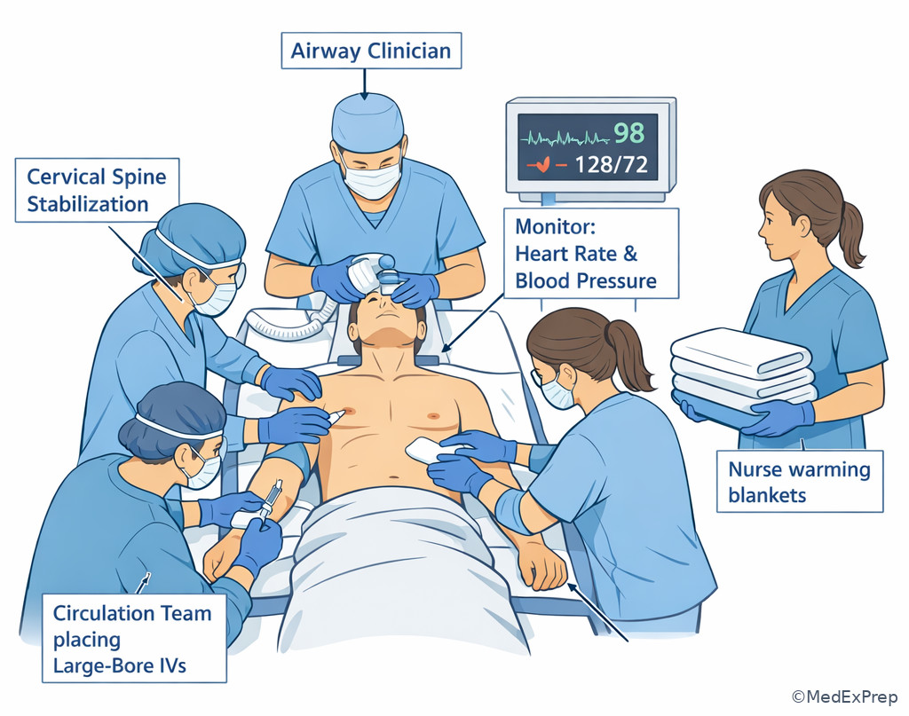
H -->|Yes| J[Adjuncts and secondary survey] Exam takeaway: Visualize parallel trauma tasks so you can identify which intervention occurs immediately at bedside.
Key Terms & Must-Know Facts
1.1.1.1 Key terms
- Primary survey — rapid search for immediately life-threatening conditions using ordered priorities.
- Triage — sorting patients by urgency and resource need.
- Airway with cervical spine protection — secure oxygenation/ventilation while assuming neck injury until cleared.
- Breathing — assess chest wall motion, breath sounds, oxygenation, and major thoracic threats.
- Circulation — evaluate perfusion and control hemorrhage.
- Disability — rapid neurologic screen, usually mental status and pupils.
- Exposure — fully inspect patient while preventing hypothermia.
- Adjuncts to primary survey — monitoring, portable chest/pelvis imaging, FAST, ABG/lactate, labs, Foley/NG when appropriate.
- Secondary survey — head-to-toe evaluation after immediate threats are addressed.
- Hemodynamic instability — physiology suggesting inadequate perfusion, often hypotension, tachycardia, cool skin, altered mentation.
- Definitive airway — cuffed tube in trachea, often after failure to maintain/protect airway.
- Mass casualty triage — prioritize salvageable patients when resources are limited.
- Golden mistake — delaying life-saving intervention for CT or detailed history.
1.1.1.2 Must-know facts
- Trauma shock is hemorrhagic until proven otherwise.
- Airway, breathing, and circulation are assessed in sequence, but resuscitation occurs continuously and in parallel.
- A patient can die from an airway or breathing problem before a CT scanner is ever reached.
- Absent breath sounds plus hypotension after trauma can justify immediate decompression without waiting for x-ray.
- Primary survey comes before a full history, medication list, or mechanism details.
- Secondary survey begins only after the patient is initially stabilized.
- Exposure without warming creates hypothermia, which worsens coagulopathy.
- Normal blood pressure does not exclude early shock.
- Repeated reassessment is built into trauma care; the sequence is not one-and-done.
| ATLS step | What you are looking for | Immediate action if abnormal | Common wrong move |
|---|---|---|---|
| Airway + C-spine | Obstruction, gurgling, stridor, inability to protect airway | Jaw thrust, suction, oxygen, definitive airway with spine protection | Removing collar and extending neck before clearance |
| Breathing | Asymmetric rise, absent sounds, severe distress, hypoxia | Bag-mask support, decompression, chest tube as indicated | Sending to CT before treating chest emergency |
| Circulation | Pulse, skin, BP, bleeding, perfusion, mental status | Direct pressure, large-bore IV access, blood products, hemorrhage control | Large workup before controlling bleeding |
| Disability | GCS trend, pupils, lateralizing deficits | Check glucose if relevant, protect airway if mental status poor | Calling it intoxication without reassessment |
| Exposure | Occult injury, back wounds, deformity, bleeding | Fully expose, logroll when appropriate, prevent hypothermia | Examining incompletely clothed patient |
Core content
1.1.1.3 Foundational sequence: what the letters really mean in exam stems
A Explanation
The key decision conflict is sequence versus distraction: do you follow the primary survey or chase the most dramatic-looking injury? The resolving rule is simple: treat the first immediately life-threatening problem in ATLS order, even if another injury looks more anatomically impressive. A mangled extremity can wait a few minutes if the patient has airway obstruction; a scalp laceration can bleed dramatically but is not the first problem in a patient with absent breath sounds and hypotension. See Fig 1.
WHY THIS IS TESTED: NBME questions hide the correct answer behind a tempting but premature test or consultation. The student must choose between “gathering more information” and “performing the step that keeps the patient alive long enough to gather more information.” Airway means patency and protection with cervical spine precautions. Breathing means oxygen delivery and ventilation, not just respiratory rate. Circulation means perfusion and bleeding control, not merely obtaining a blood pressure. Disability is a quick neurologic checkpoint, not a full neuro consult. Exposure means finding injuries you cannot treat if you do not see them, while actively preventing hypothermia.
Remember that the sequence is iterative. You do not finish A once forever. You reassess after each intervention. If intubation worsens hypotension, circulation needs immediate attention. If a patient becomes more agitated after initial stabilization, the airway and breathing portions may need to be repeated. On shelf questions, “best next step” often means the earliest step that changes mortality, not the most complete step.
B Worked example
A 27-year-old man is brought after a motorcycle collision. He has type 1 diabetes and asthma. He is shouting incoherently, has a visibly deformed open tibia fracture, HR 132/min, BP 88/56 mm Hg, RR 34/min, and oxygen saturation 86% on room air. There is blood on his face and a scalp laceration. A bystander reports he “was moving all extremities.” The intern asks whether to send him for CT because the mechanism was high energy.
Identify task: most appropriate next step in initial trauma management.
Extract key facts: hypotension, hypoxia, tachypnea, facial blood that may threaten airway, open fracture as distracting injury, no proof of stability.
Apply rule: follow A then B then C; CT is for a patient stabilized enough to leave the trauma bay.
Eliminate distractors: irrigating the fracture is not first; head CT is attractive because of scalp bleeding and confusion, but he is unstable; detailed neurologic exam waits until airway/breathing/circulation are addressed.
Verify: the best next step is immediate primary survey resuscitation with airway support, oxygenation, chest assessment, hemorrhage control, large-bore access, and trauma-team escalation.
C Exam trap
D Checkpoint
Question: A 34-year-old woman presents after a high-speed MVC. She has an obvious forearm deformity, HR 124/min, BP 92/60 mm Hg, oxygen saturation 89% on nonrebreather, and diminished breath sounds on the left. What is the most appropriate next step?
- CT chest with contrast
- Immediate assessment and treatment of breathing compromise
- Reduction and splinting of the forearm fracture
- Detailed secondary survey
- A: Tempting because major chest trauma may need imaging, but wrong because unstable respiratory compromise is treated before CT.
- B: Correct because diminished unilateral breath sounds with hypoxemia in trauma makes breathing the priority after airway assessment.
- C: Tempting because the fracture is visible and urgent, but wrong because it is not the immediate life threat.
- D: Tempting because comprehensive evaluation feels thorough, but wrong because secondary survey comes after the primary survey and initial stabilization.
1.1.1.4 Triage: who is unstable, who can wait, and who cannot leave the bay
A Explanation
The decision conflict here is stable enough for further workup versus unstable enough for immediate intervention. The single rule that resolves it: triage in trauma is driven by physiology, not mechanism alone. Mechanism matters, but a normal-appearing patient with stable vitals is triaged differently from a pale, confused, hypotensive patient after a “minor” fall. See Fig 2.
Unstable patients are the ones who cannot safely tolerate delay: hypotension, worsening tachycardia, respiratory distress, low oxygen saturation, cool clammy skin, altered mental status not otherwise explained, poor peripheral pulses, or signs of uncontrolled external bleeding. For shelf purposes, “stable” means stable enough to continue imaging and secondary survey without sacrificing survival. It does not mean perfectly normal. Elderly patients, beta-blocked patients, and athletes may not show classic tachycardia, so mental status, perfusion, and trend matter.
This is also where triage broadens beyond the single patient. In a mass-casualty setting, the best use of resources may differ from one-on-one trauma bay care. The exam usually tests the core principle: when resources are limited, priority goes to those with survivable injuries who benefit from immediate intervention. Do not let dramatic but unsurvivable injuries consume early resources while salvageable patients deteriorate.
Exam takeaway: Let perfusion, oxygenation, and mental status determine urgency rather than mechanism alone.
flowchart TD
A[Trauma arrival] --> B{Physiologically unstable?}
B -->|Yes| C[Stay in trauma bay]
C --> D[Resuscitation plus immediate bedside intervention]
D --> E[Urgent operative or procedural escalation if indicated]
B -->|No| F[Primary survey complete]
F --> G[Adjuncts: FAST, portable imaging, labs]
G --> H[Secondary survey and selective CT]
B Worked example
A 71-year-old man on metoprolol for coronary artery disease and with chronic kidney disease presents after falling down six stairs. He is alert but slow to answer. HR 82/min, BP 98/64 mm Hg, RR 24/min, oxygen saturation 95% on 2 L nasal cannula. He has left chest ecchymosis and complains of shoulder pain. His daughter says, “He always runs a low blood pressure.” Hemoglobin is 12.8 g/dL.
Identify task: triage stability and urgency.
Extract key facts: older patient, beta-blocker blunting tachycardia, borderline hypotension, tachypnea, slowed mentation, distracting shoulder pain, single normal hemoglobin.
Apply rule: physiology and trend outweigh one reassuring data point. A normal initial hemoglobin does not exclude acute bleeding.
Eliminate distractors: “Stable because HR is normal” is wrong due to beta-blockade; “safe for CT immediately” is premature if mental status and perfusion are concerning.
Verify: he requires high-priority trauma evaluation and resuscitative reassessment in the bay, not casual downgrading based on one medication-blunted vital sign.
C Exam trap
D Checkpoint
Question: Which finding most strongly upgrades a trauma patient from lower-acuity evaluation to immediate high-priority triage?
- Isolated closed ankle deformity with severe pain
- Mechanism involving vehicle rollover but normal vitals and mentation
- Cool skin and confusion with borderline blood pressure
- Large scalp laceration with normal perfusion
- A: Tempting because it is visibly dramatic and painful, but wrong because it does not by itself define physiologic instability.
- B: Tempting because rollover sounds dangerous, but wrong because mechanism alone does not outrank current physiology.
- C: Correct because perfusion changes and altered mental status indicate instability even before profound hypotension appears.
- D: Tempting because scalp wounds can bleed heavily, but wrong because the stem gives preserved perfusion and no instability.
- The trauma variable that most strongly drives triage is physiology, not mechanism.
- True or False: A normal initial hemoglobin excludes major acute bleeding. False.
- Name 3 findings that suggest instability: hypotension, altered mentation, cool clammy skin.
1.1.1.5 Airway and breathing first: when bedside action beats radiology
A Explanation
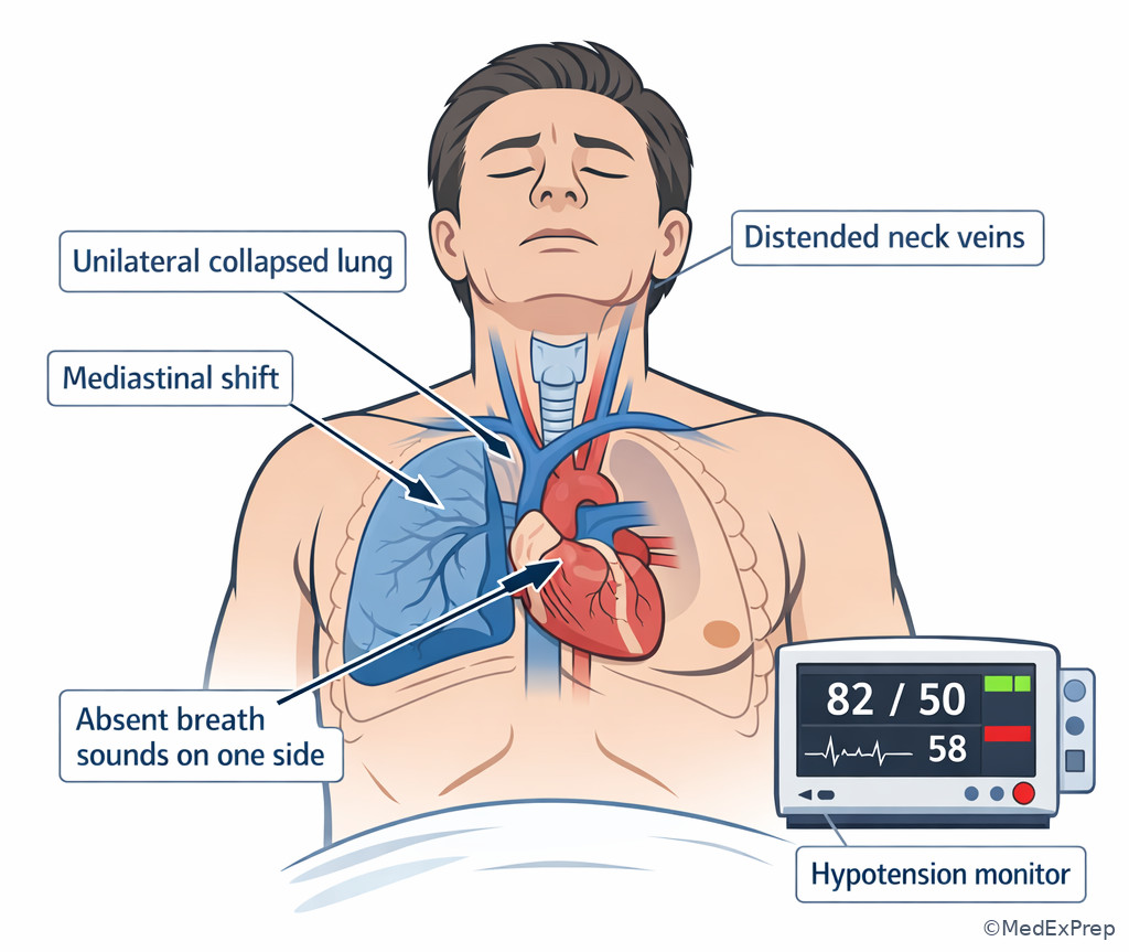
The core conflict is choosing between confirming the diagnosis and treating a clinical emergency now. The rule: if airway failure or a lethal chest problem is clinically apparent, immediate intervention comes before confirmatory imaging. See Fig 3. This is one of the most tested trauma principles because many exam stems deliberately include a tempting imaging answer.
Airway red flags include inability to speak clearly, gurgling, facial trauma with blood or swelling, stridor, severe agitation from hypoxia, progressive hoarseness, or depressed mental status with inability to protect secretions. Breathing red flags include severe dyspnea, unilateral absent breath sounds, tracheal deviation in classic cases, distended neck veins in some cases, cyanosis, or rapidly worsening oxygen saturation. A tension pneumothorax is a clinical diagnosis in an unstable patient; waiting for x-ray can be fatal. Massive hemothorax, flail chest with respiratory compromise, and open pneumothorax are also immediate priorities.
For shelf questions, think in this order: Can the patient oxygenate? Can the patient ventilate? Is there a chest problem that will kill them before the film is printed? If yes, act. Airway and breathing errors are often hidden behind distracting bleeding, fracture, or neurologic symptoms. In trauma, the chest can be the circulatory problem too: tension physiology impairs venous return and causes hypotension. This overlap explains why “B” can also rapidly worsen “C.”
Exam takeaway: Use unilateral absent breath sounds with hypotension and respiratory distress to justify immediate decompression without waiting for imaging.
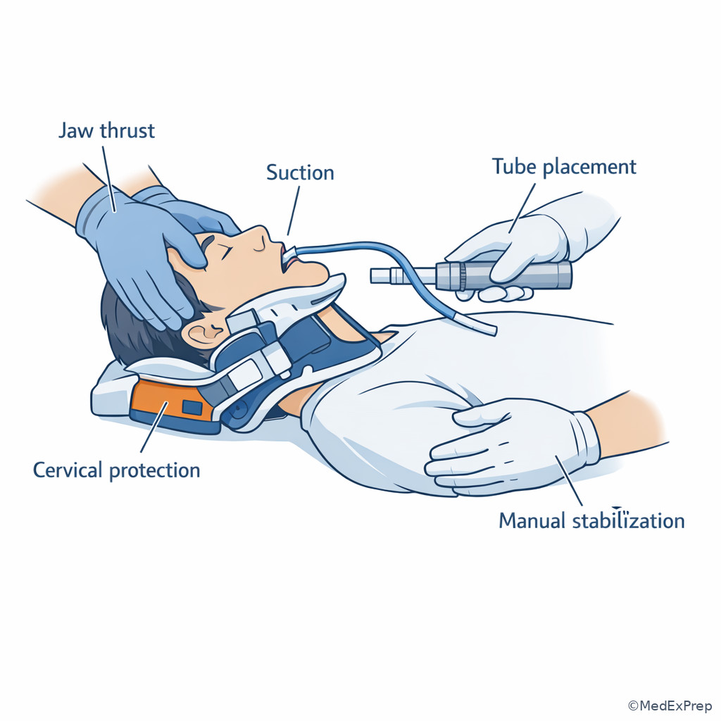
Exam takeaway: Airway management in trauma must preserve spinal precautions while restoring oxygenation and protection from aspiration.
B Worked example
A 22-year-old woman with mild scoliosis and anxiety is brought after a stabbing to the left chest. She is agitated, diaphoretic, and speaking in one-word sentences. HR 146/min, BP 78/44 mm Hg, RR 36/min, oxygen saturation 82% despite nonrebreather. The nurse notes trachea appears midline. Portable x-ray is available in 2 minutes.
Identify task: most appropriate next step.
Extract key facts: penetrating chest trauma, severe distress, hypotension, hypoxemia, likely unilateral breathing emergency; midline trachea is a distracting clue because it is neither required nor reliably present early.
Apply rule: unstable patient with likely tension physiology should undergo immediate decompression; x-ray delay is unsafe.
Eliminate distractors: CT impossible and unsafe; waiting for x-ray is wrong; fluid bolus alone does not fix obstructive physiology.
Verify: bedside decompression followed by definitive chest management and ongoing reassessment is correct.
C Exam trap
D Checkpoint
Question: A blunt trauma patient becomes increasingly agitated after arrival. He has facial abrasions, gurgling respirations, oxygen saturation 84%, and a cervical collar in place. What is the most appropriate priority?
- Remove the collar and extend the neck for better visualization
- Proceed directly to head CT because agitation suggests intracranial injury
- Address airway while maintaining cervical spine protection
- Obtain complete history from EMS before intervention
- A: Tempting because airway visualization matters, but wrong because cervical spine precautions should be maintained with manual stabilization.
- B: Tempting because trauma agitation can reflect head injury, but wrong because hypoxic airway compromise is a more immediate threat.
- C: Correct because gurgling respirations and hypoxemia indicate airway failure that must be managed immediately with spine protection.
- D: Tempting because mechanism details are useful, but wrong because bedside intervention cannot wait.
1.1.1.6 Circulation and hemorrhage control: when blood loss is the working diagnosis
A Explanation
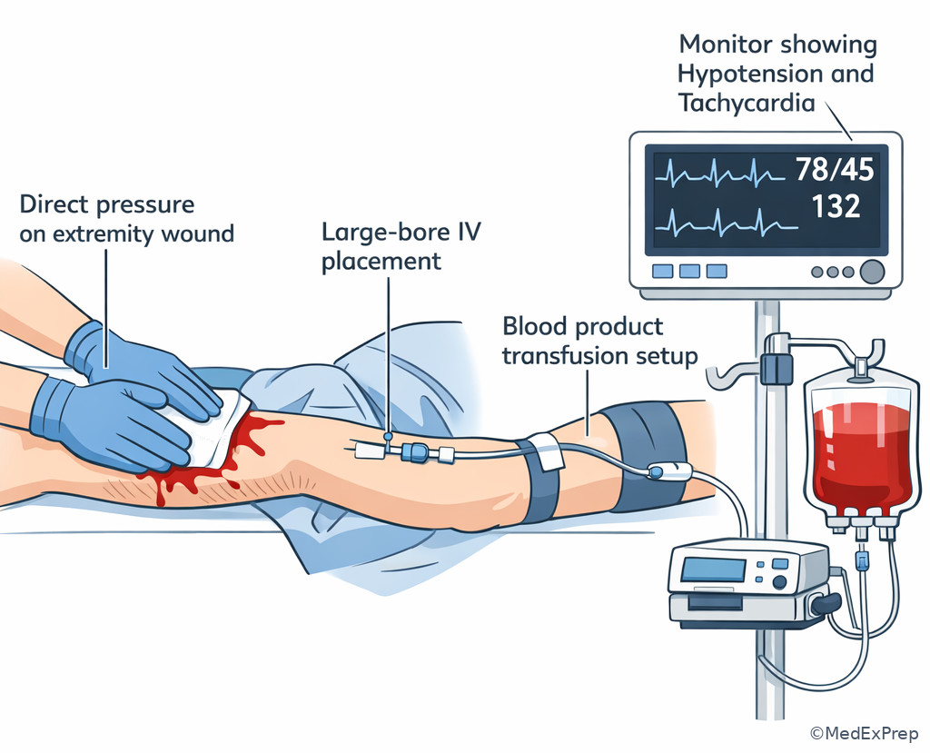
The decision conflict is whether hypotension in trauma should trigger broad diagnostic speculation or immediate hemorrhage-focused resuscitation. The resolving rule is classic: assume hemorrhage until another cause is proven and immediately treat bleeding plus poor perfusion. See Fig 4. This does not mean every patient is bleeding from the same place; it means your first circulatory framework is blood loss and hemorrhage control, not exhaustive differential expansion.
Circulation assessment includes pulse quality, skin temperature, capillary refill, blood pressure trend, mental status, and obvious external bleeding. Early shock may present with tachycardia and anxiety before hypotension appears. External hemorrhage gets direct pressure or tourniquet when appropriate. Internal bleeding is inferred from mechanism and instability and investigated with bedside tools if they do not delay care. Large-bore IV access or other rapid access, blood product resuscitation, and urgent escalation occur in parallel. Shelf questions frequently tempt you with crystalloid-heavy or imaging-first answers; the better answer often emphasizes hemorrhage control and balanced resuscitation.
Importantly, circulation in trauma is not only about liters of blood. It is about source control and perfusion restoration. If the patient remains unstable after initial blood product support, think ongoing bleed or another immediately reversible cause found earlier in the survey, such as tension pneumothorax or tamponade. The exam does not expect you to run an ICU-level hemodynamic protocol; it expects you to recognize that persistent shock means the patient has not been fixed.
Exam takeaway: In trauma hypotension, prioritize bleeding control and perfusion support before time-consuming diagnostics.
flowchart TD
A[Trauma patient with poor perfusion] --> B[Control external hemorrhage]
B --> C[Large-bore access and blood product resuscitation]
C --> D{Responding?}
D -->|Yes| E[Adjuncts and further source evaluation]
D -->|No| F[Search for ongoing hemorrhage or other reversible primary survey cause]
F --> G[Urgent operative or procedural escalation] Exam takeaway: Direct pressure and rapid bleeding control can outrank imaging in an unstable trauma patient.
B Worked example
A 39-year-old construction worker with hypertension and obesity is brought after being crushed between equipment. He is anxious, pale, and repeatedly asks for water. HR 138/min, BP 84/50 mm Hg, RR 28/min. Oxygen saturation is 98% on supplemental oxygen. There is a pelvic deformity and a forearm laceration that is oozing but not spurting. FAST has not yet been performed. A medical student suggests sending CBC, CMP, coagulation studies, and waiting for results before transfusion.
Identify task: best next step for circulation management.
Extract key facts: clear shock physiology, likely major occult hemorrhage source, oxygenation preserved, small visible bleed is distracting.
Apply rule: assume hemorrhage, control what you see, start hemorrhage-focused resuscitation now, and escalate source control.
Eliminate distractors: labs are useful but cannot delay; the forearm wound does not explain shock; stable oxygen saturation does not make him stable overall.
Verify: circulation step requires immediate hemorrhage control/resuscitation, not a wait-for-labs strategy.
C Exam trap
D Checkpoint
Question: A patient after blunt abdominal trauma has HR 130/min, BP 86/48 mm Hg, cool clammy skin, and normal oxygen saturation after airway and breathing have been addressed. What is the best next priority?
- Detailed abdominal CT scan
- Hemorrhage-focused circulatory resuscitation and bleeding control
- Formal neurologic examination
- Wait for repeat hemoglobin in one hour
- A: Tempting because abdominal injury is suspected, but wrong because unstable patients need resuscitation before CT.
- B: Correct because circulation shock after trauma is presumed hemorrhagic until proven otherwise.
- C: Tempting because neurologic status matters, but wrong because circulatory collapse is the active life threat.
- D: Tempting because serial labs are common, but wrong because treatment cannot wait for delayed laboratory change.
- The default cause of trauma shock is hemorrhage.
- True or False: A normal oxygen saturation excludes life-threatening trauma. False.
- Name the two priorities in the circulation step: control bleeding and restore perfusion.
1.1.1.7 Adjuncts, secondary survey, and when testing becomes appropriate
A Explanation
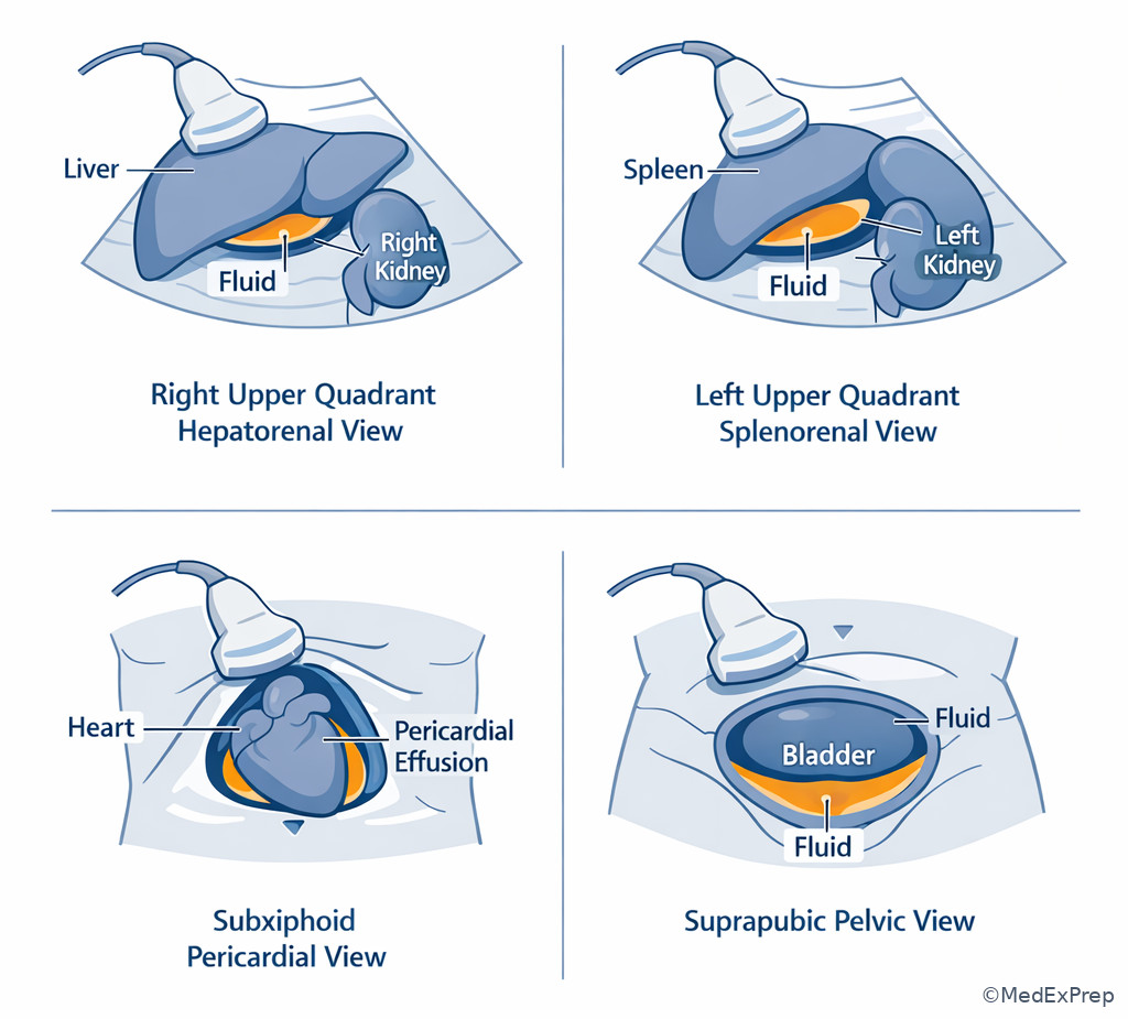
The conflict here is deciding when enough stabilization has occurred to justify adjuncts and fuller evaluation. The governing rule: adjuncts support the primary survey; they do not replace it, and secondary survey begins only after immediate threats are addressed. See Fig 5. This section matters because NBME often asks for the “best next test” and expects students to recognize whether the patient is ready for one.
Adjuncts to the primary survey commonly include ECG and pulse oximetry monitoring, blood pressure cycling, arterial blood gas or lactate, FAST examination, and portable chest and pelvis imaging in appropriate settings. Foley catheter and nasogastric tube may be considered when indicated and not contraindicated. These are useful because they are fast and bedside-based. By contrast, CT is usually reserved for the patient whose ABCs are stabilized enough to tolerate transport. Secondary survey is a head-to-toe history and exam, including AMPLE history when available, but it should not consume the first minutes of care in an unstable patient.
On exam questions, a common distinction is between a test that changes immediate management now and a test that merely adds anatomical detail later. FAST may be useful in an unstable blunt trauma patient because it can rapidly identify free fluid and help route the patient. CT abdomen provides more detail, but only after stability exists. Similarly, portable chest x-ray may be an adjunct after initial chest intervention, not a prerequisite to it.
Exam takeaway: Use bedside adjuncts early, but reserve CT and full secondary survey for patients stabilized after the primary survey.
flowchart TD
A[Primary survey complete] --> B{Immediate threats controlled?}
B -->|No| C[Continue resuscitation and bedside adjuncts only]
B -->|Yes| D[Adjuncts: FAST, portable CXR, pelvis film, labs]
D --> E{Physiology stable for transport?}
E -->|Yes| F[Secondary survey and selective CT]
E -->|No| G[Remain in trauma bay and escalate intervention] Exam takeaway: The FAST exam is a rapid bedside adjunct that helps direct unstable patients without delaying resuscitation.
B Worked example
A 46-year-old woman with rheumatoid arthritis and obesity presents after a rollover crash. After supplemental oxygen, chest decompression for a left-sided tension pneumothorax, and initiation of blood products, her blood pressure improves from 78/42 to 112/68 mm Hg and oxygen saturation rises to 98%. She now answers questions appropriately. There is abdominal tenderness without rigidity. The team asks what should happen next.
Identify task: choose the next appropriate level of evaluation.
Extract key facts: primary survey threats have been treated; physiology improved; she is now transportable; abdominal tenderness remains.
Apply rule: after stabilization, adjuncts and secondary survey guide selective imaging.
Eliminate distractors: immediate laparotomy without indication is too aggressive from the given data; repeating only the chest exam ignores new evaluation needs.
Verify: complete adjuncts/secondary survey and selective CT if stable enough is appropriate.
C Exam trap
D Checkpoint
Question: Which study is most appropriate as an early adjunct in an unstable blunt trauma patient after airway and breathing are addressed?
- CT abdomen/pelvis with IV contrast
- FAST examination
- MRI spine
- Detailed contrast esophagram
- A: Tempting because it is highly informative, but wrong because unstable patients should not leave the bay for transport-dependent imaging.
- B: Correct because FAST is rapid, bedside, and can guide immediate decisions in unstable trauma.
- C: Tempting if spine injury is a concern, but wrong because MRI is not an initial unstable-trauma adjunct.
- D: Tempting in selected penetrating injury patterns, but wrong because it does not fit early unstable blunt trauma evaluation.
1.1.1.8 Integration: choosing the best next step when several answers are partly right
A Explanation
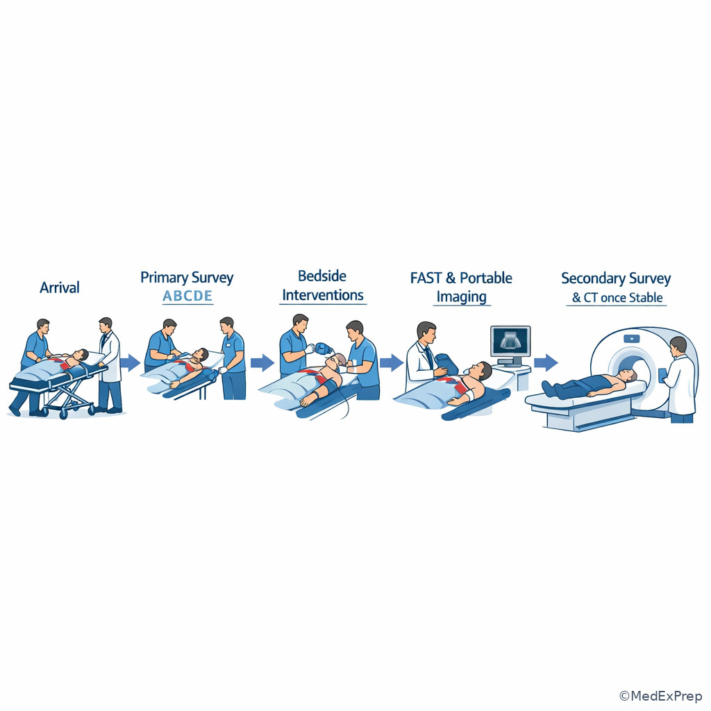
The final exam-level conflict is selecting among multiple plausible options that all seem reasonable in trauma. The single resolving rule is: choose the intervention that addresses the most immediate, reversible threat to life based on the earliest discriminating clue in the stem. This is where students must synthesize the whole sequence. One option may be a correct eventual test. Another may be a correct treatment for a different timing. The best answer is the one that fits the patient’s current physiologic problem now.
Common close-call pairs include airway intervention versus head CT, chest decompression versus portable x-ray, hemorrhage resuscitation versus waiting for labs, FAST versus CT, and primary survey action versus secondary survey detail. The best discriminator is usually one bedside clue: inability to speak, unilateral absent breath sounds, persistent hypotension, cool skin, altered mental status, or successful stabilization. Train yourself to ask, “What finding in this stem proves delay is unsafe?” When you can name that clue, the answer usually becomes obvious.
Board-style stems also include noise: comorbidities, distracting visible wounds, family comments, or one normal lab. Those details are not useless, but they must not outweigh the mortality-driving cue. Good test-taking in trauma means ignoring low-priority noise without ignoring high-risk context. The safest answer is not the broadest answer; it is the right action at the right time.
Exam takeaway: The correct answer in trauma often depends on where the patient is in the sequence, not just what injury is present.
B Worked example
A 55-year-old woman with COPD and atrial fibrillation on apixaban is brought after a pedestrian-versus-car collision. She is alert but restless. HR 118/min, BP 102/66 mm Hg, RR 30/min, oxygen saturation 90% on nonrebreather. She has extensive right chest wall bruising, a deformed wrist, and complains of abdominal pain. Breath sounds are markedly decreased on the right. A portable chest x-ray technician is entering the room. The resident asks for the best next step.
Identify task: choose the single best immediate action.
Extract key facts: respiratory compromise, unilateral decreased breath sounds, borderline perfusion, anticoagulation and abdominal pain are distracting but relevant later, wrist deformity is noise.
Apply rule: breathing threat comes before imaging and before circulation-focused imaging workup if it is the first reversible killer clue present.
Eliminate distractors: chest x-ray is tempting but later; FAST may be appropriate after breathing issue is addressed; wrist management waits.
Verify: treat the right-sided breathing emergency immediately at bedside, then reassess vitals and proceed.
C Exam trap
D Checkpoint
Question: In a trauma stem with several abnormal findings, which clue most strongly identifies the best immediate next step?
- The oldest comorbidity listed first in the stem
- The abnormality that threatens life before transport or delayed testing
- The injury that appears most dramatic on inspection
- The diagnosis with the most specific confirmatory imaging
- A: Tempting because comorbidities can shape risk, but wrong because they rarely override immediate physiology.
- B: Correct because trauma sequencing prioritizes reversible life threats over detail gathering.
- C: Tempting because visible injuries attract attention, but wrong because appearance does not equal priority.
- D: Tempting because precise diagnosis matters, but wrong because imaging confirmation is often not the first move.
Exam Traps & Differentiators
Most common wrong answer and why: CT imaging before stabilization. It is tempting because it promises diagnostic certainty, but trauma questions reward timing, not just correctness. A perfect test at the wrong moment is still the wrong answer.
| Looks similar | Why students confuse them | Single differentiator | Best exam action |
|---|---|---|---|
| Primary survey vs secondary survey | Both involve trauma assessment | Primary survey hunts immediate killers; secondary survey waits until initial stabilization | Choose primary survey action first in unstable patients |
| FAST vs CT abdomen | Both evaluate internal injury | FAST is bedside and rapid; CT requires transport and greater stability | Use FAST first in unstable blunt trauma |
| Chest decompression vs portable CXR | Both are used in thoracic trauma | Clinical tension physiology is treated before imaging | Decompress first if unstable with suggestive findings |
| Visible fracture vs occult hemorrhage | The obvious injury draws attention | Shock physiology points to circulation priority regardless of visible extremity injury | Resuscitate and control bleeding first |
| Mechanism-based concern vs physiology-based triage | High-energy mechanism sounds urgent | Vitals, perfusion, and mentation determine current urgency | Triage by physiology |
If the stem says... think...
- “Unable to speak, gurgling, blood in oropharynx” → airway first with cervical spine protection.
- “Absent unilateral breath sounds with hypotension” → immediate breathing intervention, not confirmatory imaging.
- “Cool clammy skin, confusion, borderline BP after trauma” → unstable circulation, likely hemorrhagic shock pattern.
- “Primary threats treated and vitals improved” → now consider adjuncts, secondary survey, and selective CT.
- “Normal initial hemoglobin” → do not let one lab falsely reassure you about acute bleeding.
- “Dramatic limb injury but poor oxygenation or perfusion” → the limb is not the first priority.
| Scenario category | Correct first move | Tempting but wrong alternative | Why wrong for this patient |
|---|---|---|---|
| Unstable chest trauma with absent breath sounds | Immediate decompression / chest intervention | Portable chest x-ray first | Imaging delays treatment of a clinical emergency |
| Hypotensive blunt trauma after airway/breathing addressed | Hemorrhage-focused resuscitation | Repeat CBC before acting | Lab delay does not help immediate perfusion |
| Stabilized trauma patient with abdominal tenderness | Secondary survey and selective imaging | Immediate operative assumption without indication | Now stable enough for appropriate staged evaluation |
| Visible open fracture plus hypoxia | Airway/breathing management first | Orthopedic reduction first | The fracture is not the first killer |
| Classification | Typical clues | What it means for triage |
|---|---|---|
| Immediately unstable | Hypotension, hypoxia, agitation/confusion, poor perfusion, active bleeding | Remain in trauma bay; treat life threats now |
| Transient responder | Initial improvement after intervention, then concern for recurrence | High vigilance; ongoing source may still need urgent control |
| Stable after initial survey | Improved vitals, preserved mentation, adequate oxygenation/perfusion | Proceed to adjuncts, secondary survey, selective CT |
| Mass-casualty salvage priority | Serious but treatable injuries with benefit from immediate intervention | Prioritize limited resources toward best survival yield |
Algorithm / Approach
Rapid Review
- Primary survey → ordered search for immediate killers, not a full diagnosis list
- Airway compromise → inability to speak normally or protect secretions separates urgency from observation
- Breathing emergency → unilateral absent breath sounds plus instability beats x-ray
- Circulation in trauma → hemorrhage first until proved otherwise
- Stable versus unstable → physiology outranks mechanism
- Secondary survey → only after initial threats are addressed
- FAST → bedside triage tool for unstable patients, not a replacement for CT in stable detail workup
- Visible fracture → distractor unless it is the actual life threat
- Normal early hemoglobin → does not rule out acute blood loss
- Exposure → required to find hidden injury but must be paired with warming
- Borderline vitals in elderly or beta-blocked patient → may still represent shock
- Best next step → earliest action that prevents death, not the most complete test
Self-check quiz
1. A 24-year-old man is brought after a bicycle collision. He has an obvious clavicle deformity, but he is gurgling, cannot answer questions clearly, and oxygen saturation is 85%. What is the most appropriate next step?
- Address the airway with cervical spine protection
- Obtain a clavicle x-ray
- Perform a complete secondary survey
- Send for CT angiography of the neck
2. A 63-year-old woman with hypertension presents after a ground-level fall. HR 88/min, BP 96/60 mm Hg, RR 24/min, skin is cool, and she seems confused. Which finding most strongly indicates trauma instability?
- Age older than 60 years
- Cool skin with confusion
- Mechanism limited to a fall
- Absence of severe pain complaints
3. A 29-year-old man with asthma is brought after a stab wound to the right chest. HR 142/min, BP 80/46 mm Hg, RR 34/min, oxygen saturation 81%. Breath sounds are absent on the right. What is the most appropriate next step?
- Portable chest x-ray
- Immediate right-sided chest decompression
- FAST examination
- CT chest with contrast
4. A 40-year-old woman with obesity and migraines presents after blunt abdominal trauma. Airway and breathing have been addressed. She is pale, HR 128/min, BP 82/50 mm Hg, and has diffuse abdominal tenderness. Which is the best next step in management?
- Wait for repeat hemoglobin before transfusion
- Begin hemorrhage-focused circulatory resuscitation
- Order MRI abdomen
- Perform detailed AMPLE history first
5. A 51-year-old man with diabetes and coronary disease was initially hypotensive after a motor vehicle collision but improves after airway support and resuscitation. He is now alert, BP 118/72 mm Hg, oxygen saturation 98%, and has persistent abdominal tenderness. What is the most appropriate next step?
- Proceed to adjuncts, secondary survey, and selective CT
- Assume continued instability and avoid all imaging
- Skip further evaluation and observe only
- Perform fracture reduction before any additional trauma assessment
6. A 76-year-old man on metoprolol and apixaban presents after a low-speed pedestrian collision. HR 84/min, BP 94/58 mm Hg, RR 26/min, oxygen saturation 95% on 2 L, and he is slow to answer. He has a large thigh hematoma and mild abdominal tenderness. Which is the most appropriate interpretation?
- He is stable because the heart rate is not elevated
- He is likely unstable despite normocardia and requires high-priority trauma management
- He is stable enough for MRI because oxygen saturation is preserved
- He is low risk because the mechanism is not high energy
7. A 33-year-old woman with lupus and obesity arrives after a rollover crash. She was hypotensive and hypoxic on arrival, but after decompression of a left chest emergency and transfusion, BP is 110/70 mm Hg and oxygen saturation is 99%. She remains tender over the abdomen and pelvis. Which option is now most appropriate?
- Immediate transport for selective CT and completion of secondary survey
- Delay all imaging because she was previously unstable
- Repeat only the neurologic exam and discharge if unchanged
- Ignore abdominal findings because chest trauma explained the instability
8. A 48-year-old man with COPD and alcohol use disorder is brought after a fall from a roof. He has a visibly open tibia fracture, HR 122/min, BP 90/58 mm Hg, RR 32/min, oxygen saturation 87%, and markedly decreased breath sounds on the left. Which is the best next step?
- Administer broad-spectrum antibiotics for the open fracture first
- Immediate intervention for the left-sided breathing abnormality
- CT head because alcohol use may confound mental status
- Splint the leg before any other action
Answer key
1.1.1.9 1. Answer: A — Address the airway with cervical spine protection
Why A is correct: The patient has gurgling respirations, impaired speech, and hypoxemia, which indicate airway compromise. In trauma, airway is addressed first while maintaining cervical spine precautions because oxygenation and protection from aspiration are immediate survival issues. Source: American College of Surgeons ATLS principles; Townsend et al.
Why B is tempting but wrong: A clavicle injury is visible and common after cycling trauma, but it is not the most immediate threat. The discriminating clue is the gurgling respirations and low oxygen saturation, which make airway intervention urgent. Source: ATLS; Schwartz’s Principles of Surgery.
Why C is tempting but wrong: A full secondary survey feels thorough, but it occurs only after immediate ABC threats are managed. The single clue is active airway failure. Source: ATLS.
Why D is tempting but wrong: Neck vascular injury can matter in certain mechanisms, but CT angiography is a later study for a patient stable enough for transport. The discriminating clue is current hypoxemia with poor airway protection. Source: ATLS; Sabiston.
1.1.1.10 2. Answer: B — Cool skin with confusion
Why B is correct: Cool skin and confusion are perfusion and mental-status abnormalities that strongly suggest physiologic instability, even when heart rate is not dramatic. Trauma triage is based on current physiology. Source: ATLS; American College of Surgeons trauma assessment standards.
Why A is tempting but wrong: Older age raises concern and can blunt compensatory responses, but age alone does not define instability. The discriminating clue is actual abnormal perfusion and mentation. Source: ATLS; Townsend et al.
Why C is tempting but wrong: Mechanism informs suspicion, but low-energy mechanism does not exclude severe injury and does not determine current triage by itself. The clue that matters is the physiologic abnormality. Source: ATLS.
Why D is tempting but wrong: Lack of pain complaints may occur with distraction or altered mentation, but it is not the best marker of instability. The discriminating clue is confusion with cool skin. Source: Sabiston.
1.1.1.11 3. Answer: B — Immediate right-sided chest decompression
Why B is correct: Penetrating chest trauma plus hypotension, severe hypoxemia, and absent unilateral breath sounds is classic for a life-threatening breathing emergency such as tension pneumothorax. In an unstable patient, this is treated clinically without waiting for imaging. Source: ATLS; Schwartz’s Principles of Surgery.
Why A is tempting but wrong: Portable chest x-ray is a legitimate trauma adjunct, but only when it does not delay treatment of clinically obvious tension physiology. The discriminating clue is instability with absent breath sounds. Source: ATLS.
Why C is tempting but wrong: FAST is useful for unstable trauma, but the chest finding is the earliest lethal clue and must be addressed first. FAST would be the right tool for a slightly different patient whose dominant issue is unclear circulatory collapse without obvious breathing emergency. Source: ATLS; Sabiston.
Why D is tempting but wrong: CT chest provides detail, but transport is unsafe in this unstable patient. The single clue is profound respiratory and hemodynamic compromise. Source: ATLS.
1.1.1.12 4. Answer: B — Begin hemorrhage-focused circulatory resuscitation
Why B is correct: After airway and breathing are addressed, hypotension, tachycardia, pallor, and abdominal tenderness in trauma should be treated as hemorrhagic shock until proven otherwise. Immediate resuscitation and bleeding control are the correct next priorities. Source: ATLS; Townsend et al.
Why A is tempting but wrong: Repeat hemoglobin may eventually help trend blood loss, but acute bleeding can be present before lab changes become obvious. The discriminating clue is shock physiology right now. Source: ATLS.
Why C is tempting but wrong: MRI is not an initial trauma tool for unstable patients. The clue is hypotension with likely internal bleeding. Source: Sabiston.
Why D is tempting but wrong: AMPLE history matters in secondary survey, but it cannot delay resuscitation in an unstable patient. The discriminating clue is active circulatory compromise. Source: ATLS.
1.1.1.13 5. Answer: A — Proceed to adjuncts, secondary survey, and selective CT
Why A is correct: The stem emphasizes that the patient improved after initial stabilization and is now alert with normal pressure and oxygenation. Once primary survey threats are controlled, adjuncts and more detailed evaluation become appropriate. Source: ATLS; Schwartz’s Principles of Surgery.
Why B is tempting but wrong: It is true that unstable patients should not go to CT, but this patient is now stabilized. The discriminating clue is the improvement in vitals and mental status. Source: ATLS.
Why C is tempting but wrong: Observation alone risks missing major abdominal injury; persistent tenderness still needs evaluation. The single clue is ongoing abdominal findings after stabilization. Source: Sabiston.
Why D is tempting but wrong: Fracture care is important, but persistent abdominal tenderness requires trauma evaluation first. The discriminating clue is the unresolved torso complaint after initial ABC correction. Source: ATLS.
1.1.1.14 6. Answer: B — He is likely unstable despite normocardia and requires high-priority trauma management
Why B is correct: Beta-blockers can blunt tachycardia, so older trauma patients may still be unstable despite a normal heart rate. Borderline hypotension, tachypnea, slow mentation, and a large hematoma strongly suggest significant injury and possible hemorrhage. Source: ATLS; Townsend et al.
Why A is tempting but wrong: Students often anchor on heart rate, but normocardia does not exclude shock when compensatory responses are blunted. The discriminating clue is slow mentation with low pressure and beta-blocker use. Source: ATLS.
Why C is tempting but wrong: Preserved oxygen saturation does not equal global stability, and MRI is not an initial trauma study. The clue is ongoing perfusion concern. Source: Sabiston.
Why D is tempting but wrong: Lower-mechanism injuries can still cause major bleeding, especially in anticoagulated older adults. The discriminating clue is abnormal physiology, not mechanism severity. Source: ATLS.
1.1.1.15 7. Answer: A — Immediate transport for selective CT and completion of secondary survey
Why A is correct: The patient was unstable earlier but has improved after treatment of the primary survey threats. Current stability plus persistent abdominal and pelvic findings make further staged evaluation appropriate. Source: ATLS; Schwartz’s Principles of Surgery.
Why B is tempting but wrong: It correctly recalls that unstable patients should not go to CT, but it ignores that this patient is now hemodynamically and respiratorily improved. The discriminating clue is current normalization of BP and oxygenation. Source: ATLS.
Why C is tempting but wrong: Repeating a neuro exam alone misses unresolved torso injury. The clue is persistent abdominal and pelvic tenderness. Source: Sabiston.
Why D is tempting but wrong: Earlier chest pathology may have contributed to instability, but it does not exclude simultaneous abdominal or pelvic trauma. The discriminating clue is ongoing localized tenderness after stabilization. Source: ATLS.
1.1.1.16 8. Answer: B — Immediate intervention for the left-sided breathing abnormality
Why B is correct: Hypoxemia, tachypnea, hypotension, and markedly decreased unilateral breath sounds identify a breathing emergency that outranks fracture care and diagnostic imaging. This is the earliest reversible killer clue in the stem. Source: ATLS; Townsend et al.
Why A is tempting but wrong: Open fractures do require antibiotics promptly, but not before a life-threatening breathing problem is treated. The discriminating clue is respiratory compromise with unilateral chest findings. Source: ATLS; orthopedic trauma standards.
Why C is tempting but wrong: Alcohol use can cloud mental status, and head injury is possible, but hypoxemia with decreased breath sounds is the immediate danger. The clue that separates this from a head-CT-first scenario is the chest exam. Source: ATLS.
Why D is tempting but wrong: Splinting reduces pain and soft tissue damage, but it is not first when the patient cannot oxygenate well. The discriminating clue is oxygen saturation 87% with unilateral decreased breath sounds. Source: Sabiston.
Keep going with the full Surgery Shelf Prep Course (Surg Shelf)
You just studied the way members do, in the same lesson interface. Full access unlocks every lesson, the question bank and flashcards for a full year.
One payment · 1 year of access · No auto-renewal