1.1.1 - Pediatric Vital Sign Norms
Pediatric Vital Sign Norms
Course: Pediatrics Shelf Prep Course (Peds Shelf)
- PED: Emergency/Critical Recognition → age-specific vital sign interpretation
- PED: Growth and Development → age-based normal variation
- PED: Preventive Pediatrics → well-child assessment basics
- PED: Common Ambulatory Complaints → reassurance vs testing vs escalation
- PED: Newborn Care → neonatal vs infant normal ranges
- Use age-specific heart rate, respiratory rate, blood pressure, temperature, and oxygen saturation ranges to decide whether a child is normal, concerning, or unstable. PED: Emergency/Critical Recognition - age-based triage
- Distinguish physiologic causes of abnormal vitals from pathology by integrating age, setting, fever, pain, hydration, and appearance. PED: Domain -> diagnosis from pediatric presentations
- Choose the most appropriate next step when a vital sign abnormality requires observation, repeat measurement, focused workup, or urgent escalation. PED: Domain -> next best step in management
- Avoid adult-rule traps and recognize pediatric red-flag combinations that suggest shock, respiratory failure, sepsis, dehydration, or intracranial pathology. PED: Emergency/Critical Recognition -> unstable presentation recognition
- Build a shelf-ready framework that connects vital signs to upcoming growth and developmental interpretation topics. PED: Growth and Development -> age-based interpretation
Illustrations
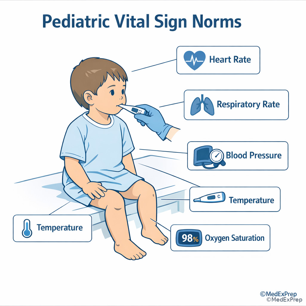
Exam takeaway: Use this visual to recall the key testable points for Pediatric Vital Sign Norms.
Why it matters
- On the shelf, the diagnosis often hides inside one age-adjusted vital sign: a “normal” respiratory rate in an infant may be tachypnea in a school-age child, and a “normal” adult pulse may be bradycardia in an infant.
- Pediatric acuity starts with vitals plus appearance. The exam repeatedly asks you to choose between reassurance, repeat assessment, targeted evaluation, or urgent escalation.
- Many wrong answers depend on applying adult thresholds to children. Pediatric vitals change rapidly with age, activity, fever, crying, and sleep state.
- Vital-sign interpretation becomes the foundation for later topics: growth, dehydration, bronchiolitis, sepsis, congenital heart disease, adolescent care, and shock recognition.
Key Terms & Must-Know Facts
1.1.1.1 Key terms
- Tachycardia: heart rate above expected range for age and state
- Bradycardia: heart rate below expected range for age; concerning in infants if persistent or associated with poor perfusion
- Tachypnea: respiratory rate above age-based norm; often earlier than hypoxemia in respiratory distress
- Hypotension: late sign of pediatric shock
- Perfusion: clinical assessment using capillary refill, pulses, mental status, skin temperature, urine output
- Pulse pressure: difference between systolic and diastolic blood pressure; narrow pulse pressure can suggest shock
- Febrile tachycardia: elevated heart rate driven by fever rather than primary cardiac pathology
- Orthostatic change: less reliable in younger children; use overall perfusion and hydration status instead
- SpO2: peripheral oxygen saturation; low value requires correlation with waveform, probe placement, and clinical appearance
- Apnea: pause in breathing, especially significant in neonates and young infants
- Age-adjusted hypotension rule: for children 1-10 years, low systolic blood pressure can be estimated by \(70 + 2 \times \text{age in years}\)
- Compensated shock: tachycardia with preserved blood pressure but poor perfusion
- Uncompensated shock: hypotension plus signs of poor perfusion
1.1.1.2 Must-know facts
- Children usually become tachycardic before they become hypotensive.
- Hypotension is a late and dangerous pediatric sign.
- Tachypnea is one of the earliest clues to respiratory illness, metabolic acidosis, or sepsis.
- Always interpret vital signs in context: age, crying, fever, pain, sleep, medications, hydration, and measurement technique.
- Oxygen saturation can appear falsely low with motion artifact, poor perfusion, cold extremities, or a bad probe signal.
- A febrile child with persistent tachycardia after antipyretics and fluids deserves a broader workup.
- Bradycardia in a sick child is preterminal until proven otherwise, especially with hypoxia.
- Normal blood pressure does not exclude shock in pediatrics.
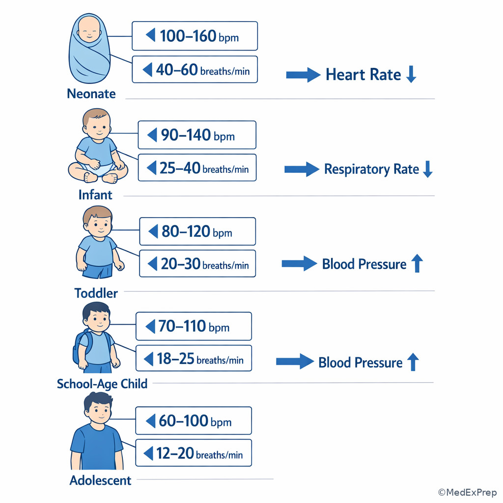
- Neonates and infants normally have faster heart and respiratory rates than adolescents.
| Age group | Heart rate awake | Respiratory rate | Systolic BP, rough lower concern threshold | Clinical pearl |
|---|---|---|---|---|
| Neonate | 100-205 | 30-60 | <60 | Brief variability is common; correlate with feeding, color, and tone. |
| Infant | 100-190 | 30-53 | <70 | Adults often undercall tachypnea in this group. |
| Toddler | 98-140 | 22-37 | <70 | Crying can transiently raise pulse and BP. |
| Preschool | 80-120 | 20-28 | <70 + 2 × age | Repeat after calm repositioning if borderline. |
| School-age | 75-118 | 18-25 | <70 + 2 × age | Persistent tachycardia deserves explanation. |
| Adolescent | 60-100 | 12-20 | <90 | Near-adult values, but clinical context still matters. |
Exam takeaway: Younger children normally have faster heart and respiratory rates, so any threshold must be age-adjusted before calling it abnormal.
flowchart LR
N[Neonate] --> I[Infant]
I --> T[Toddler]
T --> P[Preschool]
P --> S[School-age]
S --> A[Adolescent]
N --- HR1[Highest normal HR/RR]
I --- HR2[Still high]
T --- HR3[Declining]
P --- HR4[Moderate]
S --- HR5[Near adult]
A --- HR6[Adult-like range]
See Fig 1 for the single highest-yield shelf pattern in this topic: heart rate and respiratory rate steadily fall with age, while blood pressure rises.
Core content
1.1.1.3 Foundational age-based normals: what changes with age and what does not
A Explanation
The first decision conflict on exam questions is simple but decisive: Is this vital sign truly abnormal for the child’s age, or are you overcalling a normal pediatric value using adult expectations? The single rule that resolves this is age adjustment before interpretation. Neonates and infants have faster heart rates and respiratory rates than older children, while blood pressure increases with age. A respiratory rate of 44 can be normal in a 3-month-old but clearly abnormal in a 10-year-old. A heart rate of 95 may be normal in an adolescent but concerningly low in a neonate if the infant looks ill.
WHY THIS IS TESTED: Shelf items reward learners who anchor to age before choosing “reassure,” “repeat,” “work up,” or “escalate.” If you miss the age frame, the whole question collapses. The exam often places a borderline value inside a noisy vignette with fever, crying, dehydration, or mild pain to see whether you know what is expected for that age.
Use a tiered framework: first ask the child’s age group; next ask whether the measurement was obtained under calm conditions; then ask whether the child’s appearance supports or contradicts the number. In pediatrics, the appearance-perfusion-breathing triad matters as much as the absolute value. A well-appearing crying toddler with a pulse of 150 may need a repeat measurement after calming. A quiet infant with a pulse of 90 and poor feeding is more concerning despite a number that may not alarm an adult examiner. See Fig 1 to lock in the age trajectory.
Exam takeaway: Start with age, then state, then appearance before deciding that a number is normal, repeatable, or dangerous.
B Worked example
A 14-month-old boy is brought to urgent care for 1 day of rhinorrhea and decreased appetite. He has mild eczema and a history of two prior episodes of acute otitis media. He is crying throughout the examination and has a temperature of 38.4°C, heart rate 158/min, respiratory rate 32/min, blood pressure 94/58 mm Hg, and oxygen saturation 99% on room air. His grandmother reports that he “always gets a fast heartbeat when sick,” and the stem mentions mild cerumen in one ear as a distracting clue. What is the most appropriate next step?
Reasoning chain: Identify task: interpret vitals, not diagnose the ear. Determine age and acuity: toddler, not toxic, no hypoxemia, no poor perfusion. Extract key facts: fever, crying, normal oxygen saturation, normal respiratory rate for age, tachycardia that may be explained by state. Apply rule: in a crying febrile toddler, isolated tachycardia should be rechecked after antipyresis and calming rather than immediately treated as arrhythmia or shock. Eliminate distractors: ECG is tempting because of the heart rate, but the context explains it; fluid bolus is tempting because pulse is high, but perfusion is not impaired; admission is unnecessary without respiratory distress or lethargy. Verify: repeat vitals after comfort measures.
C Exam trap
D Checkpoint
Question: A well-appearing 2-month-old girl is seen for cough. Which vital sign pattern is most concerning and least likely to be normal for age?
- A. Heart rate 156/min while crying after vaccination discussion
- B. Respiratory rate 42/min while awake and afebrile
- C. Oxygen saturation 99% on room air with a good waveform
- D. Heart rate 92/min while sleepy with poor feeding and mottled skin
Answer: D
- A: Tempting because 156 sounds high, but in a crying young infant it can still be physiologic.
- B: Tempting because 42 sounds fast to adult ears, but it is within expected infant range.
- C: Tempting because oxygenation is always important, but this value is reassuring when the waveform is reliable.
- D: Correct because a relatively low heart rate in a young infant becomes concerning when paired with poor feeding and mottling; the associated perfusion clues discriminate it.
1.1.1.4 Measurement context: when to repeat, when to trust, and when technique changes the answer
A Explanation
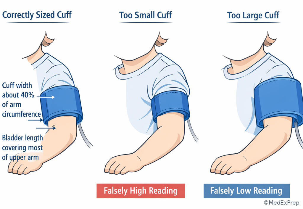
The second foundational decision conflict is choose between true pathology and a bad measurement or misleading context. The single resolving rule is that pediatric vital signs must be interpreted only after confirming the method and state. A screaming toddler can have transient tachycardia and hypertension. A sleeping infant may have a lower heart rate than when awake. A poorly fitting blood pressure cuff overestimates pressure when too small and underestimates when too large. A pulse oximeter on a cold, moving extremity may display falsely low saturations.
WHY THIS IS TESTED: Shelf writers love “false alarm” scenarios. The stem may present one abnormal number and ask for the best next step. The right answer is often not a lab or treatment but repeating the measurement correctly. This tests judgment: you are not ignoring danger; you are confirming whether danger is real.
When should you repeat instead of immediately act? Repeat when the child looks well, the abnormality is isolated, the explanation is plausible, and technique may be flawed. Do not hide behind technique when the child looks sick. A lethargic infant with delayed capillary refill and tachycardia has real physiology until proven otherwise. Similarly, a low oxygen saturation with increased work of breathing should be taken seriously even if motion artifact is possible. Measurement context refines interpretation; it does not excuse ignoring red flags.
For blood pressure, remember cuff width should be about 40% of arm circumference and bladder length should cover most of the upper arm. For oxygen saturation, assess waveform quality, probe placement, perfusion, and whether the reading correlates with the child’s color and work of breathing. For temperature, know that route matters; rectal temperature remains the most accurate core estimate in infants when a precise fever determination matters. See Fig 3 for the shelf-style repeat-versus-escalate framework.
Exam takeaway: Repeat isolated abnormal vitals in a well-appearing child when context or technique may explain them, but escalate if the number matches an ill appearance.
flowchart TD
A[Abnormal vital sign] --> B{Child ill-appearing?}
B -->|Yes| C[Trust value and escalate evaluation]
B -->|No| D{Technique or context issue likely?}
D -->|Yes| E[Repeat correctly after calming/repositioning]
D -->|No| F[Investigate persistent abnormality]
E --> G{Still abnormal?}
G -->|Yes| F
G -->|No| H[Reassure with follow-up] Exam takeaway: Before acting on a borderline value, confirm method, state, and correlation with the physical exam.
B Worked example
A 7-year-old girl with mild intermittent asthma and obesity presents for school physical clearance. She drank an energy drink on the way to clinic, is anxious about vaccines, and cries during blood pressure measurement. Her first BP is 132/86 mm Hg, pulse 124/min, temperature 36.9°C, respiratory rate 20/min, and SpO2 100%. She has no headache, no edema, and a normal cardiac exam. The stem adds a distracting family history of hypertension in her father. What is the most appropriate next step?
Reasoning chain: Identify task: decide whether this is pediatric hypertension or a repeat-measurement scenario. Determine age and acuity: school-age, asymptomatic, well appearing. Extract key facts: anxiety, stimulant drink, crying, single elevated BP, no end-organ symptoms. Apply rule: an isolated elevated BP in a child should be repeated correctly with proper cuff size after rest. Eliminate distractors: renal ultrasound is for persistent hypertension workup, not the first move; antihypertensive medication is far too premature; reassurance without repeat is also wrong because the value may still matter. Verify: repeat manual blood pressure under standardized conditions.
C Exam trap
D Checkpoint
Question: Which situation most strongly supports repeating the value before initiating a workup?
- A. A cyanotic infant with SpO2 84% and nasal flaring
- B. A lethargic toddler with pulse 170/min and delayed capillary refill
- C. A smiling school-age child with BP elevated once using a cuff that appears too small
- D. An adolescent with fever, hypotension, and petechiae
Answer: C
- A: Tempting because pulse oximeters can misread, but cyanosis plus distress means act first, not casually repeat.
- B: Tempting because tachycardia may occur with crying, but lethargy and poor perfusion override that possibility.
- C: Correct because technique error is obvious and the child appears well; repeat with proper cuff size is the best next step.
- D: Tempting because some hypotension readings are artifact, but shock signs plus petechiae require urgent evaluation.
- True or False: A single elevated BP in a crying child is enough to diagnose hypertension. False.
- The most common reason a pulse oximeter reads falsely low in a moving child is artifact/poor signal.
- Name 3 context factors that can transiently raise pediatric heart rate: fever, pain, crying.
1.1.1.5 Applied interpretation: tachycardia, tachypnea, and the difference between compensation and danger
A Explanation
The first applied decision conflict is choose between a compensated child who needs cause-directed care and a decompensating child who needs urgent escalation. The single finding that often resolves this is not the abnormal number alone but the presence of poor perfusion, altered mental status, or increased work of breathing. In pediatrics, tachycardia and tachypnea are early compensatory signs. Hypotension arrives late. This makes vital-sign interpretation fundamentally different from casual adult heuristics.
WHY THIS IS TESTED: NBME-style pediatrics asks you to spot the child whose blood pressure is still “normal” but who is already in trouble. Tachycardia with delayed capillary refill, weak pulses, dry mucous membranes, or lethargy suggests clinically significant dehydration or early shock. Tachypnea with retractions, nasal flaring, or grunting suggests respiratory distress even before oxygen saturation falls.
Use a pattern approach. Isolated tachycardia in a well-appearing febrile child may resolve with antipyretics, hydration, and reassessment. Tachycardia plus poor perfusion raises concern for hypovolemia, sepsis, or shock. Tachypnea plus clear lungs can still be important because metabolic acidosis, fever, or pain can drive it. Tachypnea plus retractions or hypoxemia points toward pulmonary disease. When evaluating a child, do not wait for hypotension to feel justified in escalating.
Exam takeaway: Tachypnea and tachycardia become dangerous when they travel with retractions, grunting, altered mental status, or poor perfusion.
{"prompt":"Realistic medical educational illustration on white background showing three pediatric patients side by side: infant with nasal flaring and subcostal retractions, toddler with delayed capillary refill and weak pulses, school-age child appearing lethargic with cool extremities; include clean labels for tachypnea, tachycardia, poor perfusion, altered mental status, retractions, and late hypotension; atlas-style pediatric emergency triage visual, not cartoon","caption":"Fig 4. Pediatric distress signs paired with abnormal vital patterns\nExam takeaway: Tachypnea and tachycardia become dangerous when they travel with retractions, grunting, altered mental status, or poor perfusion.","size":"1024x1024"} Exam takeaway: Persistent tachycardia or tachypnea matters most when paired with perfusion or respiratory distress findings.
B Worked example
A 3-year-old boy is brought to the ED for vomiting and diarrhea for 2 days. He has a history of repaired ventricular septal defect and mild speech delay. His mother mentions he “always breathes fast when anxious,” a tempting-but-wrong clue. Temperature is 37.8°C, heart rate 168/min, respiratory rate 34/min, blood pressure 92/58 mm Hg, oxygen saturation 99%, capillary refill 4 seconds, mucous membranes dry, and he is sleepy but arousable. What is the most appropriate next step?
Reasoning chain: Identify task: determine severity and next step. Determine age and acuity: toddler with multiple concerning signs. Extract key facts: tachycardia, mild tachypnea, delayed capillary refill, dry mucosa, somnolence. Apply rule: compensated shock/dehydration can present before hypotension. Eliminate distractors: outpatient oral rehydration alone is tempting because gastroenteritis is common, but perfusion is impaired; chest imaging is unnecessary despite tachypnea because the respiratory rate likely reflects compensation; reassurance is clearly unsafe. Verify: urgent fluid resuscitation and supervised escalation.
C Exam trap
D Checkpoint
Question: Which child most likely has a clinically significant abnormality requiring prompt escalation rather than simple reassessment?
- A. A 9-year-old with fever 39.2°C, pulse 128/min, warm extremities, and normal mentation after 1 day of influenza symptoms
- B. A 5-month-old with bronchiolitis, respiratory rate 62/min, nasal flaring, and subcostal retractions
- C. A 2-year-old crying during otoscopy with pulse 155/min and immediate capillary refill
- D. A 15-year-old anxious before an exam with pulse 108/min and normal BP
Answer: B
- A: Tempting because the pulse is high, but fever can explain it and there are no perfusion or breathing red flags.
- B: Correct because age-adjusted tachypnea plus retractions and flaring indicates true respiratory distress, not just transient elevation.
- C: Tempting because the number looks high, but crying with normal perfusion makes repeat assessment more appropriate.
- D: Tempting because adolescents can still have pathology, but mild tachycardia with anxiety and otherwise normal vitals is less concerning.
1.1.1.6 Blood pressure in children: recognizing late shock versus outpatient hypertension issues
A Explanation
This applied section tests a high-yield conflict: choose between blood pressure as a late emergency sign and blood pressure as a chronic outpatient screening issue. The single rule that resolves the emergency side is that hypotension is late in pediatric shock. The single rule that resolves the clinic side is that pediatric hypertension is not diagnosed from one noisy reading. These two ideas frequently appear together because learners either overreact to one reading or underreact to truly low pressure.
WHY THIS IS TESTED: Pediatrics shelf questions love contrast. The child in septic shock may still have a “normal” blood pressure, while the healthy anxious child at a checkup may have one high reading that should be repeated, not overworked. For emergency recognition, use the rough lower systolic threshold of \(70 + 2 \times \text{age in years}\) for ages 1-10 years. Below this, think hypotension and late decompensation. For chronic hypertension, formal interpretation depends on age, sex, and height percentiles over repeated visits, but the shelf-level move is usually “repeat correctly and evaluate persistence,” not “make the diagnosis immediately.”
Also recognize that a narrow pulse pressure can suggest shock even before frank hypotension. Conversely, elevated BP in a child with pain, anxiety, stimulant exposure, or crying may not reflect chronic disease. The question is always what you should do next with the information you have now.
Exam takeaway: An incorrectly sized cuff can create a false abnormal blood pressure and should be corrected before further interpretation in a stable child.
Exam takeaway: Low blood pressure in a sick child is an emergency, while one high office reading in a stable child usually needs repeat confirmation.
B Worked example
An 8-year-old boy with eczema and seasonal allergies presents with fever, abdominal pain, and vomiting. He appears sleepy, has cool extremities, pulse 156/min, respiratory rate 28/min, BP 78/46 mm Hg, capillary refill 5 seconds, and diffuse petechiae. A distracting detail notes that he was recently upset about a school test. What is the most appropriate interpretation of the blood pressure?
Reasoning chain: Identify task: interpret BP in context of a sick child. Determine age and acuity: school-age child, clearly ill. Extract key facts: hypotension, delayed refill, petechiae, tachycardia. Apply rule: hypotension in a child is late shock, not a “just repeat later” scenario. Eliminate distractors: anxiety cannot explain this degree of hypotension with poor perfusion; outpatient hypertension logic is irrelevant; observation without intervention is unsafe. Verify: this child needs immediate resuscitation and escalation for likely septic shock or meningococcemia.
C Exam trap
D Checkpoint
Question: For a 4-year-old child, which systolic blood pressure is most concerning for hypotension using a shelf-level rule of thumb?
- A. 98 mm Hg
- B. 90 mm Hg
- C. 80 mm Hg
- D. 72 mm Hg
Answer: D
- A: Tempting because it may seem low compared with adults, but it is not below the rough pediatric danger threshold.
- B: Tempting because it is lower than many clinic readings, but it is still above the emergency rule threshold.
- C: Tempting because it is borderline low; however, the rough threshold for age 4 is \(70 + 2 \times 4 = 78\), so 80 is above it.
- D: Correct because it is below the age-based lower limit and should raise concern, especially if the child is symptomatic.
- For ages 1-10 years, rough low systolic BP threshold = \(70 + 2 \times \text{age}\).
- True or False: Normal BP excludes pediatric shock. False.
- One high office BP in a stable child usually means repeat correctly and trend, not immediate diagnosis.
1.1.1.7 Integration: vital sign patterns that should trigger immediate concern on shelf questions
A Explanation
The integration-level task is choosing between local, minor illness and system-level instability when multiple vital signs point in the same direction. The single rule is pattern recognition: dangerous combinations beat isolated values. A child with tachycardia plus tachypnea plus poor perfusion has a different risk profile from a child with one abnormal number and a benign exam. Likewise, bradycardia in a sick infant, hypoxemia with increased work of breathing, or fever with hypotension and petechiae should trigger immediate concern.
WHY THIS IS TESTED: Shelf questions often present several subtle findings rather than one dramatic clue. Your job is to combine them. The exam wants you to recognize when to stop thinking “outpatient follow-up” and start thinking “urgent stabilization and supervision.” For example, tachycardia + dry mucosa + delayed refill suggests dehydration or shock; tachypnea + retractions + low saturation suggests respiratory failure risk; bradycardia + hypoxia suggests impending arrest; hypertension + bradycardia + irregular respirations suggests raised intracranial pressure.
Board-style stems may include tempting side details like family history, mild chronic disease, or one distracting lab value. Do not let these pull you away from life-threat patterns. See Fig 5 for a comparison visual that compresses these dangerous combinations.
Exam takeaway: The exam usually tests patterns of abnormal vitals plus physical findings rather than one number in isolation.
flowchart TD
A[Tachycardia + delayed refill + dry mucosa] --> A1[Think dehydration or shock]
B[Tachypnea + retractions + low SpO2] --> B1[Think respiratory distress/failure]
C[Bradycardia + hypoxia + lethargy] --> C1[Think preterminal deterioration]
D[Fever + hypotension + petechiae] --> D1[Think septic shock/meningococcemia]
E[HTN + bradycardia + irregular breathing] --> E1[Think increased ICP]
Exam takeaway: Capillary refill, skin temperature, pulses, and mental status help decide whether abnormal vital signs represent dangerous circulatory compromise.
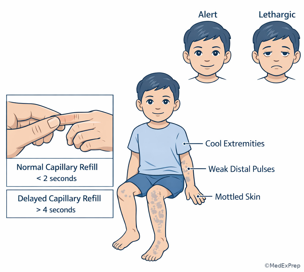
Exam takeaway: Multiple aligned abnormalities should move you quickly from interpretation to escalation.
B Worked example
A 13-year-old girl with migraine history and recent sinus infection presents with severe headache and vomiting. She has obesity and takes a combined oral contraceptive for dysmenorrhea. Vitals are BP 158/92 mm Hg, pulse 52/min, respiratory rate irregular at 10-18/min, and temperature 37.1°C. A distracting clue notes photophobia, which could pull you toward a simple migraine answer. What is the most important interpretation of this vital-sign pattern?
Reasoning chain: Identify task: interpret the vital-sign cluster. Determine age and acuity: adolescent, concerning neurologic context. Extract key facts: hypertension, bradycardia, irregular respirations. Apply rule: this triad suggests increased intracranial pressure, not uncomplicated migraine. Eliminate distractors: pain alone usually raises pulse rather than lowers it; simple anxiety does not cause this pattern; fever is absent, so sepsis is less likely. Verify: urgent neurologic evaluation and escalation are required.
C Exam trap
D Checkpoint
Question: Which combination should most strongly raise concern for impending cardiopulmonary collapse in a young child?
- A. Fever, tachycardia, warm extremities, brisk capillary refill
- B. Mild tachypnea after crying, oxygen saturation 100%
- C. Bradycardia, cyanosis, and decreased responsiveness
- D. Single elevated BP reading during a painful laceration repair
Answer: C
- A: Tempting because fever can mask serious illness, but the warm well-perfused picture is less ominous than the other options.
- B: Tempting because tachypnea always deserves attention, but the crying context and normal oxygenation are reassuring.
- C: Correct because bradycardia in a sick child, especially with cyanosis and decreased responsiveness, is a preterminal pattern.
- D: Tempting because the BP is elevated, but procedural pain commonly causes transient elevation without systemic collapse.
Exam Traps & Differentiators
Most common wrong answer and why: Applying adult normal values to infants and young children. This is tempting because adult ranges are overlearned, but pediatrics demands age-specific interpretation before every decision.
| Looks similar | Why it tempts you | Single discriminating clue | Best shelf move |
|---|---|---|---|
| Fever-related tachycardia vs shock | Both can produce a fast pulse | Shock adds poor perfusion, lethargy, weak pulses, or delayed refill | Reassess after treating fever only if perfusion is reassuring |
| Crying-related tachypnea vs respiratory distress | Both can raise respiratory rate | Respiratory distress adds retractions, flaring, grunting, or low SpO2 | Repeat if calm and well; escalate if work of breathing is present |
| Single high BP vs pediatric hypertension | One elevated number feels actionable | Diagnosis requires repeat correct measurements over time in a stable child | Repeat manually with proper cuff size |
| Low SpO2 artifact vs true hypoxemia | Pulse ox errors are common | True hypoxemia correlates with distress, cyanosis, or persistent low reading despite good signal | Check waveform and correlate clinically |
| Benign sleep-related lower pulse vs dangerous bradycardia | Sleeping children normally slow down | Dangerous bradycardia is paired with poor feeding, hypoxia, altered mental status, or poor perfusion | Escalate if symptomatic |
If the stem says... think:
- “Crying toddler, one high BP” → repeat correctly before diagnosing hypertension.
- “Normal BP but delayed cap refill and tachycardia” → compensated shock can still be present.
- “Fast breathing with clear lungs” → consider fever, pain, acidosis, or compensation, not just pneumonia.
- “Bradycardia in a sick infant” → preterminal until proven otherwise.
- “Pulse ox low on moving cold hand” → verify signal, but do not ignore if distress is also present.
- “Hypertension + bradycardia + irregular respirations” → increased intracranial pressure.
Exam takeaway: Retractions, nasal flaring, and grunting convert a fast respiratory rate from a number into a respiratory emergency clue.
{"prompt":"Realistic medical educational illustration on white background comparing a child with normal breathing to a child with increased work of breathing; labels for nasal flaring, intercostal retractions, subcostal retractions, grunting, head bobbing, normal chest movement; pediatric atlas style, clean labels, no cartoon","caption":"Normal versus increased work of breathing in a child\nExam takeaway: Retractions, nasal flaring, and grunting convert a fast respiratory rate from a number into a respiratory emergency clue.","size":"1024x1024"} Exam takeaway: Visualizing age groups helps prevent the common exam error of calling infant values abnormal by adult standards.
Tables
| Vital sign | Common benign driver | Dangerous interpretation | What resolves the choice |
|---|---|---|---|
| Tachycardia | Fever, crying, pain, anxiety | Dehydration, shock, sepsis, arrhythmia | Perfusion, persistence after calming, overall appearance |
| Tachypnea | Crying, fever, pain | Bronchiolitis, pneumonia, metabolic acidosis, respiratory failure | Work of breathing, oxygenation, lung exam, perfusion |
| Bradycardia | Sleep, athletic adolescent | Hypoxia, increased ICP, preterminal deterioration | Age, symptoms, mental status, oxygenation |
| Elevated BP | Pain, anxiety, stimulant use, wrong cuff size | Persistent pediatric hypertension | Repeated correct measurements over time |
| Low SpO2 | Motion artifact, poor perfusion, bad probe | True hypoxemia | Waveform quality plus clinical correlation |
| Pattern | Closest confounder | Shared features | Discriminator |
|---|---|---|---|
| Dehydration with compensated shock | Fever alone | Tachycardia, fatigue | Delayed refill, dry mucosa, oliguria, weak pulses |
| Respiratory distress | Crying-related tachypnea | Fast breathing | Retractions, flaring, grunting, persistent distress |
| Septic shock | Viral febrile illness | Fever, tachycardia | Poor perfusion, hypotension, petechiae, altered mental status |
| Raised intracranial pressure | Migraine | Headache, vomiting | Hypertension + bradycardia + irregular respirations |
| Physiologic infant fast HR | Pathologic tachycardia | High pulse | Age-appropriate range and reassuring exam |
| Setting | Best first move | Avoid | Why |
|---|---|---|---|
| Well-child visit with one high BP | Repeat manually with correct cuff after rest | Immediate hypertension diagnosis | Single reading may reflect anxiety or technique |
| Febrile child with isolated tachycardia | Treat fever, hydrate, reassess | Ignoring persistent elevation | Tachycardia should improve if benign |
| Infant with low SpO2 and retractions | Trust clinical distress and escalate | Blaming the probe without action | Distress corroborates true hypoxemia |
| Sick child with hypotension | Treat as late shock | Routine outpatient recheck | Pediatric hypotension is late and dangerous |
| Sleeping adolescent athlete with pulse 54 | Interpret in context | Calling all bradycardia pathologic | Symptoms and perfusion determine concern |
Algorithm / Approach
Exam takeaway: The correct next step depends on whether the abnormal value is age-adjusted, reproducible, and paired with red-flag exam findings.
See Fig 6 for the full exam-day sequence. The key is to avoid two opposite errors: overcalling normal pediatric physiology, and undercalling compensated instability because blood pressure still looks acceptable.
Rapid Review
- Infant heart rate → faster than adult; adult thresholds mislead
- Infant respiratory rate → “40s” can be normal; age decides significance
- Pediatric shock → tachycardia first, hypotension late
- Tachypnea with retractions → respiratory distress, not just fever
- Isolated office hypertension → repeat correctly before diagnosing
- Low pulse ox with bad signal → verify waveform, then correlate clinically
- Bradycardia in sick infant → dangerous until proven otherwise
- Normal BP in ill child → does not exclude shock
- Narrow pulse pressure → can support shock physiology
- Fever-related tachycardia → should improve after treatment if benign
- Persistent tachycardia after fluids/antipyretics → broaden evaluation
- HTN + bradycardia + irregular respirations → think increased ICP
- Crying-related abnormal vitals → repeat when calm if otherwise well
- Poor cap refill + tachycardia → perfusion problem beats reassuring BP
Self-check quiz
1. A healthy 3-month-old boy comes for a mild cough after daycare exposure. He is feeding well and smiling. Which respiratory rate is most likely within normal limits for age?
- A. 18/min
- B. 24/min
- C. 42/min
- D. 62/min
2. A 15-year-old girl presents for sports clearance. She has no symptoms, no medications, and a normal exam. Which resting heart rate is most consistent with an adolescent normal value?
- A. 64/min
- B. 112/min
- C. 138/min
- D. 44/min
3. A 2-year-old girl with fever and acute otitis media is crying during the exam. Temperature is 39.1°C, pulse 162/min, respiratory rate 30/min, BP 92/54 mm Hg, SpO2 100%, and capillary refill is normal. Which is the most appropriate next step?
- A. Immediate ECG for pathologic tachycardia
- B. Fluid bolus for presumed shock
- C. Antipyretics with reassessment of heart rate after calming
- D. Urgent transfer to intensive care
4. A 6-year-old boy with ADHD presents for a school physical. He drank a caffeinated soda, is anxious, and a small cuff is used for his blood pressure. The reading is 130/84 mm Hg; the rest of the exam is normal. Which is the most appropriate next step?
- A. Diagnose hypertension and begin medication
- B. Repeat blood pressure with the correct cuff after rest
- C. Order renal ultrasound immediately
- D. Ignore the reading because children do not get hypertension
5. A 5-month-old infant with 3 days of cough has respiratory rate 64/min, nasal flaring, subcostal retractions, and oxygen saturation 91% on room air. He has eczema and an older sibling with a cold. Which interpretation is most appropriate?
- A. Normal infant respiratory rate; reassure and discharge
- B. Likely artifact because pulse oximeters are unreliable in infants
- C. Respiratory distress requiring prompt escalation and supportive management
- D. Tachypnea explained by crying alone
6. A 4-year-old boy with vomiting and diarrhea for 2 days presents with pulse 158/min, respiratory rate 30/min, BP 82/50 mm Hg, cool extremities, dry mucous membranes, and capillary refill of 4 seconds. He is awake but less playful than usual. Which finding most strongly indicates clinically significant circulatory compromise rather than simple fever-related tachycardia?
- A. Age 4 years
- B. Dry mucous membranes and delayed capillary refill
- C. Recent daycare exposure
- D. Respiratory rate 30/min
7. A 12-year-old girl presents with severe headache, vomiting, and increasing somnolence after head trauma. Vital signs show BP 152/90 mm Hg, pulse 50/min, and irregular respirations. Which is the most likely interpretation of this vital-sign pattern?
- A. Anxiety-related sympathetic surge
- B. Increased intracranial pressure
- C. Septic shock
- D. Benign athletic bradycardia
8. A 10-month-old infant is brought in for poor feeding and lethargy. He is pale, has weak cries, pulse 88/min, oxygen saturation 86%, and delayed capillary refill. Which is the most appropriate interpretation of the heart rate?
- A. Normal because infants sometimes sleep deeply
- B. Expected with dehydration alone
- C. Concerning bradycardia in a sick infant requiring urgent escalation
- D. Most consistent with well-conditioned athletic physiology
Answer key
1. Answer: C
A. Tempting because 18/min is a common adult-like normal respiratory rate, but it is too low to be the best normal choice for a 3-month-old infant; the single clue is the infant age. Source: Nelson Textbook of Pediatrics; AAP pediatric assessment references.
B. Tempting because it seems more plausible than 18, but 24/min is still relatively low for a young infant and would not be the best representative normal option here; the clue is the 3-month age range. Source: Nelson.
C. Correct because a respiratory rate in the low 40s fits expected infant physiology when the infant is otherwise feeding well and well appearing; the discriminating clue is the age-adjusted norm. Source: Nelson; First Aid for the USMLE pediatrics tables.
D. Tempting because infants breathe faster than adults, but 62/min is above the usual normal range and should raise concern if persistent; the clue is that this is beyond routine infant resting rate. Source: Nelson.
2. Answer: A
A. Correct because a resting heart rate in the 60s is consistent with an adolescent normal value in an asymptomatic patient; the discriminating clue is adolescent age with no illness. Source: Nelson; AAP clinical assessment standards.
B. Tempting because mild tachycardia can be seen with anxiety, but as a resting normal value it is too high for an adolescent; the single clue is “resting” during sports clearance. Source: Nelson.
C. Tempting if you overgeneralize infant values to older children, but 138/min is too fast for a resting adolescent and suggests stress, fever, or pathology rather than normal. Source: Nelson.
D. Tempting because athletes may have lower resting rates, but 44/min is too low to be the best general normal answer in a standard asymptomatic adolescent stem; the clue is that the question asks for the most consistent typical normal value. Source: Nelson; preparticipation evaluation guidance.
3. Answer: C
A. Tempting because the pulse is high, and ECG is a common reflex for tachycardia, but this child’s fever, crying, normal perfusion, and normal oxygenation make physiologic sinus tachycardia far more likely; the discriminating clue is normal capillary refill with clear context. Source: Nelson; AAP pediatric emergency assessment guidance.
B. Tempting because tachycardia can reflect shock, but the child has no signs of poor perfusion or hypotension and is crying with fever; the single clue is normal capillary refill. Source: Nelson.
C. Correct because antipyretics and reassessment after calming are appropriate for isolated tachycardia in a febrile, crying toddler who otherwise appears stable; the mechanism is fever- and stress-mediated increased sympathetic tone. Source: Nelson; First Aid.
D. Tempting because young children can deteriorate quickly, but there are no red flags suggesting ICU-level instability; the clue is the reassuring oxygenation and perfusion. Source: Nelson.
4. Answer: B
A. Tempting because the BP appears elevated, but pediatric hypertension is not diagnosed from a single noisy reading, especially with anxiety, stimulant exposure, and wrong cuff size; the discriminator is measurement context. Source: AAP Clinical Practice Guideline for Screening and Management of High Blood Pressure in Children and Adolescents.
B. Correct because the first step in a stable child with a potentially inaccurate elevated BP is repeat measurement using the correct cuff after rest; the mechanism is avoidance of false elevation from technique and sympathetic activation. Source: AAP guideline.
C. Tempting because persistent pediatric hypertension may require renal evaluation, but that workup comes after confirmation, not before; the discriminating clue is single-reading artifact risk. Source: AAP guideline.
D. Tempting if you underappreciate pediatric hypertension, but children can have hypertension; the mistake is dismissing it entirely rather than repeating it correctly. Source: AAP guideline.
5. Answer: C
A. Tempting because infants normally breathe faster than older children, but 64/min plus flaring, retractions, and low saturation is not normal; the discriminating clue is increased work of breathing. Source: Nelson; AAP bronchiolitis guidance.
B. Tempting because pulse oximetry can be artifact-prone, but the reading is corroborated by clear respiratory distress signs, making true hypoxemia more likely; the clue is concordant physical exam findings. Source: AAP bronchiolitis guideline.
C. Correct because the infant has respiratory distress with hypoxemia and requires prompt supportive management and escalation; the mechanism is lower airway disease causing impaired ventilation and oxygenation. Source: AAP bronchiolitis guideline; Nelson.
D. Tempting because crying raises respiratory rate, but it does not explain persistent retractions and low oxygen saturation; the discriminating clue is objective work of breathing. Source: Nelson.
6. Answer: B
A. Tempting because age matters in all pediatric interpretation, but age alone does not identify circulatory compromise; the discriminating clue lies in perfusion findings. Source: Nelson; pediatric shock recognition references.
B. Correct because dry mucous membranes and delayed capillary refill directly indicate volume depletion and impaired perfusion, separating true hemodynamic compromise from simple fever-related tachycardia; the mechanism is reduced effective circulating volume. Source: Nelson; PALS concepts.
C. Tempting because daycare suggests infectious gastroenteritis, but it does not determine severity; the clue that matters is delayed refill, not exposure history. Source: Nelson.
D. Tempting because tachypnea can reflect compensation, but this value alone is less decisive than the circulatory signs; the discriminator remains perfusion status. Source: Nelson.
7. Answer: B
A. Tempting because headache and vomiting can provoke anxiety, but anxiety usually increases heart rate rather than producing bradycardia with irregular respirations; the single clue is the full triad. Source: Nelson; pediatric neurology emergency references.
B. Correct because hypertension, bradycardia, and irregular respirations are classic for increased intracranial pressure; the mechanism is Cushing response due to intracranial hypertension affecting brainstem physiology. Source: Nelson; pediatric emergency medicine texts.
C. Tempting because somnolence and abnormal vitals can occur in sepsis, but septic shock typically causes tachycardia rather than isolated bradycardia with hypertension; the discriminator is the BP-pulse pattern. Source: Nelson.
D. Tempting because athletic adolescents may be bradycardic, but that does not explain hypertension, irregular respirations, and neurologic symptoms after trauma; the clue is traumatic headache with the vital-sign triad. Source: Nelson.
8. Answer: C
A. Tempting because sleeping infants may have lower heart rates, but this infant is lethargic, poorly feeding, hypoxemic, and poorly perfused; the discriminating clue is the sick appearance. Source: Nelson; pediatric emergency assessment references.
B. Tempting because dehydration can cause abnormal perfusion, but dehydration usually causes tachycardia before bradycardia; the clue is that a low heart rate in a sick infant is not expected compensation. Source: Nelson; PALS concepts.
C. Correct because a pulse of 88/min is concerning bradycardia for a 10-month-old when paired with hypoxia and delayed refill; the mechanism is decompensation and impending deterioration, requiring urgent escalation. Source: Nelson; pediatric resuscitation standards.
D. Tempting because athletic physiology can produce low resting heart rates, but that reasoning applies to healthy older children or adolescents, not lethargic infants; the discriminator is age plus illness severity. Source: Nelson.
Keep going with the full Pediatrics Shelf Prep Course (Peds Shelf)
You just studied the way members do, in the same lesson interface. Full access unlocks every lesson, the question bank and flashcards for a full year.
One payment · 1 year of access · No auto-renewal