1.1.1 - External Ear Anatomy
External Ear Anatomy
What you’ll learn
- DOMAIN: Auditory foundations -> External ear structure recognition — identify the pinna, concha, external auditory canal, cartilaginous versus bony canal, isthmus, and lateral tympanic membrane relationship.
- DOMAIN: Audiologic assessment -> Otoscopic reasoning — connect anatomy to what can and cannot be visualized during routine otoscopy.
- DOMAIN: Interpretation -> Conductive pathway localization — decide when findings suggest an external ear contribution to conductive involvement.
- DOMAIN: Professional practice -> Scope-aware referral — recognize when external ear findings require medical referral rather than routine audiologic continuation.
- DOMAIN: Pediatric/adult management -> Age-specific anatomy cues — anticipate how infant and adult ear canal features change examination and test interpretation.
- Foundations of hearing science → outer ear anatomy and sound pathway → structure-function mapping
- Audiologic assessment and test selection → otoscopy and pretest inspection → safe test readiness
- Interpretation and site-of-lesion reasoning → conductive contribution before middle ear analysis
- Professional practice → referral, documentation, patient safety, and scope boundaries
Why it matters
- External ear findings are often the first clue that hearing difficulty is conductive before you even begin formal testing.
- Praxis-style stems commonly hide the key answer in a small anatomic clue: canal collapse, cerumen occlusion, drainage, tenderness, or congenital pinna/canal difference.
- Knowing what is normal prevents over-referral, while knowing red flags prevents unsafe continuation of testing.
- This topic sets up the next two lessons: Middle Ear Mechanics and Conductive Loss Patterns. If you miss the external ear contribution, you may misread later tympanometry or audiogram findings.
Key Terms & Must-Know Facts
1.1.1.1 Key terms
- Pinna/Auricle: visible external ear that helps collect and shape incoming sound.
- Helix: outer curved rim of the auricle.
- Antihelix: inner curved ridge parallel to the helix.
- Tragus: anterior cartilaginous prominence near canal opening.
- Antitragus: prominence opposite the tragus.
- Lobule: soft inferior portion of the auricle without cartilage.
- Concha: bowl-shaped region that funnels sound toward the canal.
- External auditory canal (EAC): passage from concha to tympanic membrane.
- Cartilaginous canal: lateral outer third of the canal; contains hair follicles and ceruminous glands.
- Bony canal: medial inner two-thirds; thinner skin, more sensitive.
- Cerumen: protective earwax produced in the cartilaginous canal.
- Isthmus: narrowest portion of the ear canal.
- Meatus: canal opening.
- Canal collapse: artificial narrowing/closure of the canal during headphone placement, especially in older adults.
- Microtia/Atresia: congenital malformation or absence/narrowing of external ear and/or canal.
1.1.1.2 Must-know facts
- The external ear includes the auricle and external auditory canal up to the lateral surface of the tympanic membrane.
- The lateral canal is cartilaginous; the medial canal is bony.
- Hair follicles and ceruminous glands are found in the cartilaginous, not bony, canal.
- The bony canal has thinner skin and is more painful when contacted by the speculum.
- Cerumen protects the canal but can also create a reversible conductive component when occluding the lumen.
- Pinna and concha help with sound collection and spectral shaping, especially for localization cues.
- External ear abnormalities can reduce sound transmission before the tympanic membrane and middle ear are even involved.
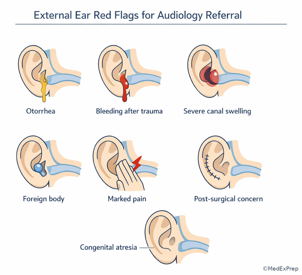
- Otorrhea, marked pain, trauma, bleeding, or suspected infection are referral-trigger findings.
- Canal collapse can mimic high-frequency hearing loss and must be checked before interpreting thresholds.
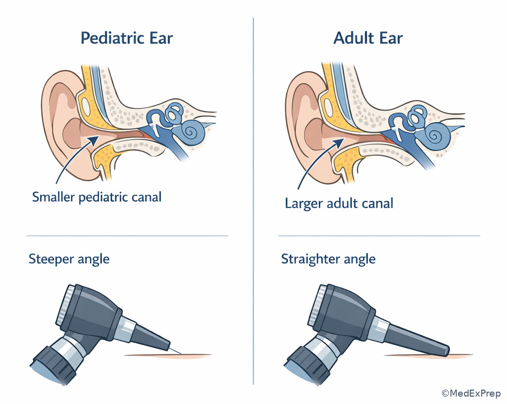
- Infant and young child canals are smaller and change the ease and angle of otoscopic visualization.
Core content
1.1.1.3 Foundational map: what structures belong to the external ear?
A Explanation
The exam decision conflict here is simple but high yield: Does a finding belong to the external ear, middle ear, or both? The single rule that resolves this is boundary recognition: the external ear extends from the auricle through the external auditory canal to the lateral surface of the tympanic membrane. If the clue is pinna shape, canal patency, cerumen, canal tenderness, stenosis, or drainage at the meatus, think external ear first. If the clue is ossicles, pressure regulation, or tympanic membrane mobility, that moves into the next topic. WHY THIS IS TESTED: stems often use one external clue to localize a conductive problem before formal results are presented.
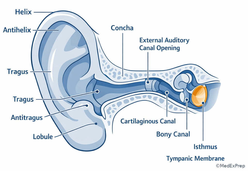
The auricle contains recognizable landmarks: helix, antihelix, tragus, antitragus, lobule, and concha. Functionally, the pinna and concha are not just cosmetic structures; they collect sound and modify spectral information that contributes to front-back and vertical localization. The canal then directs sound to the tympanic membrane. Its lateral cartilaginous segment contains glands and hair and is more flexible; its medial bony segment is narrower, more rigid, and much more sensitive. This structure-function pairing explains both normal examination behavior and patient discomfort patterns during otoscopy.
Key rule: If the problem is at the opening or within the canal lumen before the tympanic membrane, classify it as an external ear issue until proven otherwise.
See Fig 1 for the structural map and See Fig 2 for the external-to-middle boundary.
Exam takeaway: Know the auricle landmarks and canal segments so you can localize clues in stems before formal test results appear.
Exam takeaway: Distinguish canal-level clues from middle ear mechanics to avoid premature localization errors.
flowchart LR
A[Pinna and concha] --> B[External auditory canal]
B --> C[Lateral surface of tympanic membrane]
C --> D[Middle ear begins beyond membrane]
B --> E[Cerumen, stenosis, edema = external ear clues]
D --> F[Pressure, ossicles, mobility = middle ear clues]
B Worked example
A 74-year-old man presents for hearing evaluation because his family reports that he “misses speech in groups.” He has diabetes, osteoarthritis, and long-term hearing aid use in the right ear only. Otoscopy shows a narrow canal with dry cerumen laterally in the left ear and a clear view of the tympanic membrane on the right. A screening nurse note mentions “possible middle ear problem” because hearing seems reduced on the left. The most appropriate reasoning is: identify task — localize the likely anatomic contributor before accepting the label. Extract key facts — narrow canal, lateral cerumen, no middle-ear evidence given, and the clue occurs before the tympanic membrane. Apply rule — a lumen problem in the canal is an external ear issue. Eliminate distractors — do not jump to ossicular or Eustachian tube explanations without tympanic membrane or immittance clues. Verify — the left ear may have a conductive component, but the available localization is external ear first.
C Exam trap
Common wrong answer: “Middle ear disorder” based only on reduced hearing.
Why it is tempting: Many learners equate any conductive suspicion with the middle ear because ossicles are more commonly emphasized.
Single clue that eliminates it: The stem localizes the problem to the canal lumen before the tympanic membrane.
D Checkpoint
Question: Which structure-function pairing is most appropriate for the external ear?
- Concha — funnels sound toward the canal
The option is tempting because it is a visible structure that students may underappreciate; it is correct because the concha helps collect and direct sound. - Ossicular chain — matches impedance between air and cochlear fluids
This is tempting because it also affects sound transmission; it is wrong here because it belongs to the middle ear, not the external ear. - Eustachian tube — produces cerumen and traps debris
This is tempting only if the learner confuses all ear passages; it is wrong because cerumen production is a cartilaginous canal function, not Eustachian tube function. - Round window — collects localization cues from the pinna
This is tempting because localization is a hearing concept; it is wrong because the round window is an inner ear structure and does not collect sound.
Practitioner Action: ASSESS auricle landmarks, canal patency, visible cerumen, and whether the tympanic membrane can be visualized. INTERVENE only within routine audiologic pretest workflow and local policy. REFER to the appropriate medical provider if pain, trauma, active drainage, or unsafe canal status is present. DOCUMENT what was visualized, what was not visualized, laterality, and whether findings limited testing.
1.1.1.4 Foundational function: how the external ear affects sound and protection
A Explanation
The decision conflict here is choose sound-collection/protection versus pressure-transmission explanations. The single rule: the external ear primarily collects, shapes, protects, and channels sound; pressure-matching and impedance transformation belong to the middle ear. The pinna and concha add spectral shaping, which helps localization. The canal acts as a resonant tube and physical passageway, while its skin, hair, and cerumen provide protection against debris and some pathogens. When you see external canal disease or blockage, ask whether the main issue is reduced sound delivery, not ossicular motion.
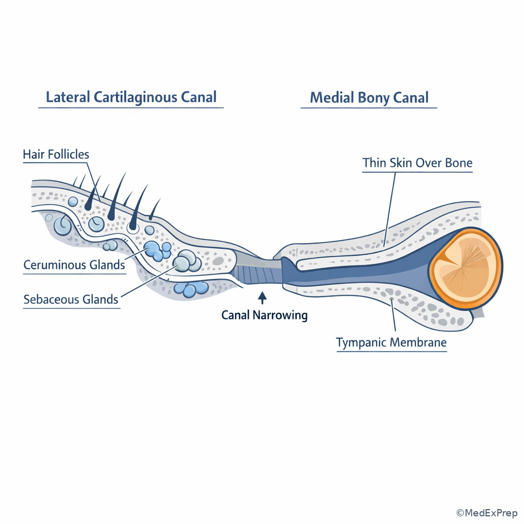
The cartilaginous canal contains ceruminous and sebaceous glands plus hair follicles; that is why wax accumulates laterally rather than medially. The bony canal, by contrast, has very thin skin adherent to bone and is easily irritated. That difference explains why deep speculum contact is painful and why instrumentation must be cautious and within scope. On the exam, if the stem emphasizes itching, discharge, canal wall tenderness, or manipulation pain, that often points to an external canal process rather than a middle ear mechanism.
Memory anchor: “C-HaG-Bare Bone” — Cartilaginous canal has Hair and Glands; the Bony canal is bare, thin, and more sensitive.
Recall: This is the first topic in the course, so build a reference point now: later in Middle Ear Mechanics, you will shift from collection/protection to impedance matching and pressure balance. Keep those jobs separate.
See Fig 3 for the cartilaginous-versus-bony comparison.
Exam takeaway: Hair and glands live laterally, while the medial bony canal is thinner-skinned and more painful when contacted.
Exam takeaway: Separate collection/protection clues from pressure/mobility clues to localize the problem correctly.
flowchart TD
A[External ear] --> A1[Collects sound]
A --> A2[Shapes localization cues]
A --> A3[Protects canal with cerumen and hair]
A --> A4[Channels sound to membrane]
B[Middle ear] --> B1[Pressure regulation]
B --> B2[Impedance matching]
B --> B3[Ossicular transmission]
B Worked example
A 29-year-old swimmer reports left ear fullness and reduced hearing after a week at the beach. He has seasonal allergies and eczema. He denies fever but says the outer ear is sore when touched. Tympanic membrane visualization is limited by canal swelling and debris. A classmate suggests Eustachian tube dysfunction because of the “fullness” complaint. Reasoning chain: identify task — decide whether the symptom cluster fits external or middle ear function. Extract key facts — swimmer exposure, outer-ear tenderness, canal swelling, debris, limited view. Apply rule — tenderness with manipulation and canal wall involvement point to external ear pathology. Eliminate distractors — allergy history makes middle ear dysfunction tempting, but it does not explain focal canal tenderness. Verify — the most appropriate audiology action is to defer assumptions about middle ear mechanics, document findings, and refer for medical evaluation if indicated by local policy and safety considerations.
C Exam trap
Common wrong answer: “Pressure imbalance from the Eustachian tube.”
Why it is tempting: Fullness is a shared symptom and allergy history nudges learners toward middle ear thinking.
Single clue that eliminates it: Pain with manipulation or focal canal tenderness is an external canal clue, not a pressure-regulation clue.
D Checkpoint
Question: Which finding most strongly supports a cartilaginous canal process rather than a middle ear problem?
- Visible lateral cerumen with hair-bearing canal skin
This is tempting because learners may dismiss it as a normal variant; it is correct because hair-bearing skin and cerumen glands identify the lateral cartilaginous canal. - Reduced tympanic membrane compliance on immittance
This is tempting because it can explain reduced hearing; it is wrong because it localizes beyond the external ear. - Negative middle ear pressure pattern
This is tempting because fullness can coexist with hearing change; it is wrong because it is a middle ear pressure finding. - Absent ossicular coupling
This is tempting because it also causes conductive involvement; it is wrong because ossicular mechanics are middle ear anatomy.
- The most common site of cerumen production is the cartilaginous lateral canal.
- True or False: The bony canal is usually less sensitive than the cartilaginous canal. False.
- Name the 3 key external ear jobs: collect, protect, channel.
Practitioner Action: ASSESS whether symptoms fit canal-level protection problems versus middle ear pressure problems. INTERVENE by adjusting test setup, using safe otoscopy technique, and following local cerumen/otoscopy policy. REFER when swelling, significant pain, drainage, or infection suspicion limits safe audiologic progression. DOCUMENT symptom triggers, visualization limits, and how findings affected test selection.
1.1.1.5 Applied anatomy: otoscopy, canal patency, and what the audiologist must decide
A Explanation
Now the decision conflict becomes practical: Do you continue routine testing, modify technique, or refer? The single rule is safety plus interpretability. If the canal is open, nonpainful, and the tympanic membrane can be adequately visualized, routine testing usually proceeds. If cerumen or anatomy limits the view but does not create acute danger, you document the limitation and determine whether testing remains interpretable. If there is active drainage, bleeding, marked edema, trauma, severe pain, or suspected foreign body, stop and refer appropriately. WHY THIS IS TESTED: Praxis questions often ask for the best next step, not just anatomy labeling.
Otoscopy is not a decorative pretest ritual; it determines whether headphones, inserts, immittance, or further measures are appropriate. Canal patency matters because a partially or fully occluded canal can create an apparent conductive pattern. Insert earphones may be preferred in some situations to reduce canal collapse from supra-aural pressure, but they are not a substitute for ignoring unsafe findings. Also, “could not visualize the tympanic membrane due to cerumen” is a clinically meaningful documentation statement, not a failure. On exam stems, the correct answer is often the one that respects scope: you identify, document, modify testing if safe, and refer when necessary rather than providing medical treatment.
Key rule: An external ear finding that makes results unreliable or the patient unsafe outranks the desire to complete the battery.
High-Yield Connection: This concept is tested alongside Conductive Loss Patterns. Know both the external ear obstruction clue and the later audiogram clue of an air-bone gap; they are frequently paired in stems.
See Fig 4 for the otoscopy decision tree.
Exam takeaway: The best next step depends on safety and test interpretability, not on finishing the battery at all costs.
flowchart TD
A[Otoscopy] --> B{Canal open and nonpainful?}
B -- Yes --> C{TM adequately visualized?}
C -- Yes --> D[Proceed with routine testing]
C -- No --> E[Document limitation and decide if results will remain interpretable]
B -- No --> F{Drainage, trauma, severe pain, foreign body, marked edema?}
F -- Yes --> G[Refer / urgent medical follow-up as indicated]
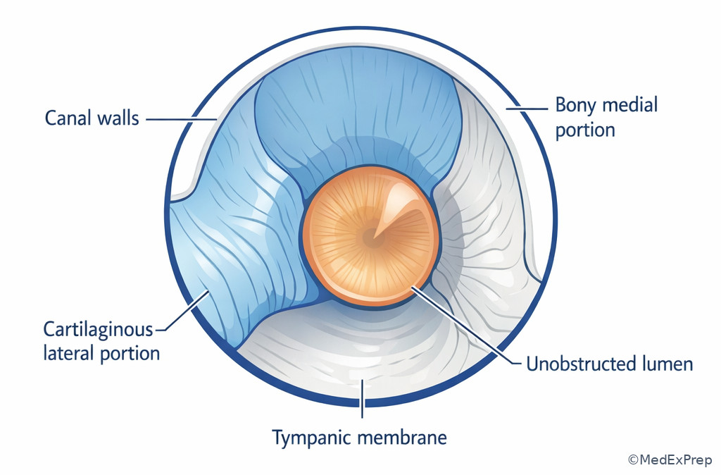
F -- No --> H[Modify plan per safety and local policy] Exam takeaway: Recognize the expected pathway and normal view so abnormal narrowing or obstruction stands out quickly.
B Worked example
A 7-year-old child is referred after a school screen. He has asthma, allergic rhinitis, and a history of recurrent cerumen buildup. He is cooperative but anxious. Otoscopy reveals the right canal is mostly occluded with dark cerumen, and the tympanic membrane cannot be visualized. The left ear is clear. A parent asks whether the child “probably just has fluid.” Reasoning chain: identify task — choose the most appropriate next audiologic step. Extract key facts — unilateral occluding cerumen, inability to visualize the tympanic membrane, no middle ear data yet, tempting history of rhinitis. Apply rule — do not label middle ear status when the external canal prevents adequate inspection and may alter test results. Eliminate distractors — immediate interpretation as otitis media is unsupported; continuing as if the ears are equally testable may reduce reliability. Verify — document occlusion, adjust test expectations, and refer/manage according to local policy for cerumen and safe follow-up.
C Exam trap
Common wrong answer: “Conclude probable middle ear effusion based on school-age history and allergies.”
Why it is tempting: The child’s age and rhinitis history fit a common pediatric middle ear story.
Single clue that eliminates it: The tympanic membrane cannot be visualized because the canal is occluded, so middle ear status has not yet been established.
D Checkpoint
Question: During pretest otoscopy, which finding most clearly requires stopping routine audiologic progression and seeking medical follow-up rather than simply documenting and continuing?
- Partial nonoccluding cerumen with adequate tympanic membrane view
This is tempting because wax can influence hearing; it is wrong because adequate visualization and safety usually allow continued testing. - Narrow canal in an older adult without pain or drainage
This is tempting because it may affect transducer choice; it is wrong because it calls for technique adjustment, not automatic referral. - Active purulent drainage from the canal with pain
This is tempting because the learner may want to continue to obtain baseline thresholds; it is correct because active drainage and pain are safety and referral triggers. - Dry lateral cerumen that partially limits the view
This is tempting because incomplete visualization is clinically relevant; it is wrong because this alone does not automatically require stopping everything if the case remains safe and policy allows next steps.
Practitioner Action: ASSESS whether the canal permits safe transducer placement and interpretable findings. INTERVENE by choosing appropriate transducer strategy or rescheduling if local protocols require clearance first. REFER to primary care/otology/urgent care as indicated for drainage, trauma, significant pain, or foreign body suspicion. DOCUMENT what prevented visualization and how that changed the plan.
1.1.1.6 Pattern recognition: common external ear abnormalities and conductive clues
A Explanation
The decision conflict is choose external obstruction/malformation versus deeper conductive pathology. The single rule: if the external ear abnormality physically reduces sound access to the tympanic membrane, expect an external conductive contribution. High-yield examples include cerumen impaction, canal stenosis, atresia, foreign body, edema from otitis externa, and collapsed canals from headphone pressure. These do not all produce identical audiometric patterns, but they share one logic: sound delivery into the canal is impaired before middle ear mechanics are tested.
Canal collapse is especially testable because it creates an artificial hearing pattern. Older adults with soft pinnae and narrow canals may show worse high-frequency air-conduction thresholds with supra-aural earphones. The best next step is not to over-interpret presbycusis or middle ear disease; it is to suspect collapse and use insert earphones when appropriate. Congenital microtia or atresia also matters because external ear differences can predict conductive pathways that bypass normal canal transmission, and these cases often point forward to bone conduction device concepts later in the course.
Memory anchor: Think “BLOCK” for external conductive clues — Buildup of wax, Lumen narrowing, Otitis externa swelling, Canal collapse, Kongenital atresia/microtia.
High-Yield Connection: This concept overlaps with Hearing Aid Fundamentals and Bone Conduction Device Concepts. External ear malformation changes both diagnosis and device pathway options.
See Fig 5 for the comparison of major external ear conductive mechanisms.
Exam takeaway: External obstruction or malformation can create a conductive pattern before middle ear pathology is considered.
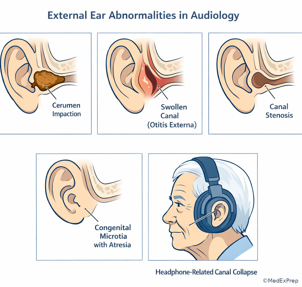
Exam takeaway: Apparent high-frequency loss in older adults may be artifactual if the ear canal collapses during testing.
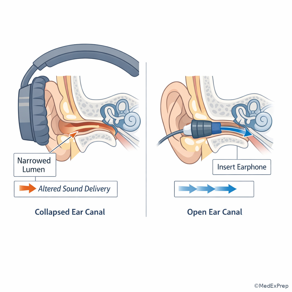
B Worked example
An 81-year-old woman presents for annual audiometry. She has hypertension, osteopenia, and mild hand tremor. She reports no ear pain and no recent infection. Using supra-aural headphones, you obtain unexpectedly poor high-frequency thresholds bilaterally compared with last year, but speech understanding in quiet seems unchanged. Otoscopy shows very soft pinnae and narrow canals without heavy cerumen. A trainee suggests rapidly progressive sensorineural decline. Reasoning chain: identify task — decide whether the pattern is true hearing change or testing artifact. Extract key facts — older adult, soft pinnae, narrow canals, high-frequency worsening, stable functional report. Apply rule — suspect canal collapse before concluding cochlear decline. Eliminate distractors — no pain or drainage for otitis externa; no middle ear evidence; no true red-flag asymmetry. Verify — repeat with insert earphones when appropriate and compare results before interpretation.
C Exam trap
Common wrong answer: “Presbycusis progression is the best explanation.”
Why it is tempting: Age plus high-frequency threshold worsening strongly resembles a familiar sensorineural pattern.
Single clue that eliminates it: The combination of soft pinnae, narrow canals, and supra-aural earphones points first to canal collapse artifact.
D Checkpoint
Question: Which scenario most strongly suggests an external ear source for a conductive contribution?
- Older adult with high-frequency threshold shift only when tested under supra-aural headphones
This is tempting because age-related sensorineural loss is common; it is correct because the transducer-specific pattern suggests canal collapse. - Adult with negative pressure tympanogram and recent upper respiratory infection
This is tempting because it also causes conductive symptoms; it is wrong because it localizes to middle ear pressure dysfunction. - Child with absent acoustic reflexes and stiff tympanic membrane pattern
This is tempting because conductive findings are present; it is wrong because the clue points to middle ear stiffness, not the external ear. - Adult with unilateral tinnitus and asymmetric sensorineural thresholds
This is tempting because it raises site-of-lesion concern; it is wrong because it is not an external ear conductive clue.
- True or False: Canal collapse can mimic high-frequency loss. True.
- The congenital pair most associated with external ear malformation is microtia/atresia.
- If supra-aural results look worse than expected in an older adult, think insert earphones.
Practitioner Action: ASSESS for obstruction, swelling, congenital difference, or transducer-related collapse. INTERVENE by repeating with a more appropriate transducer when safe and within protocol. REFER congenital or medically active abnormalities for otologic evaluation and collaborative management. DOCUMENT whether the suspected barrier is anatomical, inflammatory, occlusive, or artifactual.
1.1.1.7 Integration: age, referral triggers, and scope-aware next steps
A Explanation
The final decision conflict is when to monitor, when to adapt testing, and when to escalate. The single rule: external ear anatomy becomes an audiology management issue only as far as it affects safe assessment, interpretation, counseling, and referral. You do not prescribe, surgically manage, or independently medically diagnose beyond scope. Instead, you identify red flags and choose the safest next step. Pediatric canals are smaller and may be harder to inspect; older adult canals may collapse more easily; congenital anomalies may require interdisciplinary referral; active drainage, trauma, or sudden painful change should shift the answer toward medical follow-up.
Common red flags include otorrhea, bleeding after trauma, severe otalgia, marked swelling, suspected foreign body, post-surgical concerns, or findings inconsistent with routine audiology continuation. In exam stems, one distractor may ask you to begin medical treatment or make a surgical plan. Reject those answers because they exceed entry-level audiology scope. Another distractor may ask you to ignore the issue and continue testing “for baseline data.” Reject that when safety or interpretability is compromised. The correct answer usually combines observation, documentation, counseling, and referral.
Key rule: In ambiguous external ear cases, the best answer is the one that protects the patient and stays within audiology scope while preserving interpretable data.
Recall: In this lesson we built the first localization step. In the upcoming Middle Ear Mechanics topic, the key shift is from “Is sound getting into the ear canal?” to “What happens once sound reaches the tympanic membrane and ossicles?”
See Fig 6 for the referral-oriented summary algorithm.
Exam takeaway: External ear questions are often really scope and safety questions disguised as anatomy items.
flowchart TD
A[External ear finding] --> B{Safe and testable?}
B -- Yes --> C{Results likely interpretable?}
C -- Yes --> D[Proceed and document]
C -- No --> E[Modify technique or reschedule after management per policy]
B -- No --> F{Pain, drainage, trauma, foreign body, marked swelling?}
F -- Yes --> G[Medical referral / urgent follow-up as indicated]
F -- No --> H[Document and coordinate nonurgent follow-up] Exam takeaway: Age changes the ease of inspection and the likelihood of certain external ear testing issues.
Exam takeaway: Drainage, trauma, severe pain, and foreign body concern should shift the answer toward medical follow-up.
B Worked example
A 46-year-old woman presents for tinnitus evaluation. She has rheumatoid arthritis, migraines, and recent urgent-care treatment for “ear discomfort.” On otoscopy, the right canal contains moist debris and she reports sharp pain when the auricle is manipulated. She also mentions that tinnitus is worse on that side, making retrocochlear pathology feel like a tempting concern. Reasoning chain: identify task — choose the most appropriate next step within scope. Extract key facts — painful manipulation, moist debris, active canal finding, no formal asymmetry data yet. Apply rule — a medically active and painful external canal finding should prompt referral rather than routine completion of the battery. Eliminate distractors — do not begin steroid therapy, do not infer vestibular schwannoma from tinnitus alone, and do not force testing through pain. Verify — document the canal findings, explain the need for medical follow-up, and arrange audiologic reassessment when appropriate.
C Exam trap
Common wrong answer: “Complete the full audiologic battery first because tinnitus evaluation is the referral reason.”
Why it is tempting: Learners want complete data and may focus on the referral question instead of the safety problem.
Single clue that eliminates it: Painful manipulation and moist debris signal an active external ear condition that changes the immediate priority.
D Checkpoint
Question: Which action is most appropriate for an entry-level audiologist when otoscopy reveals active otorrhea and marked canal tenderness before a scheduled hearing test?
- Proceed quickly to get baseline pure tones before the canal worsens
This is tempting because baseline data seem useful; it is wrong because safety and interpretability come first. - Start topical treatment and return for repeat testing in 1 week
This is tempting because it sounds efficient; it is wrong because prescribing or directing medical treatment exceeds scope. - Document the finding, counsel the patient, and arrange medical referral/follow-up
This is tempting because it may feel less “complete” than testing; it is correct because it matches scope and patient safety. - Interpret the likely middle ear diagnosis and send specific surgical recommendations
This is tempting because it appears decisive; it is wrong because the finding is external and operative planning is outside scope.
Practitioner Action: ASSESS age-related anatomy, pain, drainage, trauma history, and test readiness. INTERVENE with safe modification, counseling, and deferral when needed. REFER to medical providers for active disease, trauma, congenital concerns needing interdisciplinary care, or any finding beyond audiology scope. DOCUMENT red flags, patient instructions, and the rationale for referral or delayed testing.
Exam Traps & Differentiators
- Most common wrong answer and why: Choosing a middle ear explanation for any conductive-sounding symptom. This is tempting because middle ear pathology is heavily emphasized in training, but stems that mention wax, canal swelling, canal tenderness, meatal narrowing, or headphone-related collapse are testing external ear localization.
- Looks similar but isn’t: Fullness from canal swelling can look like pressure dysfunction; the discriminator is manipulation pain or visible canal wall abnormality.
- Looks similar but isn’t: High-frequency worsening in older adults can look like presbycusis; the discriminator is transducer-dependent change suggesting canal collapse.
- Looks similar but isn’t: Pediatric reduced hearing plus allergies can suggest otitis media; the discriminator is inability to even assess the tympanic membrane because external occlusion blocks the view.
- If the stem says pain when touching or pulling the auricle, think external canal involvement.
- If the stem says hearing worsens under supra-aurals but improves with inserts, think canal collapse artifact.
- If the stem says active drainage or bleeding, think stop and refer rather than complete routine testing.
- If the stem says congenital microtia/atresia, think external pathway abnormality with conductive implications and interdisciplinary management.
- If the stem says “cannot visualize the tympanic membrane due to cerumen,” think you do not yet know the middle ear status.
| Finding in stem | Tempting interpretation | Better interpretation | Single discriminator |
|---|---|---|---|
| Fullness + allergies | Eustachian tube dysfunction | External canal disorder if tenderness/swelling present | Pain with auricle/canal manipulation |
| Older adult + high-frequency drop | Presbycusis progression | Canal collapse artifact | Occurs with supra-aurals; improves with inserts |
| Child failed screen + wax | Middle ear effusion | External occlusion may explain reduced access | TM not visualized due to canal blockage |
| Drainage + hearing complaint | Finish testing first | Medical referral/follow-up first | Active otorrhea is a red flag |
| Microtia/atresia | Cosmetic issue only | External conductive pathway abnormality | Absent or narrowed canal pathway |
High-Yield Connection: Traps here overlap with upcoming Conductive Loss Patterns. External canal obstruction and middle ear stiffness can both lower air-conduction sensitivity, but only one starts before the tympanic membrane. Keep that boundary clear on exam day.
Tables
| External ear structure | High-yield function | Exam clue if abnormal |
|---|---|---|
| Pinna/Auricle | Collects and shapes sound | Congenital malformation, trauma, localization issues |
| Concha | Funnels sound toward canal opening | Structure-recognition anatomy items |
| Cartilaginous canal | Flexible outer canal with hair and glands | Cerumen, edema, otitis externa clues |
| Bony canal | Rigid medial pathway to membrane | Pain with deep contact; thin sensitive skin |
| Isthmus | Narrow segment of canal | Retention/occlusion tendency, visualization limits |
| Cerumen | Protective barrier and lubricant | Occlusion causing conductive contribution |
| Lateral tympanic membrane boundary | Marks external-to-middle transition | Localize pathology correctly in stems |
| Comparison point | External ear | Middle ear | Why the distinction matters |
|---|---|---|---|
| Primary job | Collect, protect, channel sound | Impedance match, pressure regulate, transmit vibration | Prevents localizing all conductive clues to one system |
| Typical symptom clue | Canal tenderness, blockage, drainage at meatus | Pressure, popping, mobility change, effusion story | Supports best-next-step questions |
| Common test interference | Cerumen, collapse, stenosis | Abnormal tympanometry, reflex changes | Guides what to inspect before interpreting data |
| Key anatomic structures | Pinna, concha, canal | TM medial mechanics, ossicles, Eustachian tube | Separates topics across the course |
| Frequent referral triggers | Otorrhea, trauma, foreign body, severe canal pain | Persistent middle ear pathology patterns | Shapes scope-appropriate escalation |
| Differential / pattern | Typical external clue | Tempting wrong choice | Best next step within scope |
|---|---|---|---|
| Cerumen occlusion | Canal blockage; limited TM view | Assume otitis media | Document limitation; manage per policy/referral path |
| Otitis externa pattern | Canal tenderness, swelling, debris, drainage | Continue full test battery | Stop if unsafe; refer medically |
| Canal collapse artifact | Threshold shift with supra-aurals | Progressive SNHL | Retest with inserts if appropriate |
| Microtia/atresia | Visible congenital external anomaly | Cosmetic issue only | Coordinate audiologic assessment and referral |
| Middle ear pressure problem | No canal tenderness or occlusion; later immittance clues | External canal diagnosis | Move to middle ear reasoning in next topic |
| Age group | External ear consideration | Testing implication |
|---|---|---|
| Infant | Small canal, limited view, developmental anatomy differences | Otoscopy and probe placement require extra care |
| Child | Cerumen and cooperation may limit visualization | Do not overcall middle ear disease without view/data |
| Adult | Common canal obstruction or dermatitis history | Interpret symptoms with canal-level clues |
| Older adult | Soft pinnae and collapse risk | Consider insert earphones when pattern suggests artifact |
Algorithm / Approach
Rapid Review
- Auricle → visible collector, not pressure regulator.
- Concha → funnels sound toward the canal opening.
- Cartilaginous canal → hair and ceruminous glands live here.
- Bony canal → thinner skin, more sensitivity with contact.
- Cerumen → protective when present, conductive when occlusive.
- External ear boundary → ends at lateral tympanic membrane surface.
- Canal tenderness → think external canal, not pressure-only middle ear disease.
- Cannot visualize TM due to wax → middle ear status remains unconfirmed.
- Supra-aural high-frequency drop in older adult → suspect canal collapse before SNHL progression.
- Microtia/atresia → external pathway abnormality with conductive implications.
- Otorrhea → safety/referral issue before routine testing.
- Allergy history + fullness → not enough for middle ear diagnosis if canal clues dominate.
- Best next step questions → choose safety, interpretability, and scope awareness.
Memory anchor: “Outside before inside” — if the stem gives a canal clue before any tympanic or immittance clue, localize outside first.
Self-check quiz
1. A 23-year-old graduate student with allergic rhinitis and no prior ear surgery presents for baseline audiology. The examiner asks which portion of the ear canal contains hair follicles and ceruminous glands. Which answer is most appropriate?
- Lateral cartilaginous canal
- Medial bony canal
- Round window niche
- Eustachian tube opening
2. A 68-year-old man with osteoarthritis and hypertension is learning otoscopy technique. He asks which external ear structure most directly funnels sound into the ear canal. Which answer is most appropriate?
- Concha
- Malleus
- Oval window
- Tensor tympani
3. A 10-year-old child with asthma and seasonal allergies fails a school hearing screen. Otoscopy in clinic shows the right canal is fully occluded with cerumen and the tympanic membrane cannot be visualized; the left ear is clear. The parent asks whether this means middle ear fluid is present. Which is the most appropriate interpretation?
- The finding confirms middle ear effusion in the right ear
- The finding localizes an external ear issue that may affect hearing and limits middle ear conclusions
- The finding most strongly suggests retrocochlear pathology
- The finding proves permanent sensorineural hearing loss
4. A 79-year-old woman with diabetes and mild kyphosis reports stable communication ability, but routine testing with supra-aural headphones shows a new bilateral high-frequency drop. Otoscopy shows narrow canals and soft pinnae without drainage. Which is the most appropriate next step?
- Conclude rapid presbycusis progression and counsel immediately
- Repeat air-conduction testing with insert earphones if appropriate to evaluate canal collapse
- Begin medical treatment for otitis externa
- Assume middle ear stiffness and skip retesting
5. A 34-year-old swimmer with eczema and recent ear fullness presents for evaluation. He has canal pain with manipulation, visible edema, and moist debris in the left ear. Which action is most appropriate for the audiologist?
- Proceed with the full battery to establish a baseline before referral
- Document the external ear findings and arrange medical referral/follow-up rather than forcing routine testing
- Prescribe topical drops and schedule a repeat test
- Interpret the likely middle ear diagnosis and recommend surgery
6. A 6-year-old child with repaired cleft palate history, recurrent ear infections, and speech delay presents for hearing evaluation. On the right, the canal is narrow but patent and the tympanic membrane is visualized; on the left, the canal is blocked by cerumen and the membrane cannot be seen. The child’s teacher reports inconsistent listening. Which statement is the best interpretation at this stage?
- Bilateral middle ear disease is established because of the cleft palate history
- The left ear has an external canal limitation that must be considered before concluding middle ear status, while the right ear can proceed to further assessment
- The left ear most likely has retrocochlear pathology because the child has speech delay
- The external findings are irrelevant because teacher report outweighs otoscopy
7. A 52-year-old man with psoriasis, chronic tinnitus, and hyperlipidemia presents for hearing testing. He reports new right ear pain after using cotton swabs. Otoscopy shows scant blood in the canal and marked tenderness; he wants “just a quick hearing test today” because he took off work. Which is the most appropriate next step?
- Complete testing because tinnitus is the chief complaint and the blood is minimal
- Document the traumatic canal finding, defer routine testing if needed for safety, and direct the patient to medical evaluation
- Start oral medication and proceed with immittance only
- Interpret the likely tympanic membrane perforation and explain surgical options
8. A newborn with microtia of the right pinna, otherwise stable vitals, and no acute distress is referred for audiologic follow-up. The family asks whether the visible ear difference is purely cosmetic. Which response is most appropriate?
- Yes; external ear differences do not affect hearing if the infant is medically stable
- No; external ear malformation can affect the sound pathway and requires coordinated audiologic assessment and medical follow-up
- No; it proves cochlear damage on that side
- Yes; wait until school age before any hearing-related planning
Answer key
1.1.1.8 1. Correct answer: A. Lateral cartilaginous canal
A. Lateral cartilaginous canal — Tempting because it is a basic anatomy fact that can be forgotten under pressure; correct because the lateral cartilaginous canal contains hair follicles and ceruminous glands, explaining where wax is produced. The single stem clue is the question asking specifically about glands and hair. Authoritative support: Katz et al.; Valente et al.
B. Medial bony canal — Tempting because it is also part of the ear canal; incorrect because the bony canal has thin skin and greater sensitivity, not hair-bearing glandular tissue. The discriminator is “ceruminous glands.” Authoritative support: Katz et al.
C. Round window niche — Tempting because it is part of ear anatomy; incorrect because it is an inner ear/middle ear interface concept, not external canal skin anatomy. The discriminator is location outside the external ear. Authoritative support: ASHA-aligned foundational audiology texts.
D. Eustachian tube opening — Tempting because it also concerns ear passages; incorrect because it belongs to the middle ear/nasopharyngeal pressure pathway, not wax production. The discriminator is that the question asks about the canal. Authoritative support: Katz et al.
1.1.1.9 2. Correct answer: A. Concha
A. Concha — Tempting because the visible bowl of the auricle is easy to overlook; correct because it helps funnel sound into the external auditory canal. The discriminator is “funnels sound into the canal.” Authoritative support: Katz et al.; Musiek and Chermak.
B. Malleus — Tempting because it is involved early in sound transmission; incorrect because it is a middle ear ossicle, not an external ear collector. The clue is that the question asks about external sound funneling. Authoritative support: Katz et al.
C. Oval window — Tempting because it lies in the transmission pathway; incorrect because it is a middle-to-inner ear interface, not an external ear structure. The discriminator is structure location. Authoritative support: Valente et al.
D. Tensor tympani — Tempting because it is associated with the ear and sound mechanics; incorrect because it is a middle ear muscle. The clue is external collection versus internal mechanics. Authoritative support: Katz et al.
1.1.1.10 3. Correct answer: B. The finding localizes an external ear issue that may affect hearing and limits middle ear conclusions
A. The finding confirms middle ear effusion in the right ear — Tempting because the child has allergy history and failed a screen; incorrect because occluding cerumen prevents adequate visualization and does not establish middle ear status. The discriminator is “tympanic membrane cannot be visualized.” Authoritative support: ASHA pediatric assessment principles; Katz et al.
B. The finding localizes an external ear issue that may affect hearing and limits middle ear conclusions — Tempting because it sounds less definitive than many exam options; correct because canal occlusion is an external ear finding that can alter hearing and blocks direct middle ear assessment. The discriminator is external occlusion before the tympanic membrane. Authoritative support: Katz et al.; ASHA practice resources.
C. The finding most strongly suggests retrocochlear pathology — Tempting because school failure may prompt broad concern; incorrect because no neural asymmetry or site-of-lesion clue is given. The discriminator is the visible canal blockage. Authoritative support: Katz et al.
D. The finding proves permanent sensorineural hearing loss — Tempting because failed screening can worry families; incorrect because an external blockage may create a reversible conductive component. The discriminator is the occluding cerumen. Authoritative support: ASHA screening follow-up guidance.
1.1.1.11 4. Correct answer: B. Repeat air-conduction testing with insert earphones if appropriate to evaluate canal collapse
A. Conclude rapid presbycusis progression and counsel immediately — Tempting because the patient is older and the change is high frequency; incorrect because the transducer setup and narrow, collapsible canals create an artifactual explanation that must be checked first. The discriminator is supra-aural testing in an older adult with soft pinnae. Authoritative support: Katz et al.; ANSI/clinical audiometric best-practice teaching standards.
B. Repeat air-conduction testing with insert earphones if appropriate to evaluate canal collapse — Tempting because it requires an extra step; correct because it directly tests the most likely artifact source while remaining within audiology scope. The discriminator is the transducer-dependent collapse risk. Authoritative support: Katz et al.; audiometric procedure references.
C. Begin medical treatment for otitis externa — Tempting because canal issues can affect hearing; incorrect because there is no pain, edema, or drainage, and prescribing is outside scope. The discriminator is absence of inflammatory canal signs. Authoritative support: scope-of-practice standards; ASHA.
D. Assume middle ear stiffness and skip retesting — Tempting because conductive mechanisms are familiar; incorrect because no middle ear evidence is provided and the external ear clue is stronger. The discriminator is soft pinnae and narrow canals under supra-aurals. Authoritative support: Katz et al.
1.1.1.12 5. Correct answer: B. Document the external ear findings and arrange medical referral/follow-up rather than forcing routine testing
A. Proceed with the full battery to establish a baseline before referral — Tempting because baseline data seem useful in a symptomatic patient; incorrect because canal pain, edema, and debris indicate a medically active external ear condition that may make testing unsafe or uninterpretable. The discriminator is pain with manipulation plus visible edema. Authoritative support: ASHA scope guidance; Katz et al.
B. Document the external ear findings and arrange medical referral/follow-up rather than forcing routine testing — Tempting because it may feel less complete; correct because it prioritizes safety, stays within scope, and acknowledges the external ear pathology. The discriminator is active canal inflammation. Authoritative support: ASHA professional practice; audiology otoscopy safety teaching standards.
C. Prescribe topical drops and schedule a repeat test — Tempting because it sounds efficient and definitive; incorrect because medication management is outside the entry-level audiologist role in this exam context. The discriminator is scope boundary. Authoritative support: ASHA scope-of-practice.
D. Interpret the likely middle ear diagnosis and recommend surgery — Tempting because fullness can be confused with middle ear disease; incorrect because surgery recommendations exceed scope and the visible pathology is external. The discriminator is canal edema and manipulation pain. Authoritative support: ASHA scope-of-practice; Katz et al.
1.1.1.13 6. Correct answer: B. The left ear has an external canal limitation that must be considered before concluding middle ear status, while the right ear can proceed to further assessment
A. Bilateral middle ear disease is established because of the cleft palate history — Tempting because cleft palate history raises middle ear risk; incorrect because risk is not proof, and the left ear has a clear external barrier preventing visualization. The discriminator is that one TM is seen and one is not due to wax. Authoritative support: pediatric audiology texts; ASHA pediatric assessment guidance.
B. The left ear has an external canal limitation that must be considered before concluding middle ear status, while the right ear can proceed to further assessment — Tempting because it requires nuanced laterality-specific reasoning; correct because it respects the actual otoscopic findings rather than overgeneralizing from history. The discriminator is asymmetric visualization status. Authoritative support: Katz et al.; ASHA pediatric practice resources.
C. The left ear most likely has retrocochlear pathology because the child has speech delay — Tempting because speech delay can make learners worry about neural issues; incorrect because no neural pattern is presented, and a visible external canal limitation is the immediate issue. The discriminator is cerumen blockage. Authoritative support: pediatric differential diagnosis standards.
D. The external findings are irrelevant because teacher report outweighs otoscopy — Tempting because functional report is important; incorrect because teacher concerns do not erase a clear pretest anatomical limitation. The discriminator is that otoscopy shapes test readiness and interpretation. Authoritative support: ASHA assessment standards.
1.1.1.14 7. Correct answer: B. Document the traumatic canal finding, defer routine testing if needed for safety, and direct the patient to medical evaluation
A. Complete testing because tinnitus is the chief complaint and the blood is minimal — Tempting because the patient wants efficiency and tinnitus is the referral reason; incorrect because traumatic canal bleeding and marked tenderness create a safety issue that may limit reliable testing. The discriminator is blood plus marked tenderness after swab trauma. Authoritative support: ASHA scope and patient safety principles; Katz et al.
B. Document the traumatic canal finding, defer routine testing if needed for safety, and direct the patient to medical evaluation — Tempting because it seems less satisfying than completing the planned battery; correct because it addresses an acute external ear problem within scope and prioritizes patient safety. The discriminator is the active traumatic finding. Authoritative support: ASHA professional practice documents.
C. Start oral medication and proceed with immittance only — Tempting because it proposes treatment plus limited testing; incorrect because medication is outside scope and proceeding may remain unsafe. The discriminator is scope boundary combined with trauma. Authoritative support: ASHA scope-of-practice.
D. Interpret the likely tympanic membrane perforation and explain surgical options — Tempting because blood can make perforation seem likely; incorrect because the stem gives canal trauma, not confirmed TM perforation, and surgical counseling is outside scope. The discriminator is unconfirmed deeper pathology with clear external trauma. Authoritative support: scope-of-practice guidance; Katz et al.
1.1.1.15 8. Correct answer: B. No; external ear malformation can affect the sound pathway and requires coordinated audiologic assessment and medical follow-up
A. Yes; external ear differences do not affect hearing if the infant is medically stable — Tempting because families often separate appearance from function; incorrect because microtia can be associated with canal atresia or other conductive pathway issues. The discriminator is visible congenital external ear malformation. Authoritative support: pediatric audiology texts; ASHA pediatric evaluation resources.
B. No; external ear malformation can affect the sound pathway and requires coordinated audiologic assessment and medical follow-up — Tempting because it is broader and less absolute; correct because it accurately reflects external conductive implications and interdisciplinary management needs. The discriminator is congenital external ear anomaly in a newborn. Authoritative support: pediatric audiology practice standards; Katz et al.
C. No; it proves cochlear damage on that side — Tempting because severe congenital ear findings can make inner ear damage seem likely; incorrect because external malformation does not by itself prove cochlear involvement. The discriminator is pathway location. Authoritative support: pediatric differential diagnosis references.
D. Yes; wait until school age before any hearing-related planning — Tempting because some learners may underestimate early intervention urgency; incorrect because newborn hearing-related follow-up should not be delayed when visible external anomalies may affect access. The discriminator is newborn timing and early hearing access importance. Authoritative support: ASHA infant assessment guidance; JCIH principles.
Keep going with the full Praxis Audiology (5343) Prep Course (Praxis Aud)
You just studied the way members do, in the same lesson interface. Full access unlocks every lesson, the question bank and flashcards for a full year.
One payment · 1 year of access · No auto-renewal