1.1.1 - Eyelid Lacrimal And Orbit
Why it matters
- Eyelid and orbital findings are high-yield because many board stems test localization before diagnosis: surface, lid, lacrimal drainage, extraocular muscle cone, apex, or neurologic pathway.
- The safest answer often depends on one red-flag discriminator: pain with eye movement, decreased vision, RAPD, ophthalmoplegia, fever, or acute proptosis.
- Tear symptoms are easy to misread: epiphora can mean too much tearing or blocked drainage; the exam rewards mechanism-based interpretation, not symptom memorization.
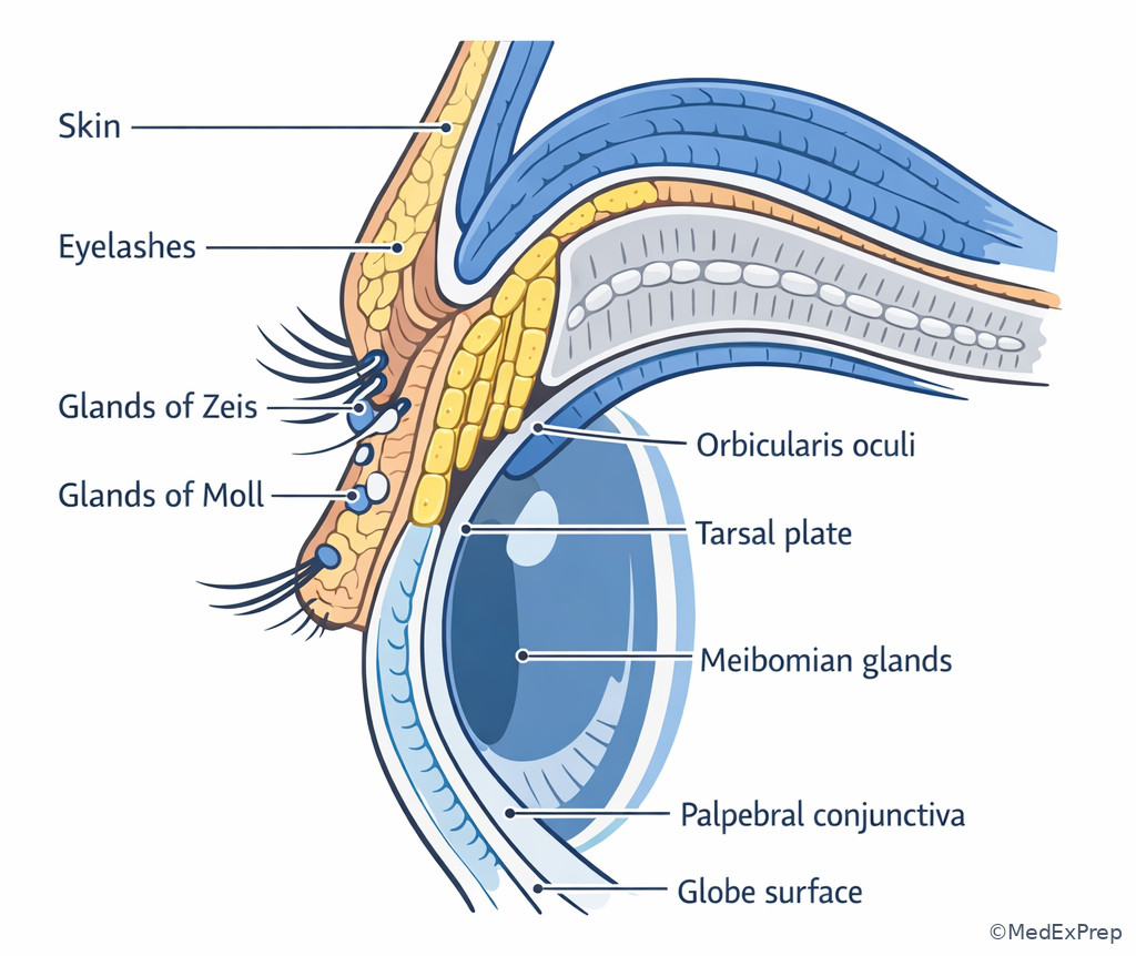
- This topic builds the framework needed for later corneal pain/reflex tearing and retinal/orbital urgency questions. See Fig 1.
Key Terms & Must-Know Facts
1.1.1.1 Key terms
- Tarsal plate — dense connective tissue giving lid structural rigidity
- Meibomian glands — sebaceous glands in tarsus producing tear-film lipid layer
- Glands of Zeis — sebaceous glands associated with eyelashes
- Glands of Moll — modified apocrine sweat glands at lash line
- Orbicularis oculi — CN VII muscle closing eyelids
- Levator palpebrae superioris — CN III skeletal muscle elevating upper lid
- Müller muscle — sympathetic smooth muscle assisting upper lid elevation
- Canaliculus — tear drainage channel from punctum to lacrimal sac
- Nasolacrimal duct — drains lacrimal sac into inferior meatus
- Orbital septum — fibrous barrier separating eyelid from deeper orbit
- Annulus of Zinn — common tendinous ring at orbital apex
- Proptosis — anterior displacement of globe
- Enophthalmos — posterior displacement of globe
- Preseptal cellulitis — infection anterior to orbital septum
- Orbital cellulitis — infection posterior to septum with orbital signs
1.1.1.2 Must-know facts
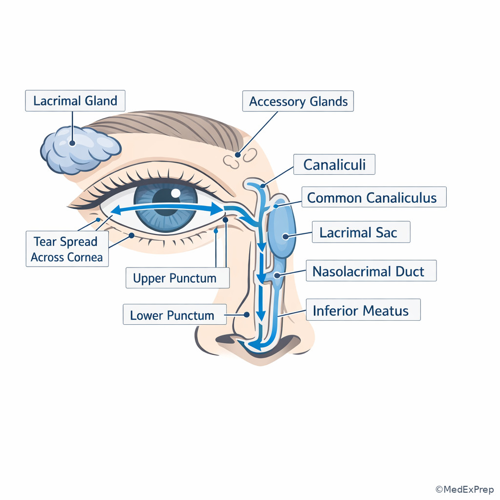
- Tears are produced mainly by the main lacrimal gland and accessory glands; they drain through puncta → canaliculi → sac → nasolacrimal duct.
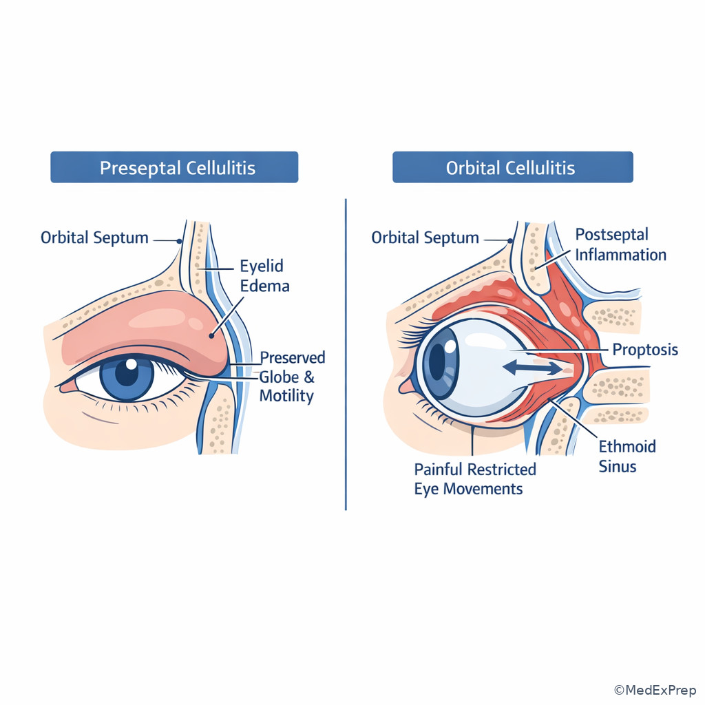
- The orbital septum is the classic exam boundary distinguishing preseptal from orbital infection.
- Pain with eye movement strongly pushes a stem toward orbital rather than purely eyelid disease.
- Meibomian dysfunction destabilizes the lipid layer and can cause reflex tearing despite underlying dryness.
- Hordeolum is acute and tender; chalazion is chronic, granulomatous, and usually nontender.
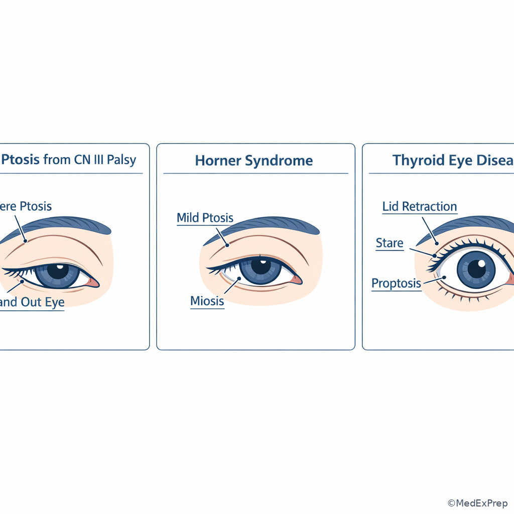
- CN III palsy causes ptosis from levator weakness; Horner syndrome causes milder ptosis from sympathetic denervation of Müller muscle.
- The orbit has 7 bones: frontal, sphenoid, zygomatic, maxillary, palatine, lacrimal, ethmoid.
- The medial orbital wall is thin; ethmoid sinus disease can spread into the orbit.
- The lacrimal gland sits in the superotemporal orbit; lacrimal sac sits inferonasal.
- Acute orbital disease with decreased vision, RAPD, color desaturation, or optic disc changes is a vision-threatening referral pattern.
| Structure | Main role | High-yield clue |
|---|---|---|
| Tarsal plate | Lid support | Contains meibomian glands |
| Orbicularis oculi | Lid closure | CN VII lesion can impair blink and exposure protection |
| Levator palpebrae | Upper lid elevation | CN III palsy causes marked ptosis |
| Müller muscle | Assists upper lid elevation | Sympathetic lesion causes mild ptosis |
| Lacrimal gland | Aqueous tear secretion | Superotemporal fullness suggests dacryoadenitis or mass |
| Lacrimal sac | Tear collection before nasal drainage | Inferonasal swelling suggests dacryocystitis |
| Orbital septum | Barrier | Key line between preseptal and orbital cellulitis |
Core content
1.1.1.3 Foundational eyelid anatomy: layers, muscles, glands, and innervation
A Explanation
The exam decision conflict here is usually: Is this a lash-margin/gland problem, a levator problem, or a facial-nerve closure problem? The single rule that resolves many stems is to identify which lid function is impaired: opening, closing, margin secretion, or protection of the ocular surface. The eyelid is built from skin, subcutaneous tissue, orbicularis, septum, tarsus, and conjunctiva. Within the tarsal plate sit the meibomian glands, which supply the outer lipid layer of the tear film. If the stem describes chronic evaporative symptoms with capped glands and frothy margin changes, choose meibomian dysfunction over aqueous deficiency.
WHY THIS IS TESTED: board questions often give similar lid swelling or ptosis presentations and ask you to choose among chalazion, hordeolum, CN III ptosis, Horner ptosis, or CN VII exposure risk. You must choose the functionally correct compartment and reject answers based only on appearance.
The upper lid is elevated mainly by levator palpebrae superioris (CN III) and secondarily by Müller muscle (sympathetic). Marked ptosis with ophthalmoplegia or pupil involvement points toward CN III pathway dysfunction, whereas mild ptosis with miosis suggests Horner syndrome. Lid closure depends on orbicularis oculi (CN VII). If blink is weak, the practical consequence is exposure and surface disease rather than a primary gland disorder.
Exam takeaway: Match the impaired lid function to the involved structure before choosing the diagnosis.
flowchart TD
A[Lid complaint] --> B{Main problem}
B --> C[Margin tenderness or focal nodule]
B --> D[Ptosis]
B --> E[Incomplete closure or weak blink]
C --> F[Acute tender lash or gland lesion -> hordeolum]
C --> G[Chronic firm nontender tarsal lesion -> chalazion]
D --> H[Severe ptosis with EOM or pupil findings -> CN III pattern]
D --> I[Mild ptosis with small pupil -> sympathetic pattern]
E --> J[CN VII weakness -> exposure risk] Exam takeaway: Start with the function that failed, then localize to gland, retractor, or closure mechanism.
Exam takeaway: Know where the tarsus, meibomian glands, orbicularis, septum, and conjunctiva sit relative to one another.
B Worked example
A 23-year-old contact lens wearer with seasonal allergies and a remote history of acne rosacea presents with a 3-week upper-lid bump. She reports occasional tearing and mild morning crusting. Visual acuity is unchanged. The lesion is firm, localized within the tarsal area, and nontender. A distracting clue is that the lid margin looks mildly erythematous, making acute infection tempting.
Reasoning chain: Identify task: localize a lid lesion. Extract key facts: chronic, firm, nontender, tarsal. Apply rule: blocked meibomian gland with lipogranulomatous inflammation gives a chalazion. Eliminate distractors: hordeolum is usually acute and painful; CN III or Horner ptosis would cause lid position change rather than a discrete nodule; dacryocystitis would be inferonasal near the lacrimal sac. Verify: rosacea and meibomian dysfunction make chalazion more likely.
C Exam trap
D Checkpoint
Question: A 64-year-old man with diabetes and hypertension presents with new ptosis of the right upper lid. He also has diplopia and limitation of adduction, elevation, and depression. The pupil is mildly enlarged. Which structure is most directly responsible for the major lid droop?
- Levator palpebrae superioris — tempting because the stem centers on ptosis; correct because marked ptosis with CN III-pattern motility deficits localizes to levator dysfunction.
- Müller muscle — tempting because sympathetic lesions can cause ptosis; wrong because Horner ptosis is usually mild and associated with miosis, not ophthalmoplegia.
- Orbicularis oculi — tempting because it is a major lid muscle; wrong because it closes the lid rather than elevating it, so weakness causes lagophthalmos rather than ptosis.
- Frontalis — tempting because patients recruit it to compensate for ptosis; wrong because it is compensatory, not the primary failed retractor in CN III palsy.
1.1.1.4 Lacrimal system and tear pathway: secretion, spread, drainage, and reflex tearing
A Explanation
The decision conflict here is: Is watery eye caused by excess reflex tearing from surface irritation or by impaired drainage? The single clue that often resolves the stem is where the abnormality sits: superotemporal gland involvement suggests secretion; medial canthus swelling and reflux suggest drainage obstruction. The main lacrimal gland lies in the superotemporal orbit and secretes aqueous tears, while accessory glands contribute baseline support. Blinking spreads tears medially across the ocular surface toward the puncta, then into canaliculi, lacrimal sac, and nasolacrimal duct.
WHY THIS IS TESTED: watery eyes are a classic distractor-heavy symptom. Exams reward choosing mechanism over symptom label. A patient can tear excessively because the eye is actually dry from evaporative instability, corneal irritation, trichiasis, or blepharitis. Conversely, persistent overflow with medial canthal swelling points to drainage failure.
Reflex tearing involves trigeminal sensory input from the ocular surface and parasympathetic output to the gland. This is a key bridge to the upcoming cornea topic: corneal irritation can drive tearing even when the complaint sounds like “too many tears.” See Fig 2. Also remember the pump mechanism of blinking: poor lid apposition or laxity can impair punctal function and worsen epiphora without a primary gland problem.
Exam takeaway: Watery eyes require you to separate reflex overproduction from outflow obstruction.
flowchart LR
A[Lacrimal gland superotemporal] --> B[Tears spread across ocular surface]
B --> C[Puncta]
C --> D[Canaliculi]
D --> E[Lacrimal sac]
E --> F[Nasolacrimal duct]
F --> G[Inferior meatus]
H[Surface irritation or dry eye] --> I[Reflex tearing]
I --> B
J[Drainage obstruction] --> K[Epiphora with overflow at lid margin]
Exam takeaway: Use location and associated signs to decide between tear overproduction and drainage failure.
Exam takeaway: Distinguish the superotemporal lacrimal gland from the inferonasal drainage structures.
B Worked example
A 71-year-old woman with rheumatoid arthritis, rosacea, and prior cataract surgery reports persistent tearing of the right eye for 6 months. She uses antihistamine drops for itching. She denies fever. Exam shows puncta in good position, mild meibomian plugging, punctate epithelial staining, and no inferonasal mass. The tempting but wrong clue is the complaint of “too many tears,” which can push you toward nasolacrimal obstruction.
Reasoning chain: Identify task: determine why the eye waters. Extract key facts: autoimmune dryness risk, evaporative risk from rosacea, punctate staining, no lacrimal sac swelling. Apply rule: a dry or irritated ocular surface triggers reflex tearing. Eliminate distractors: dacryocystitis would usually produce inferonasal tenderness/swelling; dacryoadenitis would be superotemporal; punctal malposition is not described. Verify: the surface findings explain the tearing better than drainage blockage.
C Exam trap
D Checkpoint
Question: A 35-year-old patient presents with swelling and pain in the superotemporal upper orbit with tearing and mild conjunctival injection after a recent viral illness. Which structure is most likely involved?
- Lacrimal gland — tempting because tearing is present; correct because superotemporal swelling localizes to the lacrimal gland region.
- Lacrimal sac — tempting because it is part of the tear pathway; wrong because sac disease is inferonasal near the medial canthus.
- Nasolacrimal duct — tempting because obstruction causes epiphora; wrong because the duct is not in the superotemporal orbit.
- Canaliculus — tempting because it directly receives tears; wrong because canalicular disease localizes medially near the puncta rather than superotemporally.
- The main lacrimal gland sits in the superotemporal orbit.
- True or False: Epiphora always means too much tear production. False.
- Name the tear drainage sequence: Puncta → canaliculi → sac → nasolacrimal duct.
1.1.1.5 Orbit fundamentals: walls, apex, compartments, and neurovascular relevance
A Explanation
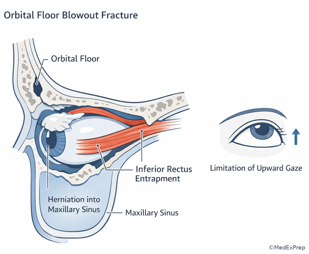
The decision conflict is: Does this stem localize to the globe surface, the eyelid, or the orbit proper? The single rule is that proptosis, motility restriction, orbital pain, sensory changes in V1/V2 distribution, or optic nerve dysfunction shift the process posterior to the septum and into orbital anatomy. The orbit is a pyramidal space with the apex posteriorly. Its seven bones matter because the thin medial wall and floor explain common spread and fracture patterns. Ethmoid sinus infection can spread medially into the orbit; blowout fractures commonly involve the floor and can trap the inferior rectus.
WHY THIS IS TESTED: stems often hide localization inside anatomy. The right answer depends less on memorizing all foramina and more on recognizing what can or cannot happen if disease is actually in the orbit. For example, eyelid cellulitis alone should not reduce vision or restrict eye movements, but orbital involvement can. At the apex, multiple cranial nerves and the optic nerve run closely together, so combined vision and motility deficits strongly suggest deep orbital localization.
The extraocular muscles form a cone around the optic nerve. Intraconal processes more readily affect optic nerve function and cause axial proptosis. Extraconal masses may displace the globe away from the lesion. Lacrimal gland lesions in the superotemporal orbit often push the globe inferonasally. See Fig 3.
Exam takeaway: Globe displacement pattern and associated deficits help localize orbital disease by compartment.
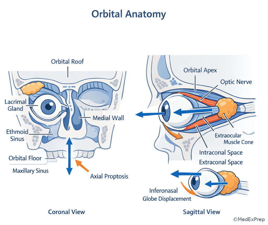
Exam takeaway: Proptosis plus motility or optic nerve findings should move you posterior to the septum.
Exam takeaway: Floor injury with inferior rectus entrapment causes vertical diplopia and limited upgaze.
B Worked example
A 19-year-old soccer player with allergic rhinitis and mild myopia is struck in the left orbit by an elbow. He has nausea, vertical diplopia, and pain on upgaze. There is minimal external bruising, which is a distracting finding that may make the injury seem minor. Visual acuity is normal, but upgaze is limited.
Reasoning chain: Identify task: localize post-traumatic diplopia. Extract key facts: trauma, vertical diplopia, limited upgaze, nausea, relatively subtle external appearance. Apply rule: orbital floor blowout fracture may trap the inferior rectus, restricting upward gaze. Eliminate distractors: simple lid edema does not explain a specific duction defect; optic nerve injury would threaten vision; isolated corneal abrasion causes pain but not restrictive motility. Verify: the orbital floor lies above the maxillary sinus and is a classic blowout site.
C Exam trap
D Checkpoint
Question: A patient with a superotemporal orbital mass develops inferonasal globe displacement. Which orbital structure is most likely the origin?
- Lacrimal gland — tempting and correct because its superotemporal position displaces the globe inferonasally when enlarged.
- Ethmoid sinus — tempting because medial disease can affect the orbit; wrong because medial lesions more often displace the globe laterally.
- Lacrimal sac — tempting because it is part of adnexal anatomy; wrong because it is medial and inferior, not superotemporal.
- Inferior rectus — tempting because muscle enlargement can affect position; wrong because the described directional displacement is most classically tied to a superotemporal extraconal lesion like the lacrimal gland.
1.1.1.6 Applied pattern recognition: ptosis, lid retraction, proptosis, and orbital inflammation
A Explanation
The decision conflict is: Which single sign best separates neurologic ptosis, sympathetic ptosis, thyroid eye disease, and inflammatory/infectious orbitopathy? The high-yield discriminator is the combination of lid position with pupil, motility, pain, and laterality. Severe ptosis plus ophthalmoplegia suggests CN III pathway involvement. Mild ptosis plus miosis suggests Horner syndrome. Lid retraction and stare with proptosis suggest thyroid eye disease. Painful proptosis with fever or decreased vision raises orbital cellulitis or other acute inflammatory orbital disease.
WHY THIS IS TESTED: the exam likes near-neighbor answer sets in which every option can affect lid position or ocular alignment. You win by choosing the sign that cannot be ignored. In thyroid eye disease, retraction and restrictive motility are more characteristic than simple conjunctival redness. In Horner syndrome, ptosis is usually subtle because only Müller muscle is lost. In CN III palsy, the lid droop is more dramatic because levator is the main elevator.
Orbital inflammation can also mimic these patterns. Pain is powerful: inflammatory or infectious orbitopathy is usually painful; neurogenic ptosis may not be. See Fig 4. If optic nerve compromise is described by decreased color vision, RAPD, or vision loss, that becomes a safety signal rather than a subtle localization puzzle.
Exam takeaway: Pair lid position with pupil, motility, and pain to separate the major orbital and neurologic patterns.
flowchart TD
A[Abnormal lid position or proptosis] --> B{Main pattern}
B --> C[Severe ptosis + ophthalmoplegia +/- large pupil]
B --> D[Mild ptosis + miosis]
B --> E[Lid retraction + stare + proptosis]
B --> F[Painful proptosis + fever or vision change]
C --> G[CN III pattern]
D --> H[Horner pattern]
E --> I[Thyroid eye disease pattern]
F --> J[Orbital cellulitis or acute orbital inflammation] Exam takeaway: One associated clue—pupil, pain, or motility—usually breaks the tie.
Exam takeaway: Visually contrast severe CN III ptosis, mild Horner ptosis with miosis, and thyroid-related lid retraction.
B Worked example
A 46-year-old woman with Graves disease, hyperlipidemia, and dry eye symptoms presents with foreign-body sensation, vertical diplopia on upgaze, and a “staring” appearance. She recently started working in a dry warehouse, which is a distracting environmental clue. Exam shows lid retraction, bilateral proptosis, and restricted elevation.
Reasoning chain: Identify task: choose the pattern behind lid and motility findings. Extract key facts: Graves history, lid retraction, proptosis, restrictive motility. Apply rule: thyroid eye disease causes lid retraction and enlargement/fibrosis of extraocular muscles, often with restrictive diplopia. Eliminate distractors: Horner causes mild ptosis, not retraction; CN III palsy causes ptosis rather than stare; isolated dry eye cannot explain proptosis and motility restriction. Verify: orbital, not surface-only, disease is being tested.
C Exam trap
D Checkpoint
Question: A 58-year-old man with long-standing smoking history and recent neck pain presents with mild right upper-lid ptosis and a smaller right pupil. Extraocular movements are full, and there is no proptosis. Which pattern is most likely being tested?
- Horner syndrome — tempting and correct because mild ptosis with ipsilateral miosis and intact motility fits sympathetic pathway disruption.
- CN III palsy — tempting because ptosis is present; wrong because CN III palsy usually causes larger motility abnormalities and often mydriasis rather than miosis.
- Thyroid eye disease — tempting because lid abnormalities are common; wrong because thyroid disease more often causes retraction or proptosis, not mild ptosis with miosis.
- Myasthenia gravis — tempting because variable ptosis is classic; wrong because the small pupil specifically favors Horner rather than neuromuscular junction disease.
- The major upper-lid elevator is levator palpebrae superioris.
- True or False: Mild ptosis with miosis is more consistent with Horner than CN III palsy. True.
- Name the 3 key thyroid eye clues: lid retraction, proptosis, restrictive motility.
1.1.1.7 Integration: preseptal vs orbital cellulitis and other urgent orbital red flags
A Explanation
The decision conflict is the classic one: preseptal cellulitis or orbital cellulitis? The single rule that resolves it is whether there are orbital signs posterior to the septum: pain with eye movement, ophthalmoplegia, proptosis, decreased vision, RAPD, or color desaturation. Lid erythema and edema alone can occur in either a superficial lid process or preseptal infection, so those findings by themselves are poor discriminators.
WHY THIS IS TESTED: this is a safety-heavy board distinction. The exam wants you to recognize when a candidate-level optometry answer must escalate urgently rather than staying in a routine eyelid framework. Ethmoid sinusitis is a major clue because the thin medial orbital wall permits spread. Fever helps, but a normal temperature does not rule out orbital disease if deep signs are present. See Fig 5.
Other urgent red flags that can masquerade as “orbital swelling” include cavernous sinus involvement, orbital apex syndrome, compressive optic neuropathy, and retrobulbar hemorrhage after trauma. The mechanism differs, but the exam-safe response pattern is similar: if vision, pupil, color vision, or motility is threatened, choose urgent escalation rather than conservative local care.
Exam takeaway: Orbital signs—not lid swelling alone—trigger urgent orbital referral logic.
flowchart TD
A[Red swollen eyelid] --> B{Orbital signs present?}
B -->|No pain with EOM, no proptosis, vision intact, full motility| C[Preseptal pattern]
B -->|Pain with EOM or ophthalmoplegia or proptosis or vision change| D[Orbital pattern]
C --> E[Anterior to septum]
D --> F[Posterior to septum]
F --> G[Urgent imaging and specialist evaluation]
E --> H[Still assess for progression and systemic clues] Exam takeaway: Screen first for the orbital signs that change the safety answer.
Exam takeaway: Compare superficial lid edema with posterior orbital involvement causing proptosis and painful restricted motility.
B Worked example
A 12-year-old boy with recent ethmoid sinusitis, asthma, and eczema presents with a swollen left upper and lower eyelid, fever, and worsening pain. His mother reports he had a “stye” last year, which is a tempting but irrelevant clue. Visual acuity is slightly reduced compared with baseline, he has pain with extraocular movements, and mild proptosis is present.
Reasoning chain: Identify task: determine urgency and localization. Extract key facts: sinusitis, fever, pain with eye movement, proptosis, mild visual reduction. Apply rule: these are orbital signs posterior to the septum. Eliminate distractors: preseptal cellulitis can cause dramatic lid swelling but should not produce proptosis and painful EOM restriction; chalazion/hordeolum are focal lid lesions without orbital signs; allergic edema is usually pruritic rather than painful with EOM limitation. Verify: urgent orbital referral logic is the safe best answer.
C Exam trap
D Checkpoint
Question: Which finding most strongly requires you to reject a preseptal-only answer in a patient with lid swelling?
- Localized lid tenderness — tempting because tenderness suggests infection; wrong because it can occur in superficial lid disease.
- Fever — tempting because systemic symptoms imply severity; wrong because fever can occur with either preseptal or orbital infection.
- Pain with extraocular movements — tempting and correct because it is a classic sign of posterior orbital involvement.
- Conjunctival injection — tempting because it suggests eye involvement; wrong because injection is nonspecific and does not alone prove orbital disease.
1.1.1.8 Integration: high-yield compare/contrast across eyelid, lacrimal, and orbit
A Explanation
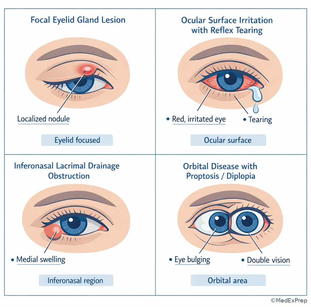
The final exam-level conflict is broad: when multiple adnexal structures could explain redness, tearing, swelling, or diplopia, which one best fits the entire pattern? The single resolving rule is to prioritize the deepest abnormal sign. If the stem has only a focal tender lid lesion, stay local. If it has punctate staining and watery eyes, think surface-driven reflex tearing. If it has inferonasal mass/tenderness, think drainage apparatus. If it has proptosis or motility restriction, think orbit. The exam often adds distracting comorbidities that fit more than one answer; the best option explains the whole pattern with the fewest assumptions.
WHY THIS IS TESTED: NBEO-style applied basic science questions reward structured elimination. You do not need specialist management; you need safe localization and the ability to recognize which finding changes urgency. This topic also connects forward to cornea and retina: exposure from lid dysfunction injures the surface, and orbital apex disease can secondarily affect optic nerve function. See Fig 6.
Exam takeaway: In mixed lid-tear-orbit stems, the deepest and most specific sign should control your answer choice.
flowchart TD
A[Swelling, tearing, pain, or diplopia] --> B{Deepest sign?}
B --> C[Focal margin lesion]
B --> D[Surface staining or irritation]
B --> E[Inferonasal sac tenderness or reflux]
B --> F[Proptosis, painful EOM, vision or pupil change]
C --> G[Lid gland disorder]
D --> H[Surface disease with reflex tearing]
E --> I[Lacrimal drainage disorder]
F --> J[Orbital disease or urgent referral pattern] Exam takeaway: Let the most dangerous specific sign outrank the most obvious superficial symptom.
Exam takeaway: Use symptom clusters to separate lid lesions, surface-driven tearing, drainage disorders, and orbital disease.
B Worked example
A 67-year-old man with diabetes, chronic blepharitis, and a recent upper respiratory infection presents with tearing, lid redness, and mild pain near the medial canthus. He mentions chronic dry eye, which is a tempting clue toward reflex tearing. Exam shows focal inferonasal tenderness and swelling below the medial canthal tendon, without proptosis or painful EOM.
Reasoning chain: Identify task: localize the source of tearing and swelling. Extract key facts: inferonasal focal swelling, medial canthal tenderness, no orbital signs. Apply rule: lacrimal sac/drainage apparatus involvement best fits. Eliminate distractors: dry-eye reflex tearing does not cause a focal inferonasal mass; orbital cellulitis would create deeper signs; chalazion is tarsal, not inferonasal. Verify: the anatomy points to dacryocystic-region disease rather than surface or orbit.
C Exam trap
D Checkpoint
Question: A patient presents with tearing, conjunctival irritation, and a new inability to close the left eyelids completely after Bell palsy. Which mechanism best explains the tearing?
- Reflex tearing from exposure-related surface irritation — tempting and correct because poor lid closure causes exposure and secondary reflex tearing.
- Primary lacrimal gland hypersecretion — tempting because the eye is watery; wrong because the stem points to an exposure trigger rather than isolated gland overproduction.
- Nasolacrimal duct obstruction — tempting because overflow suggests blocked drainage; wrong because incomplete lid closure and surface irritation provide a stronger mechanism.
- Lacrimal sac inflammation — tempting because tearing can accompany sac disease; wrong because no inferonasal swelling or focal medial tenderness is described.
Exam Traps & Differentiators
Most common wrong answer and why: Choosing the most obvious superficial symptom instead of the most localizing sign. Example: picking preseptal cellulitis because the lid is swollen, even when pain with eye movement makes orbital cellulitis the safer answer.
| Looks similar | Why it tempts you | Single best discriminator | Best board answer direction |
|---|---|---|---|
| Hordeolum vs chalazion | Both are focal lid bumps | Acute tender = hordeolum; chronic firm nontender = chalazion | Use time course and tenderness |
| Dry eye reflex tearing vs drainage obstruction | Both produce watery eye | Surface staining/irritation vs inferonasal swelling or reflux | Choose mechanism, not symptom label |
| Preseptal vs orbital cellulitis | Both have red swollen lids | Painful EOM, proptosis, vision change, or ophthalmoplegia = orbital | Escalate urgently if orbital signs |
| CN III ptosis vs Horner ptosis | Both have droopy lid | Severe ptosis + EOM deficit/large pupil vs mild ptosis + miosis | Pair ptosis with pupil and motility |
| Thyroid eye disease vs surface dry eye | Both can cause foreign-body sensation | Proptosis/lid retraction/restrictive motility = thyroid orbitopathy | Let orbital signs outrank irritation symptoms |
If the stem says...
- “Pain with eye movement” → think orbital involvement, not isolated lid disease.
- “Superotemporal swelling” → think lacrimal gland region.
- “Inferonasal tender mass near medial canthus” → think lacrimal sac/drainage pathway.
- “Mild ptosis with miosis” → think Horner pattern.
- “Lid retraction with proptosis” → think thyroid eye disease.
- “Watery eye with punctate staining” → think surface-driven reflex tearing.
Related traps from upcoming topics matter too. Corneal irritation can create reflex tearing and photophobia that mimic primary lacrimal disease, while optic nerve compromise in deeper orbital disease can later resemble neuro-ophthalmic pathology. Build the habit of localizing before naming the disease.
Tables
| Key fact/value table | High-yield answer |
|---|---|
| Main upper-lid elevator | Levator palpebrae superioris, CN III |
| Secondary upper-lid elevator | Müller muscle, sympathetic |
| Lid closure muscle | Orbicularis oculi, CN VII |
| Main lacrimal gland location | Superotemporal orbit |
| Lacrimal sac location | Inferonasal medial canthal region |
| Key anatomic boundary | Orbital septum |
| Most useful orbital cellulitis clue | Pain with extraocular movement |
| Classic thyroid eye lid clue | Lid retraction |
| Comparison | Location | Typical pain/tenderness | Best discriminator |
|---|---|---|---|
| Hordeolum | Lash follicle or lid gland | Acute, tender | Short time course with focal inflammatory pain |
| Chalazion | Tarsal meibomian gland | Usually nontender | Chronic firm nodule |
| Dacryoadenitis | Superotemporal orbit | Painful/full | Superotemporal swelling |
| Dacryocystic-region disease | Inferonasal medial canthus | Focal tenderness | Medial canthal sac localization |
| Orbital cellulitis | Posterior to septum | Painful, deep | Painful EOM/proptosis/vision change |
| Differential/classification | Key stem clue | Reject if... |
|---|---|---|
| CN III ptosis | Marked ptosis with EOM limitation, possible mydriasis | The ptosis is mild and paired with miosis only |
| Horner syndrome | Mild ptosis with miosis | There is major ophthalmoplegia or lid retraction |
| Thyroid eye disease | Lid retraction, proptosis, restrictive motility | The eye has only surface irritation without orbital signs |
| Preseptal cellulitis | Red swollen lid without orbital signs | Painful EOM, proptosis, or visual deficit appears |
| Reflex tearing from surface disease | Watering with staining/irritation/blepharitis | Medial canthal mass or sac tenderness is present |
Algorithm / Approach
Rapid Review
- Chalazion → chronic firm nontender tarsal nodule
- Hordeolum → acute tender inflammatory lash/gland lesion
- CN III ptosis → severe lid droop plus EOM deficit
- Horner ptosis → mild ptosis plus miosis
- Orbicularis weakness → incomplete closure with exposure risk, not ptosis
- Lacrimal gland disease → superotemporal swelling
- Lacrimal sac disease → inferonasal medial canthal tenderness
- Reflex tearing → watery eye plus staining/irritation
- Drainage obstruction → epiphora with medial outflow localization
- Orbital cellulitis → pain on EOM or proptosis changes the answer
- Thyroid eye disease → lid retraction beats dryness as the key clue
- Orbital floor blowout → vertical diplopia with limited upgaze after trauma
- Extraconal mass → globe displaced away from lesion
- Intraconal process → more likely axial proptosis and optic nerve risk
Self-check quiz
- Chalazion
- External hordeolum
- Preseptal cellulitis
- Dacryocystic-region inflammation
- CN III palsy
- Horner syndrome
- Thyroid eye disease
- Orbicularis oculi weakness
- Primary nasolacrimal outflow obstruction
- Reflex tearing from ocular surface irritation
- Acute lacrimal sac infection
- Superotemporal lacrimal gland mass
- Routine lid lesion localized to the tarsus
- Preseptal cellulitis anterior to the septum
- Orbital cellulitis posterior to the septum
- Allergic eyelid edema without orbital involvement
- Inferior rectus with orbital floor entrapment
- Lacrimal gland compression
- Superior oblique tendon inflammation
- Nasolacrimal duct obstruction
- Tearing in a dry environment
- Conjunctival injection
- Restrictive motility with proptosis
- Punctate epithelial staining
- Lacrimal drainage apparatus
- Intraconal orbital compartment
- Primary levator complex dysfunction
- Main lacrimal gland in the superotemporal orbit
- Müller muscle
- Orbicularis oculi
- Levator palpebrae superioris
- Frontalis
Answer key
1.1.1.9 Question 1
Correct answer: A. Chalazion
- A. Chalazion — tempting because the lesion is in the lid and linked to meibomian dysfunction; correct because the chronic, firm, painless tarsal nodule is the classic discriminator. Single stem clue: 1-month firm, painless nodule within the tarsal lid. Source: American Academy of Ophthalmology; Kanski and Bowling.
- B. External hordeolum — tempting because blepharitis and rosacea predispose to lid-margin inflammation; incorrect because hordeola are usually acute and tender, often near the lash follicle. Single stem clue: painless and chronic argues against acute infection. Source: AAO Basic and Clinical Science Course.
- C. Preseptal cellulitis — tempting because any lid swelling can suggest cellulitis; incorrect because preseptal cellulitis causes diffuse erythematous swelling, not a discrete chronic tarsal nodule. Single stem clue: localized firm nodule without fever. Source: AAO BCSC.
- D. Dacryocystic-region inflammation — tempting because adnexal disease can cause focal swelling; incorrect because dacryocystic disease localizes inferonasally near the medial canthus, not within the tarsal lid. Single stem clue: upper tarsal lid location. Source: Kanski and Bowling.
1.1.1.10 Question 2
Correct answer: B. Horner syndrome
- A. CN III palsy — tempting because ptosis is prominent in CN III lesions; incorrect because CN III palsy usually adds ophthalmoplegia and often mydriasis rather than a small pupil. Single stem clue: full extraocular movements with miosis. Source: AAO BCSC Neuro-Ophthalmology.
- B. Horner syndrome — tempting because sympathetic lesions cause ptosis and pupil change; correct because mild ptosis with ipsilateral miosis and intact motility is the classic pattern. Single stem clue: smaller pupil with mild ptosis. Source: Miller and Newman, Walsh and Hoyt’s Clinical Neuro-Ophthalmology.
- C. Thyroid eye disease — tempting because thyroid disease causes lid abnormalities; incorrect because it more often causes lid retraction, proptosis, or restrictive motility than mild ptosis with miosis. Single stem clue: small pupil. Source: AAO BCSC Orbit, Eyelids, and Lacrimal System.
- D. Orbicularis oculi weakness — tempting because CN VII disease affects the lid; incorrect because weakness causes poor closure and exposure, not ptosis with miosis. Single stem clue: ptosis rather than lagophthalmos. Source: Kanski and Bowling.
1.1.1.11 Question 3
Correct answer: B. Reflex tearing from ocular surface irritation
- A. Primary nasolacrimal outflow obstruction — tempting because constant tearing often suggests blockage; incorrect because the stem gives ocular surface burning and punctate staining without medial canthal localization. Single stem clue: punctate epithelial staining. Source: TFOS DEWS II; AAO BCSC External Disease.
- B. Reflex tearing from ocular surface irritation — tempting because rheumatoid disease and meibomian dysfunction predispose to dryness; correct because surface irritation stimulates reflex tearing despite the complaint of excess tears. Single stem clue: burning with punctate epithelial staining. Source: TFOS DEWS II.
- C. Acute lacrimal sac infection — tempting because tearing can accompany sac disease; incorrect because the stem lacks inferonasal tenderness, swelling, or discharge localizing to the sac. Single stem clue: no medial canthal swelling. Source: AAO BCSC Orbit, Eyelids, and Lacrimal System.
- D. Superotemporal lacrimal gland mass — tempting because the lacrimal gland is part of tear production; incorrect because no superotemporal fullness or mass effect is described. Single stem clue: bilateral burning/staining rather than superotemporal swelling. Source: Kanski and Bowling.
1.1.1.12 Question 4
Correct answer: C. Orbital cellulitis posterior to the septum
- A. Routine lid lesion localized to the tarsus — tempting because swollen lids can distract toward chalazion or hordeolum; incorrect because pain with EOM and proptosis are deep orbital signs. Single stem clue: pain with extraocular movements. Source: AAO BCSC; American Academy of Pediatrics guidance on orbital/preseptal infection differentiation.
- B. Preseptal cellulitis anterior to the septum — tempting because eyelid swelling and fever fit infection; incorrect because preseptal disease should not cause proptosis or painful motility limitation. Single stem clue: mild proptosis. Source: AAO BCSC Orbit.
- C. Orbital cellulitis posterior to the septum — tempting because sinusitis is a classic risk factor; correct because fever, ethmoid sinus disease, painful EOM, and proptosis localize posterior to the septum. Single stem clue: painful EOM plus proptosis. Source: AAO BCSC.
- D. Allergic eyelid edema without orbital involvement — tempting because children can have dramatic allergic swelling; incorrect because allergy does not explain painful EOM and proptosis in the setting of sinus infection. Single stem clue: ethmoid sinusitis with orbital signs. Source: AAO BCSC.
1.1.1.13 Question 5
Correct answer: A. Inferior rectus with orbital floor entrapment
- A. Inferior rectus with orbital floor entrapment — tempting because floor fractures are common after blunt orbital trauma; correct because limited upgaze and vertical diplopia after impact below the eye are classic for blowout fracture with inferior rectus entrapment. Single stem clue: limited upgaze after trauma. Source: AAO BCSC Orbit; Kanski and Bowling.
- B. Lacrimal gland compression — tempting because it is a superotemporal orbital structure; incorrect because gland injury does not explain vertical diplopia with upgaze restriction. Single stem clue: specific motility deficit. Source: Kanski and Bowling.
- C. Superior oblique tendon inflammation — tempting because vertical diplopia can involve oblique muscle disorders; incorrect because the trauma and upgaze restriction fit inferior rectus entrapment far better than a trochlear tendon process. Single stem clue: orbital trauma with floor pattern. Source: AAO BCSC.
- D. Nasolacrimal duct obstruction — tempting because trauma can injure adnexal structures; incorrect because obstruction causes tearing, not restrictive diplopia. Single stem clue: diplopia with limited upgaze. Source: AAO BCSC.
1.1.1.14 Question 6
Correct answer: C. Restrictive motility with proptosis
- A. Tearing in a dry environment — tempting because exposure symptoms occur in thyroid eye disease; incorrect because this also occurs in ordinary dry eye and is not the strongest separator. Single stem clue: nonspecific surface symptom. Source: AAO BCSC; EUGOGO guidance on Graves orbitopathy.
- B. Conjunctival injection — tempting because orbital inflammation can redden the eye; incorrect because injection is nonspecific and also common in surface disease. Single stem clue: not a localizing orbital sign. Source: AAO BCSC.
- C. Restrictive motility with proptosis — tempting because both findings directly implicate the orbit; correct because isolated ocular surface disease cannot explain proptosis plus restrictive diplopia. Single stem clue: restricted elevation with bilateral proptosis. Source: EUGOGO Clinical Practice Guidelines; AAO BCSC Orbit.
- D. Punctate epithelial staining — tempting because exposure commonly causes staining in thyroid eye disease; incorrect because staining can be secondary and does not establish the orbital process as primary. Single stem clue: surface finding is downstream, not the key localizer. Source: TFOS DEWS II; AAO BCSC.
1.1.1.15 Question 7
Correct answer: A. Lacrimal drainage apparatus
- A. Lacrimal drainage apparatus — tempting because medial tearing complaints fit the tear pathway; correct because focal inferonasal swelling near the medial canthus localizes to the sac/duct region. Single stem clue: inferonasal swelling near the medial canthus. Source: AAO BCSC Orbit, Eyelids, and Lacrimal System.
- B. Intraconal orbital compartment — tempting because deep orbital disease can produce discomfort and tearing; incorrect because intraconal processes usually create proptosis, optic nerve risk, or motility findings. Single stem clue: normal motility and color vision. Source: AAO BCSC Orbit.
- C. Primary levator complex dysfunction — tempting because adnexal disorders can involve the lid; incorrect because no ptosis or lid elevation problem is described. Single stem clue: swelling is medial/inferonasal, not retractor-related. Source: Kanski and Bowling.
- D. Main lacrimal gland in the superotemporal orbit — tempting because tearing suggests a gland issue; incorrect because the location is opposite of the gland. Single stem clue: inferonasal, not superotemporal. Source: AAO BCSC.
1.1.1.16 Question 8
Correct answer: C. Levator palpebrae superioris
- A. Müller muscle — tempting because it helps elevate the upper lid; incorrect because loss of Müller alone causes only mild ptosis, typically with miosis, not a severe CN III-pattern presentation. Single stem clue: severe ptosis with ophthalmoplegia and larger pupil. Source: AAO BCSC Neuro-Ophthalmology.
- B. Orbicularis oculi — tempting because it is a key lid muscle; incorrect because it closes the eyelid, and weakness produces incomplete closure rather than ptosis. Single stem clue: droopy lid, not poor closure. Source: Kanski and Bowling.
- C. Levator palpebrae superioris — tempting because it is the primary lid elevator; correct because CN III innervates levator, and the stem gives a classic CN III palsy pattern with severe ptosis and ophthalmoplegia. Single stem clue: adduction/elevation/depression deficits with mydriasis. Source: AAO BCSC; Walsh and Hoyt.
- D. Frontalis — tempting because patients recruit it to raise the brow in ptosis; incorrect because it is compensatory and not the main failed structure causing the lid drop. Single stem clue: true ptosis from oculomotor-pattern dysfunction. Source: Kanski and Bowling.
Keep going with the full NBEO Part II Prep Course (NBEO II)
You just studied the way members do, in the same lesson interface. Full access unlocks every lesson, the question bank and flashcards for a full year.
One payment · 1 year of access · No auto-renewal