1.1.1 - Cell Organelles And Functions
Cell Organelles And Functions
What you'll learn
- DOMAIN: General Anatomy → Cell biology → Organelle identification and function. Distinguish the major organelles and connect each to its primary cellular task.
- DOMAIN: Physiology → Membranes and cellular homeostasis → Intracellular trafficking. Predict how protein synthesis, packaging, transport, and degradation move through the cell.
- DOMAIN: Biochemistry → Metabolism → Energy production and detoxification. Choose the organelle most responsible for ATP production, lipid handling, and reactive oxygen species control.
- DOMAIN: Pathology → Cell injury and repair → Organelle-linked dysfunction. Recognize how defects in lysosomes, mitochondria, peroxisomes, and cytoskeleton create testable disease patterns.
- DOMAIN: Ocular relevance → Structure-function relationships → Vision-related cell specializations. Link organelle function to high-yield ocular tissues such as corneal epithelium, lens fibers, retina, and pigment epithelium.
Blueprint mapping
- General Anatomy → Cell structure and subcellular organization
- Physiology → Membrane systems, intracellular transport, homeostasis
- Biochemistry → Protein synthesis, metabolism, oxidative pathways
- Pathology → Cellular injury, degeneration, organelle dysfunction
- Ocular/Visual Science integration → Structure-function basis of ocular tissues
Why it matters
- NBEO stems often ask for the best explanation for a cell finding, not just a memorized label; organelles are the mechanism layer underneath anatomy and pathology.
- Many ocular tissues have extreme metabolic specialization, so organelle questions become indirect questions about corneal transparency, photoreceptor survival, lens metabolism, or retinal pigment epithelium function.
- Common distractors pair organelles with nearby but wrong jobs, such as Golgi versus rough ER or lysosome versus peroxisome.
- Early pathology, toxic injury, and inherited metabolic disease are frequently tested through which organelle fails first or which stored material accumulates.
Key Terms & Must-Know Facts
1.1.1.1 Key terms
- Nucleus
- Nucleolus
- Ribosome
- Rough endoplasmic reticulum
- Smooth endoplasmic reticulum
- Golgi apparatus
- Mitochondrion
- Lysosome
- Peroxisome
- Proteasome
- Endocytosis
- Exocytosis
- Cytoskeleton
- Microtubule
- Actin microfilament
1.1.1.2 Must-know facts
- Ribosomes make protein; free ribosomes make cytosolic proteins, bound ribosomes make secreted, membrane, and lysosomal proteins.
- Rough ER synthesizes and begins folding proteins; Golgi modifies, sorts, and packages them.
- Mitochondria generate ATP through oxidative phosphorylation and contain their own DNA.
- Lysosomes digest material in an acidic compartment; peroxisomes perform very-long-chain fatty acid oxidation and detoxify hydrogen peroxide.
- Smooth ER is important for lipid synthesis, steroid synthesis, calcium handling, and drug detoxification.
- Nucleolus is the site of rRNA synthesis and ribosomal subunit assembly.
- Microtubules support intracellular transport and cilia; actin supports cell shape, motility, and contraction-like processes.
- Golgi tags lysosomal enzymes with mannose-6-phosphate.
- Mitochondria-rich cells are metabolically active; examples relevant to optometry include photoreceptors and retinal pigment epithelium.
- Autophagy and lysosomal processing are high yield when a stem mentions recycling of damaged organelles.
| Structure | Main job | High-yield clue | Common distractor |
|---|---|---|---|
| Nucleus | Stores DNA; transcription | Chromatin, gene expression | Nucleolus |
| Nucleolus | rRNA synthesis; ribosome assembly | Prominent in rapidly protein-producing cells | Rough ER |
| Rough ER | Synthesis of secreted/membrane proteins | Basophilic cytoplasm | Golgi |
| Smooth ER | Lipids, steroids, detox, calcium | Drug metabolism | Peroxisome |
| Golgi | Modify, sort, package proteins | Mannose-6-phosphate tagging | Rough ER |
| Lysosome | Acidic digestion and recycling | Storage diseases | Peroxisome |
| Peroxisome | Oxidation, hydrogen peroxide detox | Very-long-chain fatty acids | Lysosome |
| Mitochondrion | ATP production, apoptosis signaling | Own DNA; maternal inheritance patterns in some disorders | Peroxisome |
Exam takeaway: Trace secreted and membrane proteins from nucleus to rough ER to Golgi to final destination.
flowchart LR
A[DNA in nucleus] --> B[mRNA transcription]
B --> C[Ribosome]
C --> D[Rough ER translation]
D --> E[Protein folding and initial modification]
E --> F[Golgi apparatus]
F --> G[Sorting and packaging]
G --> H[Secretion]
G --> I[Plasma membrane insertion]
G --> J[Lysosome delivery]
Exam takeaway: Separate synthesis, packaging, degradation, and detox roles to eliminate close distractors.
flowchart TD
A[Cell task in stem] --> B{What is the key job?}
B -->|Protein for secretion| C[Rough ER]
B -->|Sorting or mannose-6-phosphate tagging| D[Golgi]
B -->|Acidic digestion or recycling| E[Lysosome]
B -->|Very-long-chain fatty acid oxidation or peroxide detox| F[Peroxisome]
B -->|ATP demand or apoptosis signaling| G[Mitochondrion]
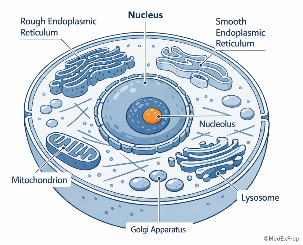
B -->|Steroid synthesis or drug detox| H[Smooth ER] Exam takeaway: Visualize the relative positions of nucleus, ER, Golgi, mitochondria, lysosomes, and cytoskeleton to answer localization stems.
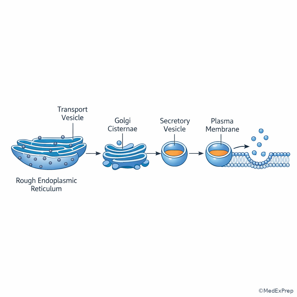
Exam takeaway: Secreted proteins follow a directional assembly-line pathway that NBEO stems often test indirectly.
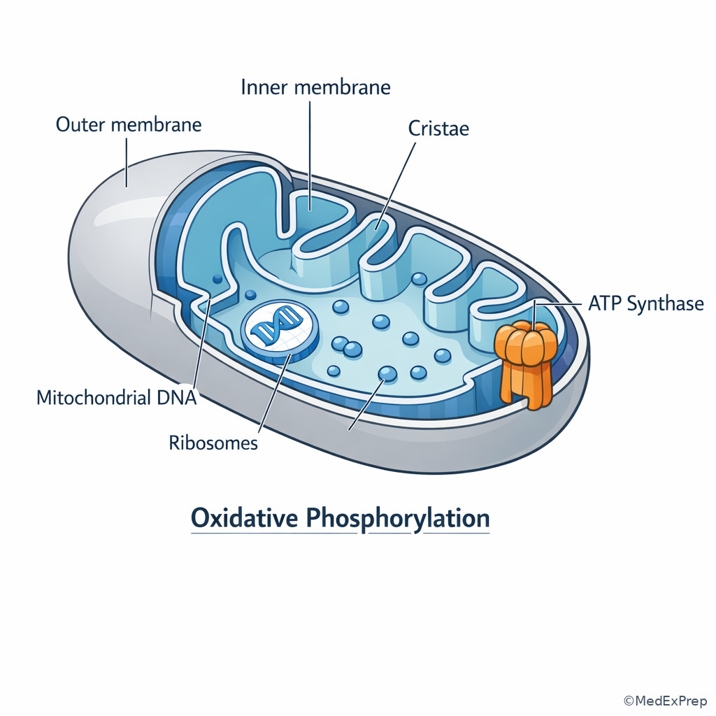
Exam takeaway: Cristae-rich mitochondria signal high aerobic energy demand and vulnerability to oxidative injury.
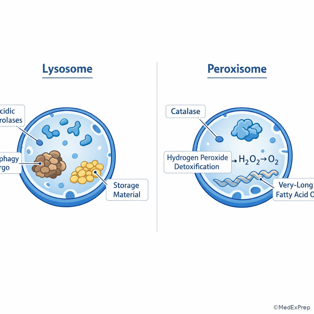
Exam takeaway: Acid hydrolase digestion belongs to lysosomes, while peroxide detox and very-long-chain fatty acid handling belong to peroxisomes.
Core content
1.1.1.3 Foundational map: who does what inside the cell?
A Explanation
Board questions at this level usually create a decision conflict between organelles with related jobs: rough ER versus Golgi, lysosome versus peroxisome, nucleus versus nucleolus. The correct move is to identify the single dominant task in the stem. If the task is making a protein that will be secreted or inserted into membrane, choose rough ER. If the task is modifying, sorting, or packaging that protein after synthesis, choose Golgi. If the task is gene transcription, choose nucleus; if the task is rRNA production and ribosome assembly, choose nucleolus. WHY THIS IS TESTED: exam writers know students memorize lists but confuse the sequence and specific handoff point.
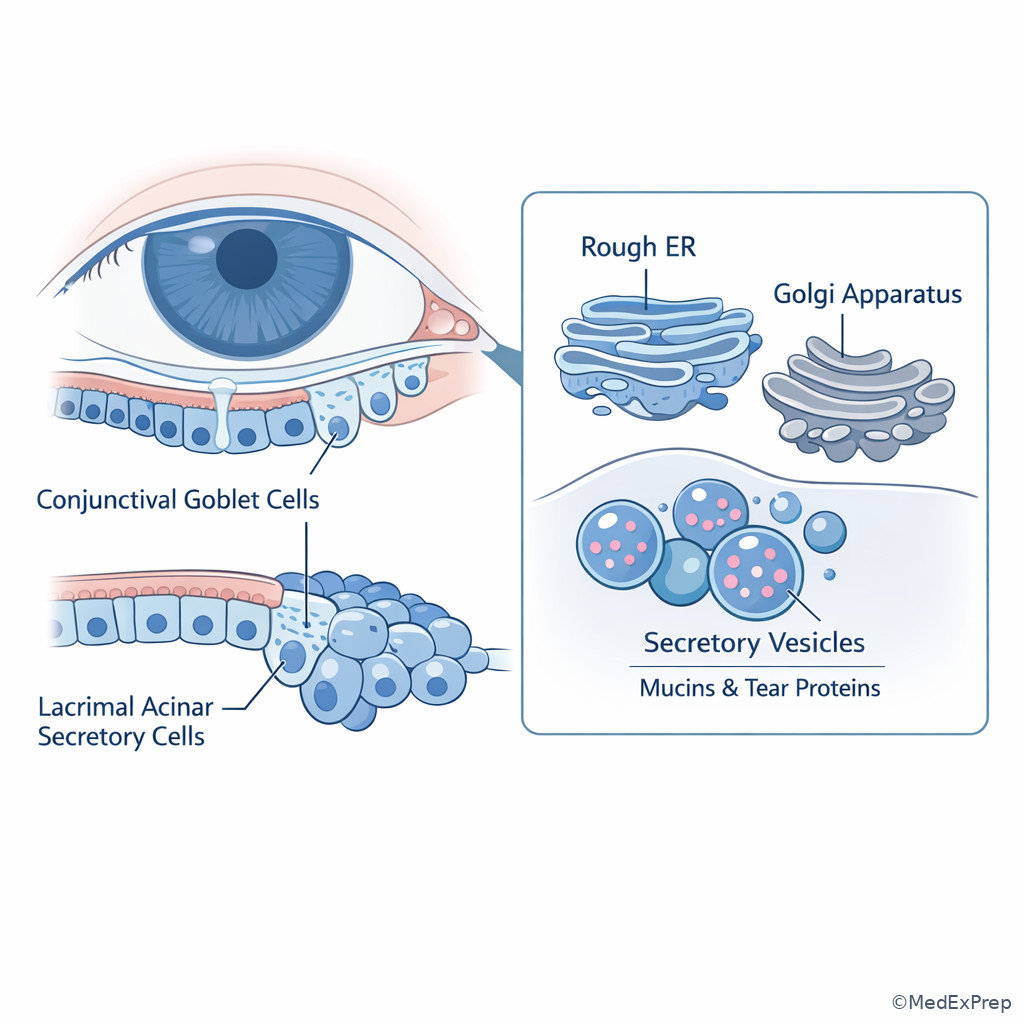
A fast mental model is to treat the cell as a factory. The nucleus holds the blueprint, the nucleolus builds the ribosome parts, ribosomes are the workers, rough ER is the protein assembly line, Golgi is the shipping and labeling center, mitochondria supply energy, lysosomes recycle waste, peroxisomes neutralize harmful oxidative byproducts, and the cytoskeleton acts as tracks and scaffolding. See Fig 1 and Fig 2.
B Worked example
A 21-year-old optometry student volunteer in a visual science lab has no ocular complaints but participates in a study of conjunctival goblet cell secretion. She has mild asthma and iron deficiency and recently started a high-protein diet. Electron microscopy of secretory cells shows abundant flattened sacs studded with ribosomes and numerous vesicles moving toward a stacked membranous complex. A distractor note reports elevated serum triglycerides. Which organelle is most directly responsible for post-translational modification and sorting of the mucin protein?
Reasoning chain: Identify task: not protein synthesis alone, but post-translational modification and sorting. Extract key facts: secretory cell, protein product, ribosome-studded sacs indicate rough ER upstream, stacked membranous complex indicates Golgi. Apply rule: RER synthesizes secreted protein; Golgi modifies and sorts it. Eliminate distractors: smooth ER is tempting because of vesicles and membranes, but it is lipid/steroid/detox focused; lysosome degrades rather than packages; nucleolus makes ribosomal subunits rather than processing mucin. Verify: goblet cells are highly secretory, so a prominent Golgi is expected.
C Exam trap
D Checkpoint
Question: A stem describes a cell that tags enzymes with mannose-6-phosphate before sending them to an acidic degradative compartment. Which organelle performs the tagging step?
- A. Golgi apparatus — Tempting because it handles trafficking; correct because mannose-6-phosphate tagging is a classic Golgi function that targets lysosomal enzymes.
- B. Rough endoplasmic reticulum — Tempting because lysosomal enzymes are proteins synthesized on bound ribosomes; wrong because synthesis occurs here, but tagging occurs later in the Golgi.
- C. Lysosome — Tempting because the enzymes end up there; wrong because the destination organelle does not perform the upstream targeting tag.
- D. Peroxisome — Tempting if the student only remembers “degradative organelle”; wrong because peroxisomes are oxidative and not part of the mannose-6-phosphate pathway.
1.1.1.4 Protein trafficking and membrane systems
A Explanation
The highest-yield pathway in cell biology is the secretory pathway. Proteins destined for secretion, lysosomes, or insertion into plasma membrane begin on ribosomes bound to rough ER. They enter the ER lumen, undergo early folding and quality control, then move in vesicles to the Golgi, where they are further modified and sorted to their destination. Free ribosomes, by contrast, make proteins that remain in the cytosol or go to organelles such as the nucleus or mitochondria by post-translational targeting. WHY THIS IS TESTED: many questions ask where a mutation would block a pathway, and the answer depends on whether the protein is secreted, membrane-bound, lysosomal, or cytosolic.
Choose rough ER when the stem describes antibody production, mucin secretion, membrane receptor synthesis, or lysosomal enzyme synthesis. Reject rough ER if the stem instead emphasizes detoxification, steroid production, or calcium storage; that points to smooth ER. Choose Golgi when the stem emphasizes glycosylation refinement, sorting, packaging, or targeting. See Fig 1. In ocular science, conjunctival goblet cells, lacrimal gland cells, and many epithelial secretory cells rely on a prominent rough ER-Golgi axis.
B Worked example
A 67-year-old man with type 2 diabetes and hyperlipidemia undergoes a research tear sample analysis because of chronic dry eye symptoms. A lab-grown lacrimal acinar cell line from his sample shows a toxin that specifically disrupts ribosomes attached to membrane-bound sacs, while free ribosomes remain intact. The cells can still produce many cytosolic enzymes, but secretion of tear proteins falls sharply. A distracting note states that the patient also has mild cataract. Which structure is most directly impaired?
Reasoning chain: Identify task: protein secretion failure with preserved many cytosolic proteins. Extract key facts: bound ribosomes on membrane sacs are affected; secretory output falls. Apply rule: secreted proteins are synthesized on rough ER-bound ribosomes. Eliminate distractors: Golgi is downstream and would impair sorting, not initial translation at bound ribosomes; smooth ER does not host ribosomes; nucleolus makes ribosomal components but the toxin specifically hits attached ribosomes already in place. Verify: lacrimal acinar cells are secretory, so rough ER dysfunction fits the phenotype.
C Exam trap
D Checkpoint
Question: Which protein is most likely synthesized on free ribosomes rather than rough ER-bound ribosomes?
- A. Secreted tear lysozyme — Tempting because it is a common ocular protein; wrong because secreted proteins use bound ribosomes and the ER-Golgi pathway.
- B. Plasma membrane ion channel — Tempting because it ends in a membrane; wrong because membrane proteins are inserted via rough ER.
- C. Lysosomal acid hydrolase — Tempting because it stays intracellular; wrong because lysosomal enzymes still enter the secretory pathway before being targeted.
- D. Cytosolic glycolytic enzyme — Tempting only if you remember free-ribosome products stay in cytosol; correct because it functions in the cytoplasm and is not secreted or membrane-inserted.
- The organelle that sorts and packages secretory proteins is Golgi apparatus.
- True or False: Lysosomal enzymes are synthesized on free ribosomes. False.
- Name the 3 common destinations after Golgi processing: plasma membrane, extracellular secretion, lysosome.
1.1.1.5 Energy, oxidative stress, and detox: mitochondria, peroxisomes, smooth ER
A Explanation
This section is about choosing among three metabolically active but commonly confused systems. Mitochondria generate ATP through the TCA cycle and oxidative phosphorylation, help regulate apoptosis, and contain their own DNA. Peroxisomes oxidize very-long-chain fatty acids and detoxify hydrogen peroxide through catalase. Smooth ER synthesizes lipids and steroids, participates in detoxification of drugs and toxins, and stores calcium in specialized cells. WHY THIS IS TESTED: exam writers often hide the answer in one metabolic phrase—ATP failure, peroxide detox, or drug metabolism.
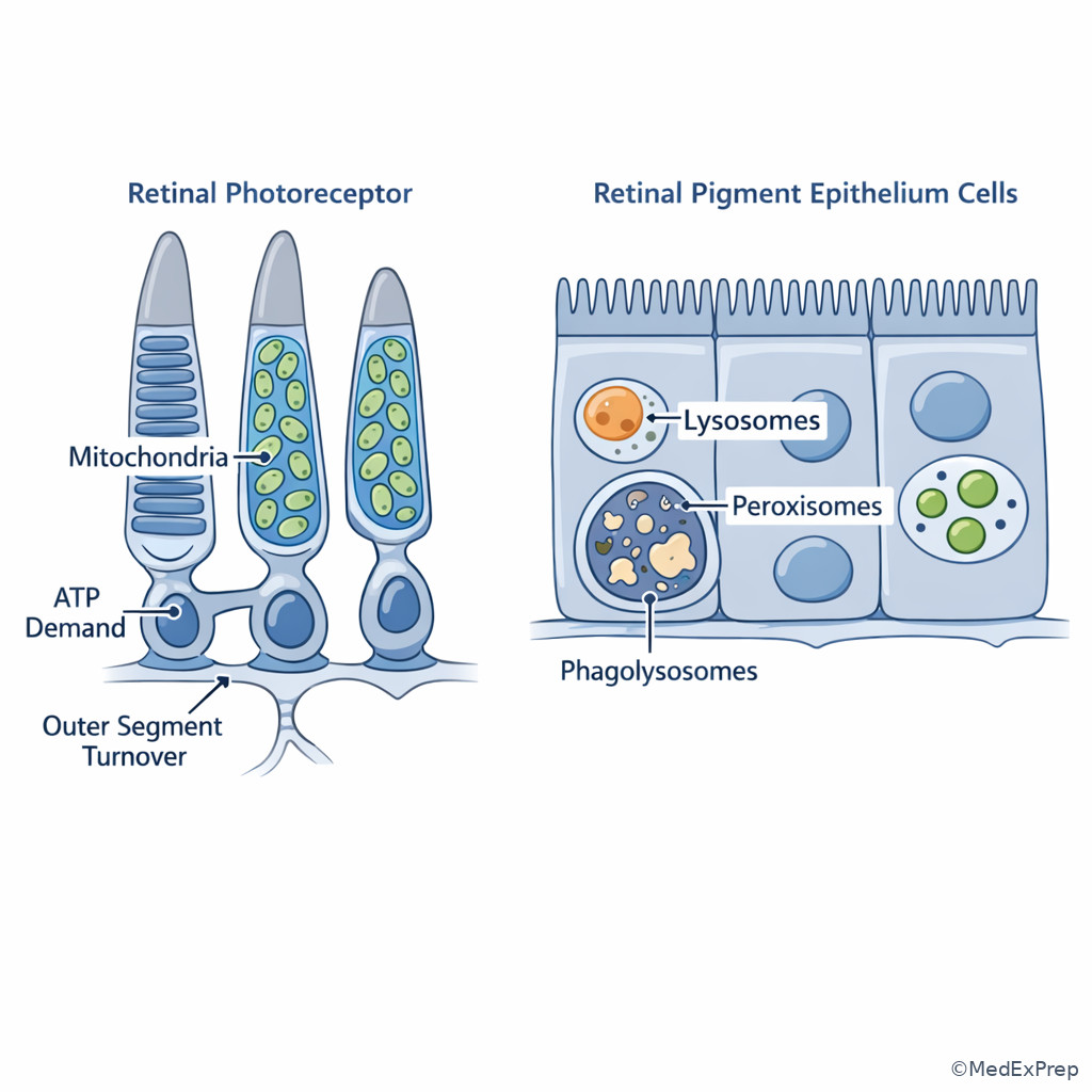
Choose mitochondria when the stem emphasizes high aerobic demand, cristae abundance, maternal inheritance, or apoptosis. Choose peroxisome when the stem emphasizes very-long-chain fatty acid accumulation, plasmalogen issues, or catalase-mediated detoxification. Choose smooth ER when the question asks about steroid-producing cells, hepatic detoxification, or intracellular calcium storage. See Fig 2. In the eye, photoreceptors and retinal pigment epithelium are metabolically intense and therefore particularly vulnerable to mitochondrial dysfunction, while oxidative stress mechanisms are central to lens and retinal injury.
B Worked example
A 13-year-old boy with developmental delay and progressive visual dysfunction is discussed in a basic science seminar. He has abnormal accumulation of very-long-chain fatty acids, hearing difficulty, and mild hepatomegaly. A classmate is distracted by his family history of exercise intolerance on the maternal side and suggests mitochondrial disease. Which organelle defect is the best explanation for the lipid abnormality?
Reasoning chain: Identify task: determine which organelle handles very-long-chain fatty acids. Extract key facts: accumulation of very-long-chain fatty acids is the major clue; maternal inheritance detail is noise designed to tempt a mitochondrial answer. Apply rule: peroxisomes carry out beta-oxidation of very-long-chain fatty acids and detoxify hydrogen peroxide. Eliminate distractors: mitochondria oxidize fatty acids too, but classically not the very-long-chain species emphasized in the stem; lysosomes degrade macromolecules, not this oxidative pathway; smooth ER handles lipid synthesis and detox but not the defining VLCFA step. Verify: peroxisomal disorders are the correct mechanism.
C Exam trap
D Checkpoint
Question: A pharmacology experiment shows marked proliferation of a membrane network after repeated exposure to a detoxified xenobiotic. Which structure is most likely increased?
- A. Smooth endoplasmic reticulum — Tempting because detoxification is a classic hepatic and cellular smooth ER function; correct for drug metabolism expansion.
- B. Rough endoplasmic reticulum — Tempting because it is also a membrane network; wrong because ribosome-studded protein synthesis does not explain xenobiotic detox adaptation.
- C. Peroxisome — Tempting because it handles oxidative detox; wrong because the question frames repeated xenobiotic metabolism, a more classic smooth ER function.
- D. Golgi apparatus — Tempting because drugs can affect trafficking; wrong because Golgi sorting is not the main detox platform.
1.1.1.6 Degradation and recycling: lysosomes, proteasomes, autophagy
A Explanation
When a stem asks how the cell disposes of material, first decide what kind of material is being removed. Lysosomes digest extracellular material brought in by endocytosis, old organelles delivered by autophagy, and macromolecules that need acidic hydrolases. Proteasomes degrade ubiquitin-tagged proteins in the cytosol and nucleus, especially misfolded or short-lived regulatory proteins. WHY THIS IS TESTED: students know both are degradative, but the exam separates vesicle-based acidic digestion from targeted cytosolic protein turnover.
Lysosomal storage diseases are a classic clue to lysosomal failure. Accumulation of undegraded substrate in membrane-bound inclusions strongly favors lysosomes. By contrast, if a question emphasizes ubiquitin tagging, proteasome inhibition, or failure to clear misfolded intracellular proteins without mentioning vesicles or acidic compartments, think proteasome. Autophagy is the bridge concept: damaged organelles can be enclosed in autophagosomes and delivered to lysosomes for degradation. In retinal cells and retinal pigment epithelium, lysosomal and autophagic pathways matter because these cells handle substantial metabolic waste and phagocytic burden.
B Worked example
A 6-year-old child in a genetics vignette has progressive neurodevelopmental decline and accumulation of undegraded sphingolipids in membrane-bound intracellular inclusions. The stem adds that the child recently had an uncomplicated otitis media and a normal complete blood count. A peer proposes a defect in peroxisomal oxidation because the disease involves lipids. Which cellular structure is most directly dysfunctional?
Reasoning chain: Identify task: determine the degradative compartment. Extract key facts: undegraded sphingolipids, membrane-bound inclusions, storage phenotype. Apply rule: lysosomal acid hydrolases degrade many complex macromolecules; failure causes storage diseases. Eliminate distractors: peroxisomes handle oxidation of very-long-chain fatty acids, not the classic acidic storage compartment; Golgi sorts lysosomal enzymes but is not the final defective digestion site unless specifically framed as targeting failure; proteasomes do not create membrane-bound storage inclusions. Verify: the pattern fits lysosomal dysfunction.
C Exam trap
D Checkpoint
Question: A researcher blocks ubiquitin-mediated destruction of misfolded cytosolic proteins. Which structure is most directly inhibited?
- A. Lysosome — Tempting because it is the best-known degradative organelle; wrong because the stem emphasizes ubiquitin-tagged cytosolic proteins rather than vesicle-based acidic digestion.
- B. Proteasome — Tempting because proteasomes are specialized for ubiquitin-tagged intracellular proteins; correct for selective cytosolic protein turnover.
- C. Golgi apparatus — Tempting if the student associates misfolding with protein processing; wrong because the degradative mechanism named is ubiquitin-mediated destruction, not sorting.
- D. Peroxisome — Tempting if oxidative damage is assumed; wrong because no peroxide or VLCFA clue is present.
- The organelle that degrades endocytosed material in an acidic compartment is the lysosome.
- True or False: Proteasomes primarily digest extracellular material taken up by phagocytosis. False.
- Name the process that sends damaged organelles to lysosomes: autophagy.
1.1.1.7 Cytoskeleton, cilia, transport, and cell shape
A Explanation
The cytoskeleton is high yield because it converts cell biology into visible structure-function findings. Microtubules are hollow tracks built from tubulin; they support mitotic spindles, intracellular vesicle transport, and cilia. Microfilaments are actin-based and support cell shape, contraction-like movement, and microvilli. Intermediate filaments provide tensile strength and are useful in tissue identification. WHY THIS IS TESTED: stems often present a structure—cilia, vesicle transport, spindle failure, cell motility—and ask for the underlying component.
Choose microtubules when the stem mentions dynein, kinesin, cilia, flagella, or vesicles moving along defined tracks. Choose actin microfilaments when it describes cortical support, microvilli, shape changes, or contractile rings in cytokinesis. Choose intermediate filaments when the focus is mechanical stability. This matters for eye-related tissues because epithelial polarity, intracellular transport, and ciliary structures are all clinically relevant; upcoming tissue lessons will build directly on these structural rules. See Fig 3.
B Worked example
A 24-year-old graduate student with chronic sinus congestion and recurrent respiratory infections is discussed in a cell biology review. He also reports a sibling with reduced fertility. Nasal epithelial sampling shows impaired ciliary motion. The stem includes a distracting note that his serum transaminases are mildly elevated after a weekend of alcohol use. Which intracellular component is most directly responsible for the defective movement?
Reasoning chain: Identify task: ciliary motility mechanism. Extract key facts: recurrent infections, impaired ciliary motion, fertility issues suggest dysfunctional cilia. Apply rule: cilia are microtubule-based structures using dynein arms for movement. Eliminate distractors: actin supports microvilli rather than motile cilia; intermediate filaments give strength, not motility; smooth ER and mitochondria may support cell life but are not the structural basis of ciliary beating. Verify: microtubules are the correct cellular component.
C Exam trap
D Checkpoint
Question: Vesicles move from Golgi to the plasma membrane along intracellular tracks using motor proteins. Which cytoskeletal element provides the track?
- A. Intermediate filaments — Tempting because they form a network throughout the cytoplasm; wrong because their role is strength, not directional transport.
- B. Actin microfilaments — Tempting because cortical actin supports membrane-associated processes; wrong because long-range organelle and vesicle transport classically uses microtubules.
- C. Microtubules — Tempting because kinesin and dynein move cargo along them; correct for intracellular vesicle transport.
- D. Extracellular collagen fibers — Tempting only if the student confuses tissue-level support with intracellular structure; wrong because the transport is happening inside the cell.
1.1.1.8 Integration: organelles in ocular cells and board-style pattern recognition
A Explanation
Integration questions stop asking “What does this organelle do?” and instead ask “Which organelle is emphasized in this tissue and why?” For NBEO, the useful pattern is to connect cell type to dominant task. Secretory epithelial cells such as goblet cells or lacrimal acinar cells are rich in rough ER and Golgi. High-energy cells such as photoreceptors and retinal pigment epithelium rely heavily on mitochondria. Cells involved in phagocytic or degradative processing emphasize lysosomal pathways. Drug-metabolizing cells emphasize smooth ER. WHY THIS IS TESTED: it forces you to move from memorized organelle lists to structure-function reasoning.
In ocular science, this means: if a stem describes continual outer segment processing in retinal support cells, think lysosomal/autophagic burden; if it emphasizes extreme ATP demand, think mitochondria; if it centers on mucin or protein secretion to support the ocular surface, think rough ER plus Golgi. Looking forward, tissue lessons will add epithelial polarity and connective-tissue support on top of this cellular base. See Fig 1, Fig 2, and Fig 3.
B Worked example
A 72-year-old woman with hypertension and well-controlled hypothyroidism enrolls in a retinal aging study. Imaging researchers discuss retinal pigment epithelial cells that continually process photoreceptor outer segment material and face chronic oxidative burden. The stem adds a distracting detail about her borderline elevated LDL and prior history of dry eye. Which pair of organelles is most important to this cell’s recycling and oxidative-defense workload?
Reasoning chain: Identify task: recycling plus oxidative defense. Extract key facts: continual processing of outer segment material suggests degradative machinery; chronic oxidative burden suggests reactive oxygen species handling. Apply rule: lysosomes support degradative processing, and peroxisomes contribute peroxide detoxification; mitochondria are important for energy but do not best match the recycling plus peroxide-defense pair asked. Eliminate distractors: Golgi plus rough ER fit secretion more than waste handling; mitochondria plus Golgi miss the degradation clue; smooth ER plus ribosome miss the phagolysosomal burden. Verify: lysosome with peroxisome is the best paired answer for this stem framing.
C Exam trap
D Checkpoint
Question: Which cell type would be expected to show the most prominent rough ER and Golgi apparatus?
- A. Lacrimal acinar cell secreting tear proteins — Tempting because secretion requires robust protein synthesis and packaging; correct for abundant rough ER and Golgi.
- B. Photoreceptor inner segment specialized for ATP demand — Tempting because it is highly active; wrong because mitochondria are the dominant specialization.
- C. Cell overloaded with very-long-chain fatty acids — Tempting because lipid handling suggests organelles; wrong because peroxisomal pathways are more relevant than secretory machinery.
- D. Cell digesting old organelles by autophagy — Tempting because active turnover implies many vesicles; wrong because lysosomes, not rough ER/Golgi, are the key emphasis.
Exam takeaway: Match tissue workload to organelle specialization rather than memorizing isolated definitions.
flowchart TD
A[Ocular cell in stem] --> B{Main workload}
B -->|Protein secretion| C[Lacrimal or goblet cell -> RER + Golgi]
B -->|High ATP demand| D[Photoreceptor or active epithelium -> Mitochondria]
B -->|Waste digestion or autophagy| E[RPE or phagocytic cell -> Lysosomes]
B -->|Oxidative detox or VLCFA handling| F[Peroxisomes]
B -->|Drug metabolism or lipid synthesis| G[Smooth ER] Exam takeaway: Goblet and lacrimal secretory cells are classic examples of rough ER and Golgi prominence.
Exam takeaway: Photoreceptors and retinal support cells are tested by linking high ATP demand and waste processing to the right organelles.
Exam Traps & Differentiators
- Most common wrong answer and why: Rough ER instead of Golgi. Students see a secreted protein and stop too early in the pathway. If the stem says sorting, packaging, glycosylation refinement, or mannose-6-phosphate, think Golgi.
- Looks similar but isn't: Lysosome versus peroxisome. Both are small intracellular organelles involved in breakdown, but lysosomes use acidic hydrolases for digestion and storage disease patterns, whereas peroxisomes handle peroxide detox and very-long-chain fatty acids.
- If the stem says “bound ribosomes” think rough ER.
- If the stem says “ubiquitin-tagged protein” think proteasome.
- If the stem says “very-long-chain fatty acids” think peroxisome.
- If the stem says “prominent nucleolus” think active ribosome production, not transcription of all proteins.
- If the stem says “cilia or dynein” think microtubules.
| Confusable pair | Why they seem similar | Single best discriminator | Exam move |
|---|---|---|---|
| Rough ER vs Golgi | Both handle proteins for secretion | Synthesis/folding versus sorting/packaging | Ask which step the stem describes |
| Lysosome vs Peroxisome | Both participate in intracellular breakdown | Acid hydrolases/storage versus peroxide/VLCFA metabolism | Look for substrate and chemistry clue |
| Nucleus vs Nucleolus | Both are nuclear structures | DNA transcription versus rRNA/ribosome assembly | Find whether the stem asks about genes or ribosomes |
| Microtubules vs Actin | Both support movement | Cilia/vesicle tracks versus cortical shape/microvilli | Identify the structure being moved |
| Lysosome vs Proteasome | Both degrade cellular material | Acidic vesicles versus ubiquitin-tagged proteins | Look for “ubiquitin” or storage inclusions |
| Stem clue | Think | Why |
|---|---|---|
| Goblet cell mucin secretion | Rough ER + Golgi | Heavy secretory protein production and packaging |
| Photoreceptor inner segment ATP demand | Mitochondria | High oxidative metabolism |
| Outer segment waste processing in retinal support cells | Lysosome | Degradation and recycling burden |
| VLCFA accumulation | Peroxisome | Characteristic oxidative defect |
| Drug detoxification adaptation | Smooth ER | Xenobiotic metabolism machinery expands |
| Ciliary motility disorder | Microtubules | Cilia are tubulin-based |
Algorithm / Approach
Rapid Review
- Golgi apparatus → packaging/sorting beats protein synthesis as the discriminator
- Rough ER → bound ribosomes and secreted or membrane proteins
- Free ribosome → cytosolic protein destination
- Nucleolus → ribosome assembly, not broad DNA transcription
- Smooth ER → steroid synthesis and xenobiotic detox, not ATP generation
- Mitochondrion → cristae-rich high-energy demand and apoptosis signaling
- Peroxisome → very-long-chain fatty acids and hydrogen peroxide detox
- Lysosome → acidic hydrolases and storage disease pattern
- Proteasome → ubiquitin-tagged intracellular proteins
- Microtubule → cilia, spindle, vesicle tracks
- Actin microfilament → microvilli and cortical shape change
- Secretory ocular cell → rough ER plus Golgi prominence
- Photoreceptor energy demand → mitochondrial emphasis
- RPE waste handling → lysosomal burden
Self-check quiz
1. A conjunctival goblet cell is producing a large amount of mucin for secretion onto the ocular surface. Which organelle is most directly responsible for packaging and sorting that protein before release?
- A. Golgi apparatus
- B. Rough endoplasmic reticulum
- C. Smooth endoplasmic reticulum
- D. Nucleolus
2. A laboratory stem describes accumulation of very-long-chain fatty acids in cultured cells from a child with progressive neurologic dysfunction and reduced vision. Which organelle is the best match for the defective pathway?
- A. Lysosome
- B. Peroxisome
- C. Mitochondrion
- D. Golgi apparatus
3. A 64-year-old woman with Sjogren-like dry eye symptoms and hypothyroidism undergoes research sampling of lacrimal gland cells. A toxin selectively impairs ribosomes attached to a membranous network, while free ribosomes continue to function. Tear protein secretion falls, but many cytosolic enzymes remain normal. Which structure is most likely affected?
- A. Rough endoplasmic reticulum
- B. Golgi apparatus
- C. Lysosome
- D. Peroxisome
4. A 19-year-old man with chronic sinusitis and infertility is discussed in a cell motility module. Nasal epithelial biopsy shows impaired ciliary beating. A distracting note states that he has mild vitamin D deficiency. Which intracellular component best explains the motility defect?
- A. Actin microfilaments
- B. Intermediate filaments
- C. Microtubules
- D. Collagen fibrils
5. A research stem describes a cell unable to degrade endocytosed material within an acidic, membrane-bound compartment. Undegraded substrate accumulates over time. Which organelle is most directly implicated?
- A. Proteasome
- B. Lysosome
- C. Peroxisome
- D. Smooth endoplasmic reticulum
6. A 71-year-old woman with hypertension, early cataract, and controlled diabetes is enrolled in a retinal aging study. Investigators emphasize that a support cell continuously processes shed photoreceptor outer segment material and must also limit oxidative byproducts. Which pair of organelles is the most appropriate match for this workload?
- A. Rough ER and Golgi apparatus
- B. Mitochondria and Golgi apparatus
- C. Lysosomes and peroxisomes
- D. Nucleolus and microtubules
7. A 28-year-old research participant with no systemic disease undergoes a cell labeling experiment. A newly synthesized enzyme is produced on bound ribosomes, enters a membranous lumen, receives a mannose-6-phosphate tag, and is delivered to an acidic compartment. Which organelle performs the tagging step that determines the enzyme’s final destination?
- A. Rough endoplasmic reticulum
- B. Golgi apparatus
- C. Lysosome
- D. Proteasome
8. A 15-year-old boy with developmental delay, hearing difficulty, and progressive visual dysfunction has elevated very-long-chain fatty acids. His maternal aunt has migraine, creating a tempting clue toward a mitochondrial disorder. Which detail most strongly supports a peroxisomal defect over a mitochondrial defect?
- A. High ATP demand in neural tissue
- B. Family history on the maternal side
- C. Accumulation of very-long-chain fatty acids
- D. Presence of oxidative stress in affected cells
Answer key
1.1.1.9 1. Answer: A. Golgi apparatus
A. Golgi apparatus: Tempting because secreted proteins need a shipping center; correct because packaging and sorting before secretion are Golgi functions. The single stem clue is packaging and sorting before release. Source: Junqueira’s Basic Histology; Alberts et al.
B. Rough endoplasmic reticulum: Tempting because goblet cells synthesize secreted proteins on bound ribosomes; incorrect here because synthesis is not the same as packaging and sorting. The discriminating clue is the downstream step in the pathway. Source: Alberts et al.
C. Smooth endoplasmic reticulum: Tempting if the learner overfocuses on membranes and secretion; incorrect because smooth ER is associated with lipid synthesis, steroid synthesis, and detoxification rather than mucin packaging. The discriminating clue is protein secretion. Source: Guyton and Hall.
D. Nucleolus: Tempting because rapidly secretory cells often have a prominent nucleolus; incorrect because the nucleolus assembles ribosomal subunits and does not package mucin. The discriminating clue is the specific task of sorting secretory product. Source: Junqueira’s Basic Histology.
1.1.1.10 2. Answer: B. Peroxisome
A. Lysosome: Tempting because lipid accumulation suggests a storage disorder; incorrect because the specific biochemical clue is very-long-chain fatty acids, which points to peroxisomal oxidation failure rather than lysosomal substrate digestion. Source: Robbins and Cotran; Alberts et al.
B. Peroxisome: Tempting and correct because peroxisomes oxidize very-long-chain fatty acids and detoxify hydrogen peroxide. The single discriminating clue is very-long-chain fatty acid accumulation. Source: Robbins and Cotran.
C. Mitochondrion: Tempting because mitochondria are strongly linked to metabolism and fatty acid oxidation generally; incorrect here because the stem names the chain length class classically tied to peroxisomes. Source: Guyton and Hall.
D. Golgi apparatus: Tempting if a learner associates all inherited cell disorders with trafficking defects; incorrect because the stem gives a metabolic oxidation clue, not a sorting clue. Source: Alberts et al.
1.1.1.11 3. Answer: A. Rough endoplasmic reticulum
A. Rough endoplasmic reticulum: Tempting and correct because bound ribosomes on membrane sacs synthesize secreted tear proteins. The single stem clue is preserved cytosolic protein synthesis with impaired secretion after damage to attached ribosomes. Source: Alberts et al.; Junqueira’s Basic Histology.
B. Golgi apparatus: Tempting because secretion falls; incorrect because the toxin injures attached ribosomes, identifying the rough ER step rather than the sorting step. The discriminating clue is the explicit mention of ribosomes. Source: Alberts et al.
C. Lysosome: Tempting if secretion failure is misread as vesicle failure; incorrect because the stem is about synthesis of tear proteins, not degradation of cellular contents. The discriminating clue is secretory protein production. Source: Robbins and Cotran.
D. Peroxisome: Tempting if dry eye and oxidative stress are overinterpreted; incorrect because no peroxide or VLCFA clue is given. The discriminating clue is bound ribosomes. Source: Guyton and Hall.
1.1.1.12 4. Answer: C. Microtubules
A. Actin microfilaments: Tempting because actin contributes to cell movement and shape; incorrect because motile cilia are built on a microtubule axoneme rather than actin. The single clue is impaired ciliary beating. Source: Junqueira’s Basic Histology; Alberts et al.
B. Intermediate filaments: Tempting because they support structural integrity; incorrect because they do not generate ciliary motion. The discriminating clue is motility, not tensile strength. Source: Alberts et al.
C. Microtubules: Tempting and correct because cilia require tubulin-based structure and dynein-associated movement. The single discriminating clue is the ciliary finding paired with recurrent infections and infertility. Source: Robbins and Cotran.
D. Collagen fibrils: Tempting only if extracellular support is confused with intracellular movement; incorrect because collagen is extracellular matrix, not the internal motility scaffold. The discriminating clue is intracellular ciliary function. Source: Junqueira’s Basic Histology.
1.1.1.13 5. Answer: B. Lysosome
A. Proteasome: Tempting because it also degrades proteins; incorrect because the stem specifies endocytosed material in an acidic membrane-bound compartment, which is lysosomal. The discriminating clue is the acidic vesicle. Source: Alberts et al.; Robbins and Cotran.
B. Lysosome: Tempting and correct because lysosomes digest endocytosed material using acid hydrolases. The single stem clue is acidic, membrane-bound compartment. Source: Robbins and Cotran.
C. Peroxisome: Tempting if intracellular breakdown is generalized; incorrect because peroxisomes perform oxidative metabolism, not acidic digestion of endocytosed cargo. The discriminating clue is endocytosed substrate accumulation. Source: Guyton and Hall.
D. Smooth endoplasmic reticulum: Tempting because membrane systems are involved; incorrect because smooth ER is not the cellular digestive compartment. The discriminating clue is degradative acidity. Source: Alberts et al.
1.1.1.14 6. Answer: C. Lysosomes and peroxisomes
A. Rough ER and Golgi apparatus: Tempting because the retinal support cell is very active; incorrect because the stem emphasizes recycling of shed material and oxidative byproducts, not protein secretion. The discriminating clue is outer segment processing. Source: Adler’s Physiology of the Eye; Alberts et al.
B. Mitochondria and Golgi apparatus: Tempting because retinal cells are metabolically active; incorrect because the question asks for recycling plus oxidative defense, making lysosomal degradation central and peroxisomal detox supportive. The discriminating clue is continual processing of shed material. Source: Adler’s Physiology of the Eye.
C. Lysosomes and peroxisomes: Tempting and correct because lysosomes handle degradation of phagocytosed outer segment material, while peroxisomes help manage oxidative byproducts. The single best clue is the combination of recycling workload and oxidative burden. Source: Adler’s Physiology of the Eye; Robbins and Cotran.
D. Nucleolus and microtubules: Tempting if one overthinks high activity and intracellular movement; incorrect because neither directly matches the paired functions asked. The discriminating clue is degradative and detoxifying function. Source: Alberts et al.
1.1.1.15 7. Answer: B. Golgi apparatus
A. Rough endoplasmic reticulum: Tempting because the enzyme is synthesized on bound ribosomes and enters the ER lumen first; incorrect because the question asks about the mannose-6-phosphate tagging step, which occurs in the Golgi. The discriminating clue is the explicit tag. Source: Alberts et al.; Junqueira’s Basic Histology.
B. Golgi apparatus: Tempting and correct because Golgi sorting includes mannose-6-phosphate tagging for lysosomal targeting. The single clue is mannose-6-phosphate. Source: Alberts et al.
C. Lysosome: Tempting because the enzyme ends up in an acidic compartment; incorrect because destination is not the same as the site of tagging. The discriminating clue is the word “tagging step.” Source: Robbins and Cotran.
D. Proteasome: Tempting if learners confuse intracellular processing with degradation; incorrect because proteasomes degrade ubiquitin-tagged proteins and are unrelated to lysosomal enzyme targeting. The discriminating clue is lysosomal delivery rather than ubiquitin turnover. Source: Alberts et al.
1.1.1.16 8. Answer: C. Accumulation of very-long-chain fatty acids
A. High ATP demand in neural tissue: Tempting because mitochondria are central to ATP production; incorrect because high ATP demand is nonspecific and does not distinguish peroxisomal from mitochondrial disease. The discriminating clue must be more specific. Source: Guyton and Hall; Robbins and Cotran.
B. Family history on the maternal side: Tempting because maternal inheritance is a classic mitochondrial clue; incorrect here because the stem explicitly frames it as distracting noise. The discriminating clue is the biochemical abnormality, not the family-history trap. Source: Robbins and Cotran.
C. Accumulation of very-long-chain fatty acids: Tempting and correct because VLCFA accumulation is the classic peroxisomal discriminator. The single clue is the named substrate class. Source: Robbins and Cotran.
D. Presence of oxidative stress in affected cells: Tempting because both mitochondria and peroxisomes intersect oxidative metabolism; incorrect because oxidative stress is too broad and nondiscriminating. The discriminating clue remains VLCFA accumulation. Source: Alberts et al.
Keep going with the full NBEO Part I Prep Course (NBEO I)
You just studied the way members do, in the same lesson interface. Full access unlocks every lesson, the question bank and flashcards for a full year.
One payment · 1 year of access · No auto-renewal