1.1.1 - Tooth Morphology Landmarks
Tooth Morphology Landmarks
What you’ll learn
- DOMAIN: Biomedical Foundations - Head and Oral Structures -> Tooth crown landmark recognition. Distinguish cusp, ridge, fossa, groove, pit, cingulum, and marginal ridge in board-style stems.
- DOMAIN: Biomedical Foundations - Head and Oral Structures -> Root and surface landmark orientation. Use line angles, point angles, heights of contour, CEJ curvature, and root form to identify tooth surfaces and likely tooth groups.
- DOMAIN: Patient Assessment - Oral Examination and Charting -> Surface description accuracy. Translate morphology into precise charting, instrumentation planning, and communication.
- DOMAIN: Dental Hygiene Care - Preventive and Therapeutic Services -> Anatomy-guided care modification. Predict where biofilm, calculus, and caries tend to occur based on anatomy.
- DOMAIN: Radiographic Interpretation Basics -> Correlate anatomy with appearance. Connect external landmarks to likely radiographic and clinical findings without overstepping into dentist-only diagnosis.
- Biomedical Foundations → Head and Oral Structures → Tooth morphology, landmark identification, and tooth orientation
- Patient Assessment → Oral Examination and Charting → Surface naming and recognition of normal anatomical findings
- Dental Hygiene Care → Instrumentation and Nonsurgical Care → Adaptation based on crown and root anatomy
- Radiology and Safety → Radiographic Interpretation Basics → Recognize anatomy that influences image interpretation
Why it matters
- Tooth morphology is the language behind charting, instrumentation, sealant selection, and patient education.
- NBDHE stems often hide the answer inside one landmark clue: a cingulum, a transverse ridge, a cusp of Carabelli, a mesiolingual groove, or a CEJ curvature pattern.
- Dental hygiene care depends on anatomy-guided decisions: where deposits collect, where explorers catch, where radiographic overlap misleads, and where adaptation must change.
- Knowing normal landmarks helps you avoid overcalling pathology and helps you recognize findings that require referral rather than unsupported conclusions.
Key Terms & Must-Know Facts
1.1.1.1 Key terms
- Cusp: Elevated point or mound on a crown.
- Ridge: Linear elevation on the crown surface.
- Marginal ridge: Border ridge on occlusal surfaces of posterior teeth and lingual surfaces of anterior teeth.
- Triangular ridge: Ridge descending from a cusp tip toward the center of the occlusal surface.
- Transverse ridge: Union of two triangular ridges crossing the occlusal table.
- Oblique ridge: Ridge crossing maxillary molar occlusal surface obliquely.
- Fossa: Shallow depression.
- Sulcus: Long valley between ridges and cusps.
- Developmental groove: Linear depression marking junction of developmental lobes.
- Supplemental groove: Extra shallow groove not marking lobe junction.
- Pit: Pinpoint depression, often at groove junctions.
- Cingulum: Cervical lingual prominence on anterior teeth.
- Mamelon: Developmental enamel elevations on newly erupted incisors.
- Height of contour: Greatest crown convexity on a given surface.
- Line angle / point angle: Junction of two surfaces / three surfaces.
1.1.1.2 Must-know facts
- Anterior teeth have no occlusal surface; they have an incisal edge or ridge.
- Lingual surfaces of anterior teeth often show cingulum, marginal ridges, and lingual fossa.
- Premolars usually have one buccal cusp; maxillary premolars usually have two cusps, mandibular second premolars may have two or three cusps.
- Maxillary molars commonly have an oblique ridge; this is a favorite exam clue.
- Mandibular first molars often show five cusps; maxillary first molars usually have four major cusps and may have a cusp of Carabelli.
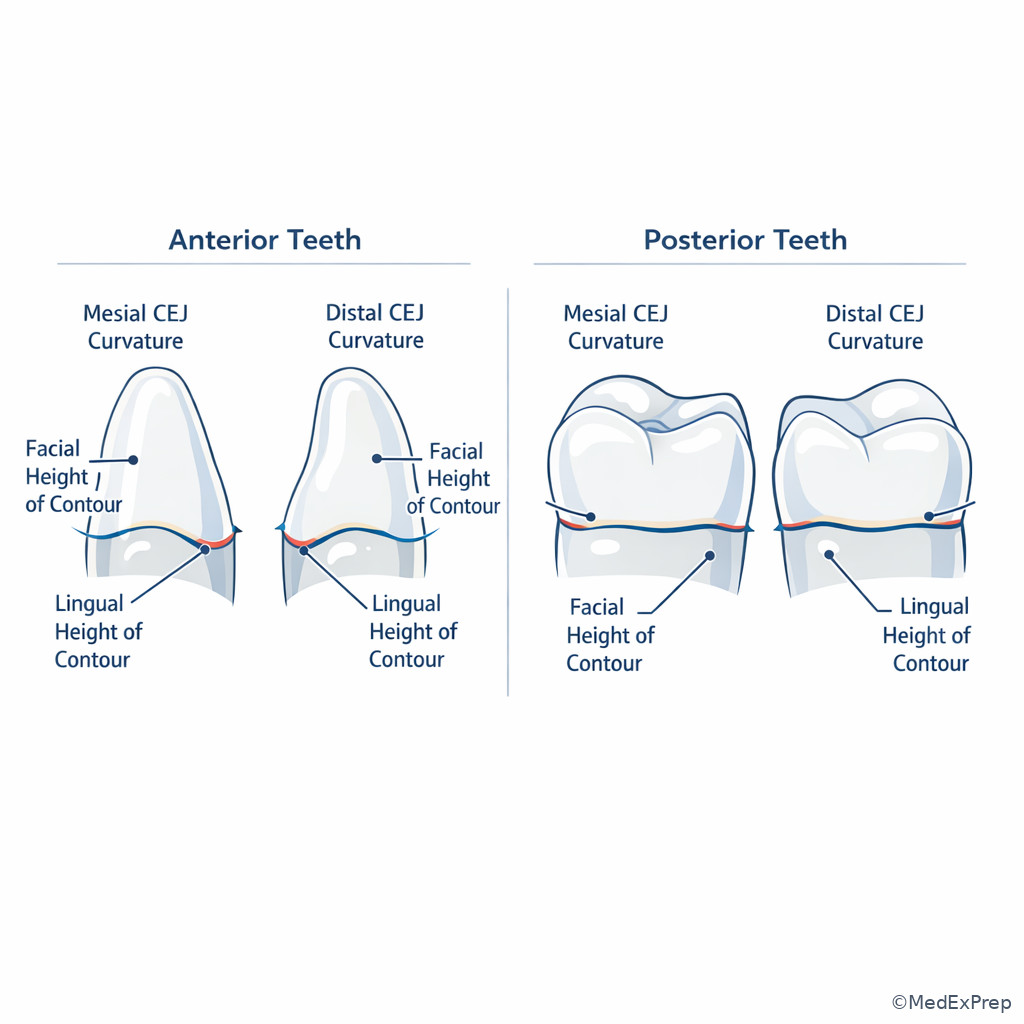
- The CEJ curvature is generally greater on mesial than distal.
- Posterior teeth have broader contact areas in posterior direction and contacts move more cervically from anterior to posterior.
- Heights of contour matter for self-care instruction and instrument adaptation because overcontoured and undercontoured areas change plaque retention and tissue protection.
| Landmark | Where commonly found | High-yield exam use |
|---|---|---|
| Cingulum | Lingual cervical third of anterior teeth | Helps identify lingual surface and anterior tooth type |
| Oblique ridge | Maxillary molars | Separates maxillary molars from mandibular molars |
| Transverse ridge | Premolars, especially mandibular first premolar | Can divide mesial and distal fossae |
| Pit | Occlusal groove junctions, lingual pits | Common plaque and caries-retentive site |
| Marginal ridge | Posterior occlusal border; anterior lingual border | Helps orient crown surface and contact area |
| Height of contour | All surfaces | Important for adaptation and plaque control |
Core content
1.1.1.3 Foundational landmark vocabulary: what the stem is really asking you to recognize
A Explanation
The first exam conflict is usually choose the correct landmark name vs reject a similar but wrong term. If a stem describes a linear elevation, choose ridge; if it describes a shallow depression, choose fossa; if it describes a narrow linear depression between developmental parts, choose developmental groove; if it describes a pinpoint defect at a groove intersection, choose pit. The single rule that resolves many beginner errors is this: elevations and depressions are not interchangeable, and location matters more than memorized lists.
WHY THIS IS TESTED: NBDHE items commonly pair almost-correct terms from the same category and ask you to identify the one landmark that best explains plaque retention, explorer catch, or tooth orientation.
Think in clusters. On anterior lingual surfaces, the cluster is cingulum + marginal ridges + lingual fossa. On posterior occlusal surfaces, the cluster is cusp + triangular ridge + groove + pit + fossa. A sulcus is broader than a groove and sits as a valley between ridges/cusps; a developmental groove often lies within a sulcus. If the question asks what makes a posterior tooth high risk for pit-and-fissure retention, reject “marginal ridge” unless the stem describes a border structure; choose “pit” or “developmental groove” when the clue is a narrow plaque-retentive depression.
See Fig 1 for the foundational map of elevations versus depressions and how they are grouped for exam recognition.
Exam takeaway: Separate elevations from depressions first, then use surface location to choose the correct landmark term.
flowchart TD
A[Unknown landmark in stem] --> B{Elevation or depression?}
B -->|Elevation| C[Cusp or ridge family]
B -->|Depression| D[Fossa groove sulcus pit]
C --> E{Anterior lingual cervical?}
E -->|Yes| F[Cingulum]
E -->|No| G{Linear elevation?}
G -->|Yes| H[Ridge]
G -->|No| I[Cusp]
D --> J{Pinpoint at groove junction?}
J -->|Yes| K[Pit]
J -->|No| L{Broad shallow depression?}
L -->|Yes| M[Fossa]
L -->|No| N[Developmental groove or sulcus] Exam takeaway: Visual grouping of cusp, ridge, groove, fossa, and pit prevents terminology mix-ups on single-best-answer questions.
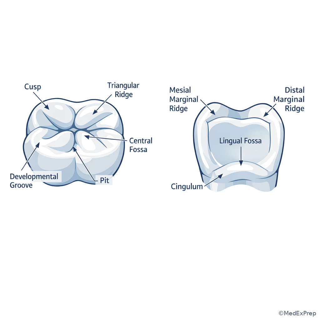
B Worked example
A 19-year-old college student presents for preventive care. She wears clear aligners, snacks frequently on dried fruit, and reports no pain. A distractingly prominent stain line is visible on a mandibular molar occlusal surface. The hygienist notes a narrow linear depression joining a small pinpoint depression in the occlusal table. The question asks for the most likely plaque-retentive landmark.
Identify task: Name the anatomy, not the disease. Extract key facts: posterior tooth, occlusal surface, narrow linear depression plus pinpoint depression. Apply rule: linear depression at developmental junction = groove; pinpoint at junction = pit. Eliminate distractors: fossa is broader and shallow; marginal ridge is elevated border; cusp is elevated, not depressed. Verify: the most plaque-retentive named landmark here is the pit, because the stem highlights the pinpoint depression where material accumulates.
C Exam trap
Common wrong answer: Fossa.
Why it is tempting: Students remember that posterior occlusal anatomy contains fossae and assume any depression on the chewing surface must be a fossa.
Single clue that eliminates it: The stem says pinpoint depression at a groove junction; that points to a pit, not a broad shallow fossa.
D Checkpoint
Question: A chart note describes a “linear elevation running from a cusp tip toward the center of the occlusal surface.” Which landmark is being documented?
- Triangular ridge — Tempting because it is a named ridge on posterior teeth, and it is correct because it descends from a cusp tip toward the center.
- Marginal ridge — Tempting because it is also a ridge on occlusal surfaces, but it is wrong because it forms the mesial or distal border rather than running from a cusp tip inward.
- Transverse ridge — Tempting because it involves triangular ridges, but it is wrong because it requires union of two triangular ridges, not just one ridge.
- Oblique ridge — Tempting because it is a prominent posterior ridge, but it is wrong because it is specific to maxillary molars and crosses the surface obliquely between defined cusps.
Practitioner Action: Assess: Surface form and location before naming anatomy. Intervene: Use correct terms in charting, OHI, and deposit description. Refer: If anatomy appears defective or unusually deep and suspicious for pathology or structural concern, communicate findings to the dentist. Document: Exact surface, landmark, and whether it is normal anatomy or a plaque-retentive site.
1.1.1.4 Crown orientation and surface identification: how to know mesial, distal, buccal, lingual, and occlusal clues
A Explanation
The next decision conflict is choose the correct surface or side of a tooth vs reject the mirror-image distractor. The best single rule is this: morphology is asymmetrical in predictable ways. Mesial contacts are usually more incisal/occlusal than distal contacts, CEJ curvature is usually greater on the mesial, and many crowns taper differently toward lingual or distal. The exam often gives one orientation clue and asks you to identify the tooth or the surface.
On anterior teeth, the lingual surface is recognized by the cingulum, marginal ridges, and lingual fossa. On posterior teeth, the occlusal surface is recognized by cusps, grooves, and fossae. Buccal surfaces are usually smoother and more convex; lingual anatomy can be either simpler or more complex depending on tooth type. For example, the mandibular first premolar has a strong transverse ridge and a small nonfunctional lingual cusp, while maxillary molars may show a cusp of Carabelli on the lingual of the mesiolingual cusp.
The exam-friendly habit is to ask three questions: What surface am I looking at? Which side is more mesial? Which landmark is unique enough to narrow the tooth group? Use CEJ curvature, contact position, cusp size, and cervical prominence together rather than relying on one fact. See Fig 2 for a compact orientation flow.
Exam takeaway: Use surface family first, then mesial-distal asymmetry, then signature landmarks to orient a tooth rapidly.
flowchart TD
A[Unknown tooth view] --> B{Anterior or posterior?}
B -->|Anterior| C[Look for cingulum lingual fossa incisal edge]
B -->|Posterior| D[Look for cusps grooves fossae]
C --> E{Which side has greater CEJ curve?}
D --> F{Which side contact is more occlusal?}
E --> G[That side is mesial]
F --> G
G --> H{Unique clue present?}
H -->|Oblique ridge| I[Maxillary molar]
H -->|Strong transverse ridge tiny lingual cusp| J[Mandibular first premolar]
H -->|Cusp of Carabelli| K[Maxillary first molar] Exam takeaway: Surface identification becomes faster when cingulum, CEJ curvature, contact location, and cusp pattern are seen together.
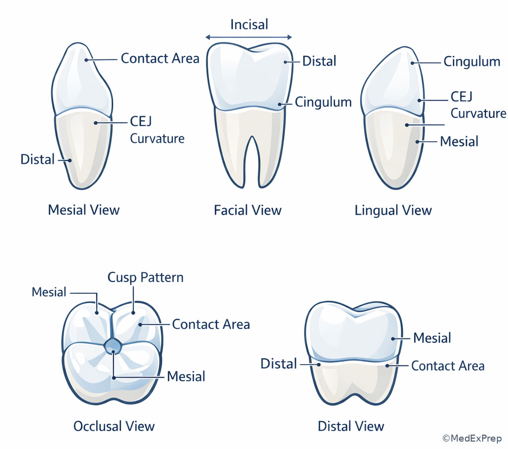
B Worked example
A 27-year-old patient with orthodontic history and mild generalized gingivitis presents for assessment. A study tooth image in the exam stem shows a crown with a prominent cingulum and a sharper incisal angle on one side. There is also a distracting mention of coffee staining and a recent whitening product use. The question asks which side is mesial.
Identify task: Orientation, not color change. Extract key facts: anterior tooth, cingulum indicates lingual aspect, one incisal angle is sharper, mesial CEJ curvature is usually greater, mesioincisal angle on maxillary incisors is typically sharper than distal. Apply rule: the sharper incisal angle and greater CEJ curvature identify the mesial side. Eliminate distractors: staining and whitening history do not affect morphology; cingulum alone tells lingual, not mesial. Verify: choose the side with the sharper angle and greater CEJ curve as mesial.
C Exam trap
Common wrong answer: Use the larger contact area to call the side mesial.
Why it is tempting: Students remember mesial contacts are often more incisal or occlusal and overgeneralize size instead of position.
Single clue that eliminates it: The tested discriminator is position and curvature, not simply “larger-looking contact.” Mesial usually shows greater CEJ curvature.
D Checkpoint
Question: Which landmark most strongly identifies the lingual surface of an anterior tooth?
- Cingulum — Tempting because it is a classic anterior feature, and it is correct because it is the cervical convexity on the lingual surface.
- Central groove — Tempting because grooves help identify surfaces, but it is wrong because central grooves are posterior occlusal landmarks.
- Oblique ridge — Tempting because ridges can help orient teeth, but it is wrong because it belongs to maxillary molars, not anterior teeth.
- Buccal pit — Tempting because pits can be distinctive, but it is wrong because it is not the defining lingual landmark of anterior teeth.
- The surface with a cingulum on an incisor is the lingual surface.
- True or False: CEJ curvature is usually greater on the distal surface. False.
- Name the 3 key anterior lingual features: cingulum, marginal ridges, lingual fossa.
Practitioner Action: Assess: Surface orientation before recording findings or planning adaptation. Intervene: Use correct mesial/distal and facial/lingual language in charting and patient education. Refer: If form appears anomalous or suggests developmental irregularity beyond routine recognition, alert the dentist. Document: Tooth number, surface, and landmark-based description.
1.1.1.5 Tooth-group signatures: incisors, canines, premolars, and molars
A Explanation
Once you know landmark vocabulary and orientation, the next conflict is choose the correct tooth group vs reject the near-neighbor that shares one feature. The single rule is: do not identify a tooth from one landmark alone when the tooth group has a landmark pattern. Incisors are built for cutting, canines for tearing, premolars for transition, and molars for grinding. The exam rewards pattern recognition, not isolated facts.
Incisors have an incisal edge, no cusp, and prominent lingual anatomy in varying degrees. Canines have one cusp, a strong labial ridge, and the longest root in the dentition. Premolars have transitional morphology: usually one buccal cusp plus one or more lingual cusps, shorter occlusal tables than molars, and common transverse ridges. Molars have broad occlusal surfaces with multiple cusps and well-developed groove/fossa patterns. If the stem says oblique ridge, think maxillary molar. If it says five cusps and two buccal grooves, think mandibular first molar. If it says mesiolingual groove with two lingual cusps, think mandibular second premolar three-cusp type. See Fig 3.
For NBDHE reasoning, these identifiers matter because they predict where deposits accumulate and how you adapt instruments. Broad molar anatomy creates more occlusal plaque-retentive zones; canines and premolars create root prominence and line-angle access challenges. In upcoming Oral Histology and Tissues, these external landmarks connect directly to enamel thickness, dentin support, and pulp form.
Exam takeaway: Identify tooth groups by landmark patterns rather than one isolated clue.
flowchart LR
A[Incisor] --> A1[Incisal edge]
A --> A2[No cusp]
A --> A3[Lingual fossa and cingulum]
B[Canine] --> B1[One cusp]
B --> B2[Labial ridge]
B --> B3[Longest root]
C[Premolar] --> C1[Buccal cusp]
C --> C2[One or more lingual cusps]
C --> C3[Transitional occlusal form]
D[Molar] --> D1[Multiple cusps]
D --> D2[Large occlusal table]
D --> D3[Grooves fossae pits]
Exam takeaway: Side-by-side comparison of tooth groups helps separate look-alike distractors under timed conditions.
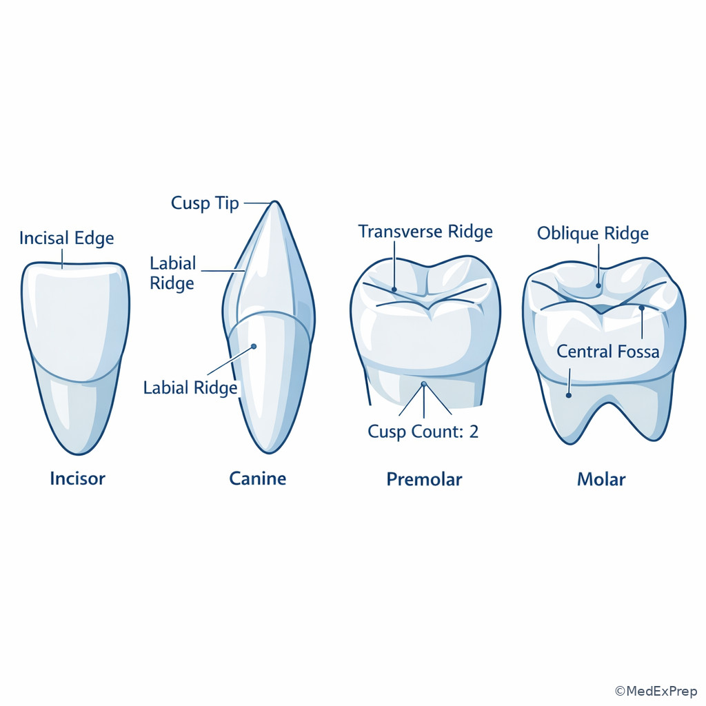
B Worked example
A 34-year-old patient with xerostomia from antidepressant use and a history of orthodontic retention presents for recare. An exam item describes an extracted study tooth with one buccal cusp, a very small lingual cusp, and a prominent transverse ridge. A distracting note mentions mild attrition and stain. The stem asks for the most likely tooth.
Identify task: Tooth-group and likely specific tooth. Extract key facts: premolar pattern, one buccal cusp, tiny nonfunctional lingual cusp, strong transverse ridge. Apply rule: these are classic clues for the mandibular first premolar. Eliminate distractors: maxillary first premolar usually has two more functional cusps and often a mesial concavity; mandibular second premolar often has larger lingual cusp(s); canine has one cusp but no occlusal table. Verify: the transverse ridge plus small lingual cusp is the deciding clue.
C Exam trap
Common wrong answer: Maxillary first premolar.
Why it is tempting: Students hear “premolar with two cusps” and jump to a maxillary premolar without checking lingual cusp function.
Single clue that eliminates it: A very small, nonfunctional lingual cusp with strong transverse ridge points to the mandibular first premolar.
D Checkpoint
Question: Which feature most strongly supports identification of a maxillary molar rather than a mandibular molar?
- Oblique ridge — Tempting because many molars have ridges, and it is correct because the oblique ridge is a signature maxillary molar feature.
- Five cusps — Tempting because cusp count helps identify molars, but it is wrong because five cusps classically favors the mandibular first molar.
- Two buccal grooves — Tempting because groove count feels specific, but it is wrong because this points toward the mandibular first molar.
- Small distal cusp — Tempting because distal morphology can distinguish teeth, but it is wrong because this is not the best discriminator between maxillary and mandibular molars.
Practitioner Action: Assess: Tooth-group form before selecting adaptation and self-care emphasis. Intervene: Tailor OHI to anatomy-driven retention sites such as molar pits and deep lingual areas. Refer: If unusual morphology affects eruption, occlusion, or suspected pathology, inform the dentist. Document: Tooth and surface landmarks that influenced care decisions.
1.1.1.6 Landmark-based caries and deposit risk: where morphology changes preventive strategy
A Explanation
This is the first higher-level application conflict: choose the most at-risk surface or preventive focus vs reject a surface that sounds plausible but is less vulnerable in the scenario. The single rule is: deep, sheltered, poorly self-cleansing anatomy retains biofilm longer. Pits, fissures, developmental grooves, lingual pits, and cervical contour changes are commonly tested because they change disease risk and hygiene access.
Posterior occlusal pits and fissures are classic plaque-retentive sites, especially in newly erupted teeth where enamel maturation and eruption status affect self-cleansing. Lingual pits of maxillary incisors can be tested as localized anatomy-linked risk sites. Cervical areas near the height of contour can also trap plaque when self-care is poor, xerostomia is present, or the crown contour complicates access. For the hygienist, the practical decision is not “restore or not restore” but rather recognize the at-risk morphology, implement preventive measures within scope, educate, document, and refer as indicated.
See Fig 5 for a risk map of common plaque-retentive landmarks. This also previews the upcoming eruption lesson: newly erupted molars are especially vulnerable because morphology and eruption stage together reduce effective natural cleansing.
Exam takeaway: Pits, fissures, and sheltered contours predict where prevention and instrumentation should be targeted first.
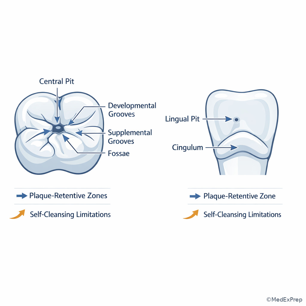
Exam takeaway: Combine morphology with patient risk factors rather than treating every groove pattern as equally urgent.
flowchart TD
A[Retentive anatomy found] --> B{Patient risk factors present?}
B -->|Yes xerostomia frequent sugar poor OH| C[Higher prevention priority]
B -->|No major risk factors| D[Routine prevention with monitoring]
C --> E[Targeted OHI fluoride communication and documentation]
D --> E
E --> F[Refer or consult if suspicious lesion or need exceeds scope]
B Worked example
A 14-year-old patient presents for recare 8 months after eruption of permanent second molars. Medical history includes asthma managed with inhaled corticosteroids and seasonal allergies; the patient also drinks sports beverages daily. The distractor is that interproximal contacts appear tight, which may make proximal surfaces seem like the main issue. The stem asks which landmark warrants the most immediate preventive focus.
Identify task: Prioritize prevention based on anatomy and risk. Extract key facts: newly erupted molars, deep occlusal anatomy, xerostomia tendency from mouth breathing and medication effects, frequent sugar exposure. Apply rule: sheltered occlusal pits and fissures on erupting molars are high priority. Eliminate distractors: proximal contacts matter, but the stem emphasizes recent eruption and occlusal anatomy; buccal height of contour is not the most retentive site here. Verify: select the occlusal pits/fissures of the erupting molars as the primary preventive focus.
C Exam trap
Common wrong answer: Tight interproximal contact.
Why it is tempting: Students know proximal caries risk is real and may overread the mention of contact tightness.
Single clue that eliminates it: The stem centers on newly erupted molars with deep occlusal anatomy, which makes pits and fissures the immediate anatomy-based target.
D Checkpoint
Question: A hygienist is prioritizing OHI for a patient with poor plaque control. Which landmark is most likely to be described as a pinpoint plaque-retentive site on a posterior occlusal surface?
- Pit — Tempting because the description is specific, and it is correct because a pit is a pinpoint depression often located where grooves meet.
- Fossa — Tempting because it is a depression on the occlusal surface, but it is wrong because it is broader and shallower than a pinpoint site.
- Triangular ridge — Tempting because ridges shape occlusal anatomy, but it is wrong because it is elevated rather than retentive as a pinpoint depression.
- Marginal ridge — Tempting because it borders the occlusal table, but it is wrong because it is not a pinpoint depression.
- The most common anatomy-based retentive sites on posterior teeth are pits and fissures.
- True or False: A broad shallow depression is a pit. False.
- Name 3 anatomy-linked plaque traps: pit, developmental groove, sheltered cervical contour.
Practitioner Action: Assess: Retentive anatomy in relation to age, eruption stage, xerostomia, and diet. Intervene: Provide scope-appropriate preventive education and services. Refer: Suspicious lesions, cavitation concerns, or restorative needs go to the dentist. Document: Specific tooth landmarks, patient risk factors, preventive recommendations, and follow-up plan.
1.1.1.7 Root morphology, CEJ, and contour: why external form changes instrumentation and charting
A Explanation
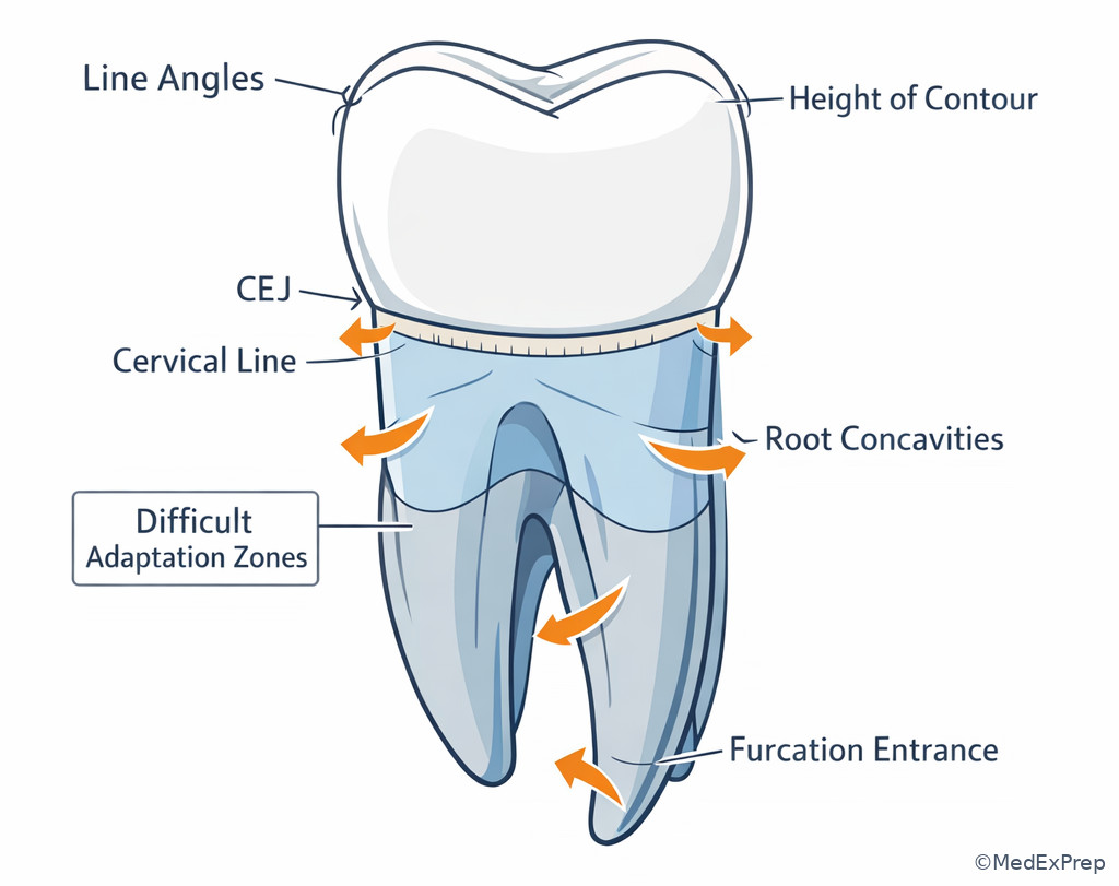
The final integration conflict is choose the safest hygiene action based on form vs reject a one-size-fits-all approach. The single rule is: crown and root morphology dictate access, adaptation, and where deposits hide. Root concavities, furcation entrances, line angles, cervical bulges, and CEJ relationships change what the hygienist can feel and how to adapt instruments safely.
Root morphology matters even in a foundational lesson because exam stems may ask which area is more difficult to debride or where an instrument should be adapted more carefully. Posterior teeth often present line-angle and root-trunk complexity; premolars may show root depressions; molars create furcation-related access issues. On crowns, heights of contour protect gingiva when normal, but excess contour can worsen plaque retention and distort self-cleansing. The CEJ is also a key boundary in charting, sensitivity discussion, and recognizing cervical anatomy versus defect.
For the NBDHE, the hygienist role is to recognize how anatomy modifies care, not to perform out-of-scope definitive treatment decisions. If the stem presents unusual anatomy plus a suspicious defect, your answer should emphasize recognition, adaptation, documentation, and referral. See Fig 6 for the anatomy-to-action map.
Exam takeaway: External form predicts where instrumentation becomes more difficult and where deposits are likely to remain if adaptation is poor.
Exam takeaway: Mesial-distal CEJ differences and height-of-contour positions are exam-relevant for orientation and clinical description.
B Worked example
A 52-year-old patient with type 2 diabetes, controlled hypertension, and a history of smoking cessation 3 years ago presents for periodontal maintenance. A distracting clue in the stem is mild enamel craze lines on incisors. During instrumentation of a maxillary molar, the clinician encounters recurrent deposit detection near a complex root area despite visible crown cleanliness. The question asks for the most important morphology-based explanation.
Identify task: Connect anatomy to difficult deposit removal. Extract key facts: posterior maxillary molar, recurrent hidden deposits, root complexity. Apply rule: molar roots and furcation-related anatomy create difficult access and adaptation zones. Eliminate distractors: incisor craze lines are irrelevant; crown surface shine does not rule out root deposits; generalized plaque control is not the morphology-based explanation asked. Verify: choose complex root contour and furcation-associated access limitations as the best explanation.
C Exam trap
Common wrong answer: Increase lateral pressure because the deposit is “stubborn.”
Why it is tempting: Students may think persistence equals more force.
Single clue that eliminates it: The stem asks for a morphology-based explanation; the issue is adaptation around complex anatomy, not force deficiency.
D Checkpoint
Question: Which statement best guides safe adaptation when anatomy is complex?
- Modify adaptation to line angles and root contour rather than increasing force uniformly — Tempting because it sounds technique-focused, and it is correct because anatomy, not brute force, should guide the approach.
- Ignore crown contour once the gingival margin is crossed — Tempting because root instrumentation feels separate, but it is wrong because crown-root transitions and CEJ orientation matter clinically.
- Use the same adaptation on all posterior teeth because molars are similar — Tempting because it simplifies care, but it is wrong because root and furcation patterns vary significantly.
- Proceed with definitive correction of abnormal anatomy — Tempting because it sounds proactive, but it is wrong and out of scope for the hygienist candidate.
Practitioner Action: Assess: CEJ, contour, line angles, and likely root complexity before instrumentation. Intervene: Adapt strokes and patient education to anatomy-guided risk areas. Refer: Unusual structural defects, suspicious cervical changes, or care needs beyond dental hygiene scope require dentist evaluation. Document: Difficult-access areas, response to care, and communication with supervising providers.
Exam Traps & Differentiators
Common traps & distractors: The exam rarely tests a landmark in isolation. It usually pairs a near-match from the same category and expects you to notice one location clue, one shape clue, or one tooth-group clue.
1.1.1.8 Most common wrong answer and why
The most common error is choosing a broad category instead of the specific landmark. Example: selecting “ridge” when the correct answer is “oblique ridge,” or selecting “depression” language such as fossa when the stem specifically describes a pinpoint pit. This happens because the surface anatomy sounds familiar, but the stem is asking for the narrowest accurate label.
| Looks similar | But isn’t | Single discriminator | Why the wrong answer is tempting |
|---|---|---|---|
| Pit | Fossa | Pit is pinpoint; fossa is broad and shallow | Both are depressions on posterior teeth |
| Triangular ridge | Marginal ridge | Triangular ridge runs from cusp tip inward; marginal ridge forms the border | Both are ridges on occlusal surfaces |
| Transverse ridge | Oblique ridge | Transverse ridge is union of two triangular ridges; oblique ridge is specific to maxillary molars | Both cross part of the occlusal table |
| Cingulum | Height of contour | Cingulum is anterior lingual cervical prominence; height of contour is a general concept on any surface | Both involve convexity |
| Mandibular first premolar | Maxillary first premolar | Tiny nonfunctional lingual cusp and strong transverse ridge favor mandibular first premolar | Both are premolars with cusp-based occlusal anatomy |
| Maxillary molar | Mandibular molar | Oblique ridge strongly favors maxillary molar | Both have multiple cusps and central fossae |
1.1.1.9 If the stem says ___, think ___
- If the stem says pinpoint depression at a groove junction, think pit.
- If the stem says cervical lingual prominence on an anterior tooth, think cingulum.
- If the stem says ridge crossing a maxillary molar occlusal surface obliquely, think oblique ridge.
- If the stem says five cusps with two buccal grooves, think mandibular first molar.
- If the stem says small nonfunctional lingual cusp with strong transverse ridge, think mandibular first premolar.
- If the stem says greater CEJ curvature, think mesial.
| Commonly confused pair | Best clue to choose correctly | Cross-topic tie-in |
|---|---|---|
| Normal deep groove vs suspicious defect | Normal anatomy follows expected landmark pattern; suspicious change is irregular or inconsistent | Related to Oral Histology and Tissues; know what intact enamel anatomy should look like |
| Erupting molar occlusal risk vs smooth-surface risk | Recent eruption plus deep pits/fissures elevates occlusal prevention priority | Related to Dentition Development and Eruption |
| Anatomy-based retention vs poor technique | If the site is a furcation, root depression, or line angle, anatomy may be the key barrier | Related to Instrumentation and Nonsurgical Care |
Tables
| Key fact / value pattern | High-yield takeaway |
|---|---|
| Mesial CEJ curvature | Usually greater than distal; useful for orientation |
| Anterior lingual landmark cluster | Cingulum + marginal ridges + lingual fossa |
| Posterior occlusal landmark cluster | Cusps + ridges + grooves + fossae + pits |
| Maxillary molar clue | Oblique ridge; possible cusp of Carabelli on first molar |
| Mandibular first molar clue | Usually 5 cusps and 2 buccal grooves |
| Mandibular first premolar clue | Strong transverse ridge and small lingual cusp |
| Canine clue | Single cusp with longest root |
| Most plaque-retentive pinpoint site | Pit |
| Comparison | Incisor | Canine | Premolar | Molar |
|---|---|---|---|---|
| Primary working form | Incisal edge | Single cusp | Transitional cusp form | Multiple cusp grinding surface |
| Signature lingual/occlusal clue | Cingulum and lingual fossa | Pronounced lingual ridge | Transverse ridge common | Fossae, grooves, pits |
| Frequent exam discriminator | Mamelons or incisal angles | Longest root | Lingual cusp size pattern | Oblique ridge or cusp count |
| Plaque-retentive anatomy emphasis | Lingual pit possible | Cervical contour / line angles | Grooves and small lingual access zones | Pits and fissures |
| Finding in stem | Likely classification / interpretation | What to do with it as a hygienist candidate |
|---|---|---|
| Pinpoint occlusal depression | Pit | Note plaque-retentive risk and tailor prevention |
| Broad shallow occlusal depression | Fossa | Recognize normal anatomy and associated groove pattern |
| Cervical lingual prominence on anterior tooth | Cingulum | Use to orient surface and explain plaque-retentive zones |
| Ridge across maxillary molar occlusal surface | Oblique ridge | Use as a maxillary molar identifier |
| Very small lingual cusp on premolar | Mandibular first premolar pattern | Anticipate challenging lingual adaptation and documentation accuracy |
| Recurrent hidden deposits near molar roots | Root contour/furcation access issue | Modify adaptation, document, and communicate if needed |
Algorithm / Approach
Exam takeaway: A repeatable identify-orient-classify-risk sequence improves speed and reduces anatomy distractor errors.
Use this five-step method every time you see a tooth diagram or morphology-heavy stem. First, decide whether the item asks for naming, orientation, classification, or clinical implication. Next, classify the shape and location. Then orient the surface and side. Finally, link the landmark to the in-scope action: recognition, documentation, prevention, or referral. This prevents a common NBDHE error—answering a dentist-level treatment question when the real tested skill is anatomy recognition plus safe hygiene judgment. See Fig 7.
Rapid Review
- Cingulum → cervical lingual convexity of anterior teeth, not a general contour term
- Pit → pinpoint depression at groove junction, more specific than fossa
- Fossa → broad shallow depression, not a narrow line
- Triangular ridge → cusp tip toward center, not border ridge
- Marginal ridge → mesial or distal border of occlusal or anterior lingual surface
- Oblique ridge → maxillary molar clue
- Mandibular first molar → usually 5 cusps, often 2 buccal grooves
- Mandibular first premolar → small lingual cusp plus strong transverse ridge
- Anterior lingual surface → cingulum + lingual fossa + marginal ridges
- Mesial surface → greater CEJ curvature than distal
- Canine → single cusp plus longest root
- Molars → broad occlusal table with pits/fissures as key retentive sites
- Height of contour → contour concept affecting tissue protection and plaque retention
- Root complexity → adaptation problem, not an invitation to use more force
Self-check quiz
1. A 16-year-old patient with fixed orthodontic retainers and high sports drink intake presents for recare. On a permanent molar, the hygienist identifies a pinpoint depression where developmental grooves meet. Which landmark is the most likely finding?
- Pit
- Fossa
- Marginal ridge
- Triangular ridge
2. A 20-year-old patient with mild mouth breathing and no significant medical history has an anterior tooth image shown on the lingual view. Which landmark best identifies the lingual surface of an anterior tooth?
- Cingulum
- Central groove
- Buccal pit
- Oblique ridge
3. A 29-year-old patient with generalized plaque-induced gingivitis, ADHD treated with stimulant medication, and frequent snacking presents for preventive care. A study model shows a tooth with one buccal cusp, a very small lingual cusp, and a strong transverse ridge. Which tooth is most likely represented?
- Mandibular first premolar
- Maxillary first premolar
- Mandibular canine
- Mandibular second premolar, three-cusp type
4. A 13-year-old patient presents 6 months after eruption of first premolars and second molars. Medical history includes asthma and seasonal allergies; the parent reports frequent juice sipping. Which surface requires the most appropriate preventive focus based on morphology alone?
- Deep occlusal pits and fissures of recently erupted molars
- Incisal edges of maxillary incisors
- Middle third of facial surfaces on canines
- Distal height of contour of mandibular incisors
5. A 24-year-old patient with a history of whitening product use and moderate coffee stain has a single-tooth image on exam. The crown shows an anterior lingual surface with one side demonstrating greater CEJ curvature. Which interpretation is most appropriate?
- The side with greater CEJ curvature is mesial
- The side with greater CEJ curvature is distal
- The cingulum always indicates the mesial side
- CEJ curvature is not useful for orientation
6. A 55-year-old patient with controlled type 2 diabetes, stage 1 hypertension, and past tobacco use presents for periodontal maintenance. During instrumentation, deposits repeatedly persist in a maxillary posterior tooth despite good visibility of the crown. The stem asks for the best explanation for the difficult access. Which answer is most appropriate?
- Complex root contour and furcation-associated access limitations
- Increased need for heavier lateral pressure on all strokes
- The presence of a cingulum on the lingual surface
- The mesial contact being more cervical than distal
7. A 32-year-old patient with xerostomia related to antidepressant therapy and a history of irregular dental visits presents with a study tooth image. The occlusal surface shows multiple cusps and a ridge running obliquely from the mesiolingual toward the distobuccal portion of the occlusal table. Which interpretation is most likely?
- Maxillary molar identified by an oblique ridge
- Mandibular first molar identified by two buccal grooves
- Mandibular first premolar identified by a transverse ridge
- Maxillary canine identified by a labial ridge
8. A 17-year-old patient with recent orthodontic debonding, high carbohydrate snacking, and inconsistent flossing presents for evaluation. An exam item asks the hygienist to choose the safest next step within scope after identifying a deep retentive occlusal pit with suspicious discoloration on a newly erupted molar. Which choice is best?
- Document the anatomy and findings, provide preventive guidance within scope, and communicate the suspicious site to the dentist for evaluation
- Independently diagnose occlusal caries and schedule restorative treatment
- Ignore the site because deep pits are always normal anatomy
- Reduce the pit depth mechanically during the hygiene visit
Answer key
1.1.1.10 1. Correct answer: A. Pit
A. Pit: Tempting because the stem says “pinpoint depression,” and it is correct because a pit is a small pinpoint depression often occurring where developmental grooves meet. Single stem clue: pinpoint depression at a groove junction. Authoritative support: standard dental anatomy texts describe pits as minute depressions commonly found at the junction of developmental grooves on occlusal surfaces (Fehrenbach and Popowics; Wheeler).
B. Fossa: Tempting because it is also a depression on the occlusal surface, but it is incorrect here because a fossa is broader and shallower, not pinpoint. Single stem clue: “pinpoint” excludes fossa. Source support: Wheeler’s Dental Anatomy, Physiology and Occlusion.
C. Marginal ridge: Tempting because it is common posterior anatomy, but it is incorrect because it is an elevated border, not a depression. Single stem clue: depression language rules it out. Source support: Wheeler.
D. Triangular ridge: Tempting because it is a named occlusal landmark, but it is incorrect because it is an elevation descending from a cusp tip toward the center. Single stem clue: the described structure retains plaque as a depression. Source support: Fehrenbach and Popowics.
1.1.1.11 2. Correct answer: A. Cingulum
A. Cingulum: Tempting because it is a classic landmark, and it is correct because it identifies the cervical convexity on the lingual surface of anterior teeth. Single stem clue: anterior tooth lingual view. Source support: Wheeler; Nield-Gehrig.
B. Central groove: Tempting because grooves help orient teeth, but it is incorrect because central grooves are posterior occlusal landmarks. Single stem clue: anterior tooth. Source support: Wheeler.
C. Buccal pit: Tempting because pits can be surface-specific, but it is incorrect because it does not define the lingual surface of anterior teeth. Single stem clue: lingual identification was requested. Source support: Fehrenbach and Popowics.
D. Oblique ridge: Tempting because it is distinctive, but it is incorrect because it is a maxillary molar feature, not an anterior landmark. Single stem clue: anterior tooth image. Source support: Wheeler.
1.1.1.12 3. Correct answer: A. Mandibular first premolar
A. Mandibular first premolar: Tempting because the clue cluster sounds classic, and it is correct because a small nonfunctional lingual cusp plus a strong transverse ridge strongly identifies this tooth. Single stem clue: very small lingual cusp with strong transverse ridge. Source support: Wheeler; Nield-Gehrig.
B. Maxillary first premolar: Tempting because it is also a premolar with two cusps, but it is incorrect because the lingual cusp is not typically described as tiny and nonfunctional in the same way. Single stem clue: disproportionately small lingual cusp excludes it. Source support: Wheeler.
C. Mandibular canine: Tempting because it has one dominant cusp, but it is incorrect because a canine lacks the premolar-style occlusal table and transverse ridge. Single stem clue: strong transverse ridge on an occlusal surface. Source support: Fehrenbach and Popowics.
D. Mandibular second premolar, three-cusp type: Tempting because mandibular premolars are often confused, but it is incorrect because the three-cusp type has two lingual cusps, not one tiny lingual cusp. Single stem clue: one very small lingual cusp. Source support: Wheeler.
1.1.1.13 4. Correct answer: A. Deep occlusal pits and fissures of recently erupted molars
A. Deep occlusal pits and fissures of recently erupted molars: Tempting because eruption timing and anatomy both point there, and it is correct because sheltered occlusal anatomy on newly erupted molars is a high-yield preventive target. Single stem clue: recently erupted molars. Source support: ADA prevention principles and standard dental anatomy texts.
B. Incisal edges of maxillary incisors: Tempting because those surfaces are visible and can show wear or stain, but it is incorrect because they are not the classic retentive anatomy described by the stem. Single stem clue: morphology-based risk on erupting molars is the issue. Source support: Fehrenbach and Popowics.
C. Middle third of facial surfaces on canines: Tempting because facial surfaces collect plaque when hygiene is poor, but it is incorrect because it lacks the sheltered pit-and-fissure anatomy emphasized in the scenario. Single stem clue: deep occlusal anatomy beats smooth-surface concern here. Source support: standard preventive dentistry references.
D. Distal height of contour of mandibular incisors: Tempting because contour affects plaque retention, but it is incorrect because this is not the highest-yield anatomy-based target in a newly erupted molar scenario. Single stem clue: age and eruption status. Source support: ADA and oral anatomy references.
1.1.1.14 5. Correct answer: A. The side with greater CEJ curvature is mesial
A. The side with greater CEJ curvature is mesial: Tempting because it matches a known orientation rule, and it is correct because mesial CEJ curvature is generally greater than distal. Single stem clue: greater CEJ curvature on one side. Source support: Wheeler.
B. The side with greater CEJ curvature is distal: Tempting because students may reverse the rule under test pressure, but it is incorrect because distal usually has less curvature. Single stem clue: the word “greater” points to mesial, not distal. Source support: Wheeler; Nield-Gehrig.
C. The cingulum always indicates the mesial side: Tempting because the cingulum is useful for orientation, but it is incorrect because it identifies the lingual surface, not mesial versus distal by itself. Single stem clue: the question asks side orientation, not surface orientation. Source support: Fehrenbach and Popowics.
D. CEJ curvature is not useful for orientation: Tempting because multiple features should be used together, but it is incorrect because CEJ curvature is indeed a useful orientation clue. Single stem clue: one side had greater curvature. Source support: Wheeler.
1.1.1.15 6. Correct answer: A. Complex root contour and furcation-associated access limitations
A. Complex root contour and furcation-associated access limitations: Tempting because the scenario names a maxillary posterior tooth with persistent deposits, and it is correct because molar root anatomy commonly creates difficult access and residual deposit risk. Single stem clue: repeated persistence in a maxillary posterior root area despite visible crown cleanliness. Source support: Nield-Gehrig, Fundamentals of Periodontal Instrumentation and Advanced Root Instrumentation; periodontal instrumentation standards.
B. Increased need for heavier lateral pressure on all strokes: Tempting because stubborn deposits can make more force seem logical, but it is incorrect because the stem asks for the explanation, and heavy uniform pressure does not solve anatomy-driven access issues. Single stem clue: anatomy, not force, is the limiting factor. Source support: instrumentation texts emphasize adaptation and fulcrum modification over indiscriminate force.
C. The presence of a cingulum on the lingual surface: Tempting because it is a named landmark, but it is incorrect because cingulum applies to anterior teeth, not the maxillary posterior tooth in this stem. Single stem clue: maxillary posterior location. Source support: Wheeler.
D. The mesial contact being more cervical than distal: Tempting because contact position helps orientation, but it is incorrect because it does not explain difficult root-area deposit access. Single stem clue: deposit persistence in root complexity, not crown contact height. Source support: Wheeler; Nield-Gehrig.
1.1.1.16 7. Correct answer: A. Maxillary molar identified by an oblique ridge
A. Maxillary molar identified by an oblique ridge: Tempting because the stem directly describes a ridge crossing obliquely, and it is correct because the oblique ridge is a classic maxillary molar hallmark. Single stem clue: ridge running obliquely across the occlusal table. Source support: Wheeler.
B. Mandibular first molar identified by two buccal grooves: Tempting because mandibular first molars are cusp-rich and highly testable, but it is incorrect because the stem describes an oblique ridge, not two buccal grooves. Single stem clue: explicit ridge pattern excludes the groove-based identification. Source support: Wheeler.
C. Mandibular first premolar identified by a transverse ridge: Tempting because transverse ridge is a favorite distractor, but it is incorrect because the described tooth has multiple cusps and a molar-type occlusal table. Single stem clue: multiple cusps plus oblique ridge pattern. Source support: Fehrenbach and Popowics.
D. Maxillary canine identified by a labial ridge: Tempting because canines have a named ridge, but it is incorrect because a canine does not have a molar-style multi-cusp occlusal surface. Single stem clue: multiple cusps and occlusal table. Source support: Wheeler.
1.1.1.17 8. Correct answer: A. Document the anatomy and findings, provide preventive guidance within scope, and communicate the suspicious site to the dentist for evaluation
A. Document the anatomy and findings, provide preventive guidance within scope, and communicate the suspicious site to the dentist for evaluation: Tempting because it sounds careful and role-appropriate, and it is correct because the hygienist candidate should recognize suspicious anatomy-linked findings, act preventively within scope, and refer or consult when definitive diagnosis or restorative decisions are needed. Single stem clue: “safest next step within scope.” Source support: NBDHE-aligned hygiene scope principles; ADA standards for patient-centered preventive care and communication.
B. Independently diagnose occlusal caries and schedule restorative treatment: Tempting because discoloration plus deep pit can suggest caries, but it is incorrect because definitive restorative treatment planning exceeds routine hygienist scope. Single stem clue: the question asks for an in-scope next step. Source support: exam scope guardrails and standard dental hygiene role boundaries.
C. Ignore the site because deep pits are always normal anatomy: Tempting because pits can be normal anatomy, but it is incorrect because suspicious discoloration in a high-risk site warrants recognition, documentation, and communication rather than dismissal. Single stem clue: “suspicious discoloration” on a high-risk site. Source support: preventive and screening standards.
D. Reduce the pit depth mechanically during the hygiene visit: Tempting because it sounds like direct prevention, but it is incorrect and outside routine entry-level dental hygiene scope because altering tooth structure is not the proper response. Single stem clue: mechanical alteration of anatomy exceeds the intended role. Source support: scope-of-practice principles and standard of care.
Keep going with the full NBDHE Prep Course (NBDHE)
You just studied the way members do, in the same lesson interface. Full access unlocks every lesson, the question bank and flashcards for a full year.
One payment · 1 year of access · No auto-renewal