1.1.1 - CABG Postoperative Priorities
CABG Postoperative Priorities
What you’ll learn
- Immediate Postoperative Assessment: Prioritize the first CABG ICU checks that separate expected recovery from early deterioration.
- Hemodynamics and Perfusion: Distinguish hypovolemia, low cardiac output, bleeding, and tamponade using bedside trends rather than isolated numbers.
- Bleeding, Tamponade, and Coagulation: Recognize when chest tube output is reassuring, concerning, or falsely low.
- Pulmonary and Ventilatory Care: Identify respiratory findings that are common after CABG versus findings that require immediate escalation.
- Professional Judgment and Safety: Choose the highest-priority nursing action, what to monitor, what can be delegated, and when to notify the surgeon or intensivist now.
- Immediate Postoperative Assessment → Initial CABG arrival assessment and early trend interpretation
- Hemodynamics and Perfusion → Perfusion data synthesis, preload-output relationships, shock pattern recognition
- Bleeding, Tamponade, and Coagulation → Chest tube trend interpretation and urgent complication recognition
- Pulmonary and Ventilatory Care → Early postoperative oxygenation and ventilation priorities
- Professional Judgment and Safety → Best-next-action reasoning, escalation, scope, and reassessment
Why it matters
- Most early CABG complications are first detected at the bedside through trend recognition, not through a single dramatic lab value.
- Board stems often force a choice between two reasonable nursing actions; the correct answer is the one that addresses the most immediate threat to circulation, oxygenation, or graft perfusion.
- After CABG, “normal postoperative findings” and “early decompensation” overlap; exam success depends on recognizing the one clue that changes the priority.
- Chest tube output, hemodynamics, rhythm, oxygenation, temperature, urine output, and mental status must be interpreted together.
Key Terms & Must-Know Facts
1.1.1.1 Key terms
- CABG: Coronary artery bypass grafting
- Low cardiac output syndrome: Inadequate perfusion despite postoperative support
- MAP: Mean arterial pressure; key perfusion target variable
- CVP: Central venous pressure; trend marker of right-sided filling, not a stand-alone volume diagnosis
- Chest tube patency: Whether mediastinal/pleural drainage is flowing freely
- Tamponade: Cardiac filling impairment from pericardial pressure
- Graft ischemia clue: New ST changes, ventricular dysfunction, low output, malignant dysrhythmia
- Vasoplegia: Low SVR state with warm vasodilated profile and hypotension
- Temporary epicardial pacing wires: Postoperative wires for rhythm support if needed
- Atelectasis: Common early postoperative cause of mild hypoxemia
- Mixed venous oxygen saturation: Global balance of oxygen delivery and consumption when available
- Urine output: Practical bedside perfusion marker
1.1.1.2 Must-know facts
- High chest tube output suggests bleeding; suddenly low output with hemodynamic collapse raises concern for tamponade or tube obstruction.
- Tachycardia after CABG is not automatically pain or agitation; first ask whether it is compensating for falling stroke volume.
- Hypotension plus rising filling pressures plus narrowing pulse pressure is more concerning than hypotension alone.
- Low urine output is often an early perfusion warning before creatinine rises.
- Mild hypothermia, low-grade leukocytosis, and transient atelectatic hypoxemia can be expected early; worsening instability is not.
- A new arrhythmia after CABG can be the problem or the consequence of ischemia, electrolyte disturbance, hypoxia, or low output.
- Local protocols vary; confirm with your institution for vasoactive titration ranges, pacing practices, and blood product thresholds.
- In CSC-style questions, the best answer usually starts with the most immediate reassessment and stabilization step within nursing scope, followed by prompt escalation when red flags persist.
Core content
Exam takeaway: Use a circulation-first trend framework to decide whether the next action is reassessment, intervention, or urgent escalation.
flowchart TD
A[Post-CABG ICU arrival] --> B[Check airway ventilation oxygenation]
B --> C[Check MAP rhythm pulse quality]
C --> D[Review chest tubes and drainage trend]
D --> E[Correlate CVP urine output skin temperature mental status]
E --> F{Stable trend?}
F -->|Yes| G[Continue protocol monitoring and wean support]
F -->|No| H[Identify bleeding vs low output vs tamponade vs respiratory cause] Exam takeaway: Visually anchor the first scan of lines, drains, pacing wires, sternotomy, and perfusion clues before focusing on any single monitor number.
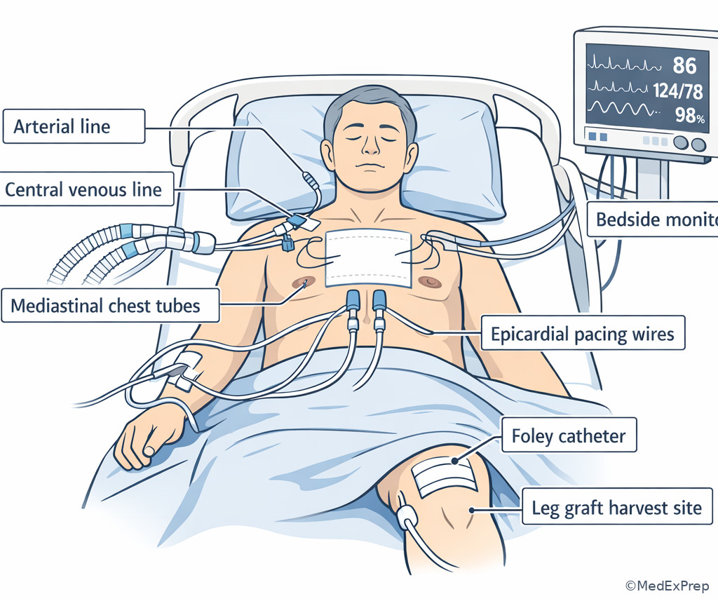
Exam takeaway: Trace drainage amount, color, tubing patency, and sudden changes because the trend matters more than one isolated chamber reading.
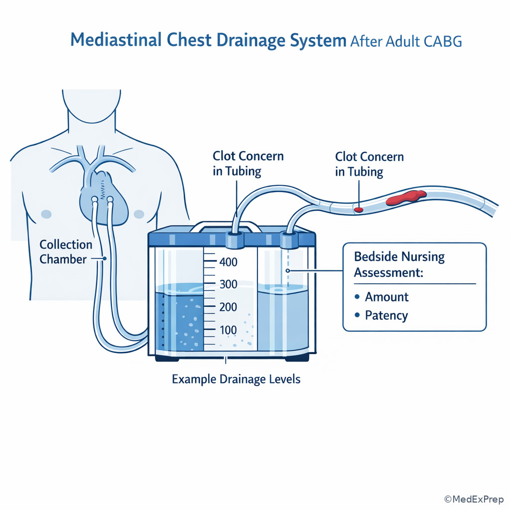
1.1.1.3 Foundational bedside priorities in the first postoperative hours
A Explanation
The first CABG question is rarely “What diagnosis is this?” It is usually “What do you assess first, and what finding changes your priority?” The decision conflict is between treating a monitor number versus evaluating the whole postoperative pattern. The single rule that resolves this: after CABG, circulation and oxygen delivery are judged by trends across hemodynamics, drainage, rhythm, urine output, and clinical perfusion together. See Fig 1 and Fig 2.
In the first ICU hours, some findings are common: intubation, mild hypothermia, modest vasopressor need, serosanguineous chest drainage, transient metabolic acidosis correction, and mild atelectatic hypoxemia. What is not reassuring is a pattern of worsening perfusion: falling MAP, rising lactate, cool clammy skin, decreasing urine output, altered mentation, rising filling pressures, new ST changes, or escalating pressor requirement. The exam often gives one distracting detail such as agitation, mild fever, or incisional pain; do not let that distract from circulation.
WHY THIS IS TESTED: CABG stems force you to choose between a familiar but lower-priority postoperative action and the assessment finding that signals impending shock or graft-related compromise.
Choose the action that answers the highest-risk question first: Is the patient oxygenating? Is the patient perfusing? Are the drains functioning? Is there evidence of bleeding, tamponade, or low output? A bedside RN does not independently decide operative management, but must identify unstable patterns early, intervene within protocol, and escalate promptly.
B Worked example
A 68-year-old man is 45 minutes post-CABG x3. History includes diabetes, hypertension, and CKD stage 3. He is intubated, lightly sedated, warm blankets in place. HR 104/min, MAP 63 mm Hg on low-dose vasopressor, CVP 10 mm Hg, chest tube output 90 mL in the last 30 minutes, urine output 20 mL since arrival, lactate mildly elevated, oxygen saturation 96% on mechanical ventilation. The stem mentions that he is “restless when stimulated” and has a temperature of 35.7°C.
Identify task: Most appropriate priority assessment/intervention.
Extract key facts: Borderline MAP, low urine output, modest but not extreme drainage, tachycardia, CKD as noise because baseline renal reserve is lower, mild hypothermia/restlessness are common postoperative distractions.
Apply rule: Focus on global perfusion and trend synthesis, not isolated agitation. Reassess hemodynamics, drainage trend, waveform quality, peripheral perfusion, and response to current support.
Eliminate distractors: Giving analgesia or warming measures may be appropriate, but neither is the top priority until circulation is clarified. Assuming low urine output is only baseline CKD misses an early perfusion clue.
Verify: The correct next step is targeted circulation reassessment with prompt escalation if MAP, output, and urine output remain poor or worsen.
C Exam trap
D Checkpoint
Question: A patient 1 hour after CABG has HR 108/min, MAP 61 mm Hg, chest tube output 70 mL in the past 30 minutes, urine output 15 mL since ICU arrival, oxygen saturation 97% on the ventilator, and cool extremities. What is the most appropriate next nursing action?
- Increase environmental warming and reassess in 30 minutes.
- Perform focused perfusion reassessment, verify line/drain patency and trends, and notify the provider if instability persists.
- Administer PRN analgesia for likely incisional pain causing tachycardia.
- Document the findings as expected for the immediate post-bypass period.
Answer: B. A is tempting because hypothermia is common, but it does not address the active perfusion concern. B is right because the hemodynamic and urine-output pattern may represent early low output or volume loss and requires circulation-focused reassessment. C is tempting because pain can cause tachycardia, but cool extremities and low urine output make perfusion the priority. D is tempting because some instability is expected early, but the low perfusion pattern prevents simple observation.
1.1.1.4 Distinguishing expected findings from bleeding and low cardiac output
A Explanation
After CABG, the bedside challenge is to choose between “continue routine monitoring” and “treat as evolving circulatory failure.” The single rule that resolves this: expected postoperative findings should trend toward stability; bleeding and low output do not. See Fig 3.
Bleeding usually presents with increasing chest tube output, falling hemoglobin, tachycardia, hypotension, increasing vasopressor requirement, and hypovolemia signs. Low cardiac output may present with hypotension, cool extremities, oliguria, rising lactate, low mixed venous saturation if available, and often rising filling pressures if ventricular function is poor. The key is that both can produce hypotension and tachycardia. The separator is the drainage/perfusion relationship. Heavy ongoing mediastinal drainage points toward blood loss. Minimal drainage with shock does not reassure you; it may mean tamponade or obstructed tubes rather than stability.
Do not over-read a single CVP. A “normal” or elevated CVP does not exclude poor output. Similarly, a modest chest tube number can still be dangerous if it suddenly drops in a patient who is deteriorating. Many exam distractors rely on the assumption that more drainage is always worse and less drainage is always better. That is false after cardiac surgery.
WHY THIS IS TESTED: CSC-style stems test whether you can prioritize circulation patterns before lab confirmation and avoid being misled by one superficially reassuring value.
Exam takeaway: A worsening perfusion pattern with either excessive drainage or suddenly absent drainage should change your priority immediately.
flowchart TD
A[Hypotension after CABG] --> B{Chest tube trend}
B -->|Increasing output| C[Bleeding more likely]
B -->|Sudden drop or minimal output| D[Tamponade or tube obstruction concern]
B -->|Stable modest output| E[Consider low cardiac output or vasoplegia]
C --> F[Check Hgb trend, pulses, volume response, notify]
D --> G[Check patency, rising filling pressures, narrow pulse pressure, urgent escalation]
E --> H[Check perfusion markers, ventricular function clues, vasoactive response] B Worked example
A 74-year-old woman 3 hours after CABG x4 has obesity, COPD, and chronic anemia. HR 116/min, MAP 58 mm Hg, CVP 6 mm Hg, chest tube output has been 180 mL, then 220 mL, then 250 mL in successive hours. Skin is cool, urine output has fallen from 40 mL/hr to 15 mL/hr. The stem also notes coarse breath sounds and a low-grade fever of 38.0°C.
Identify task: Determine the most likely priority problem and best immediate nursing action.
Extract key facts: Progressive drainage increase, hypotension, tachycardia, declining urine output, cool skin. COPD and coarse breath sounds are distracting but not the primary circulation threat.
Apply rule: Increasing chest tube output with worsening hemodynamics is bleeding until proven otherwise.
Eliminate distractors: Treating breath sounds first misses circulation. Attributing fever to infection is incorrect in this immediate window and does not explain the drainage trend. Waiting for the next hemoglobin misses active deterioration.
Verify: Immediate focused reassessment, support per protocol, preparation for blood products if ordered, and urgent provider notification are appropriate.
C Exam trap
D Checkpoint
Question: Which postoperative CABG trend most strongly supports bleeding rather than isolated low cardiac output?
- MAP 62 mm Hg, cool feet, urine output 20 mL/hr, chest tube output 30 mL/hr and unchanged
- MAP 60 mm Hg, tachycardia, chest tube output increasing each hour, and falling urine output
- MAP 58 mm Hg, rising CVP, narrowed pulse pressure, and suddenly decreased chest tube output
- MAP 64 mm Hg, low-grade fever, mild metabolic acidosis, and diminished bibasilar breath sounds
Answer: B. A is tempting because it shows hypoperfusion, but unchanged low drainage does not specifically point to bleeding. B is right because increasing drainage plus worsening perfusion is the strongest bleeding pattern. C is tempting because it is dangerous, but it points more toward tamponade or tube obstruction. D is tempting because acidosis and fever sound serious, but neither pattern localizes the problem to bleeding.
- The most dangerous “low chest tube output” pattern is low output plus hemodynamic worsening.
- True or False: A single normal CVP rules out low cardiac output. False.
- Name the 3 key bedside perfusion features besides blood pressure: urine output, skin/temperature, mental status.
1.1.1.5 Recognizing tamponade and obstructed drainage as a circulation emergency
A Explanation
The classic decision conflict is whether a patient with worsening hypotension and less chest tube drainage is “improving” or actually developing tamponade. The single rule that resolves it: after CABG, less drainage is dangerous when it is paired with rising filling pressures, narrowing pulse pressure, worsening output, or shock. See Fig 4.
Tamponade is a mechanical filling problem. The ventricle cannot fill adequately because external pressure impairs diastolic filling. Bedside clues may include hypotension, tachycardia, rising CVP, narrowing pulse pressure, low urine output, restlessness, cool skin, and suddenly reduced chest tube output or evidence of clot burden/obstruction in the system. Postoperative tamponade does not always present with textbook Beck triad. CSC questions often give a more realistic ICU picture: a patient on some vasoactive support whose numbers gradually worsen while chest tubes stop draining.
The nursing choice is not to diagnose independently but to recognize the pattern, ensure tubing is not kinked or dependent, rapidly reassess, support hemodynamics per protocol, and escalate urgently. Waiting for a dramatic hemoglobin drop or for severe hypoxemia delays recognition. Tamponade is a circulation emergency even when oxygen saturation still looks acceptable.
WHY THIS IS TESTED: Many learners are conditioned to equate more drainage with more danger. CABG questions reverse that assumption and reward recognition of obstructive shock clues.
Exam takeaway: Sudden loss of chest tube drainage with worsening perfusion should trigger concern for retained blood or tamponade rather than reassurance.
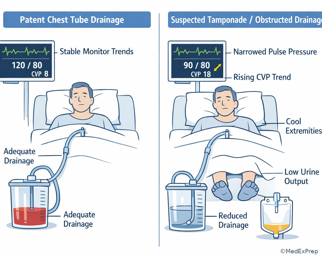
Exam takeaway: Identify clots, kinks, dependent loops, and abrupt drainage changes because mechanical problems can mimic clinical improvement.
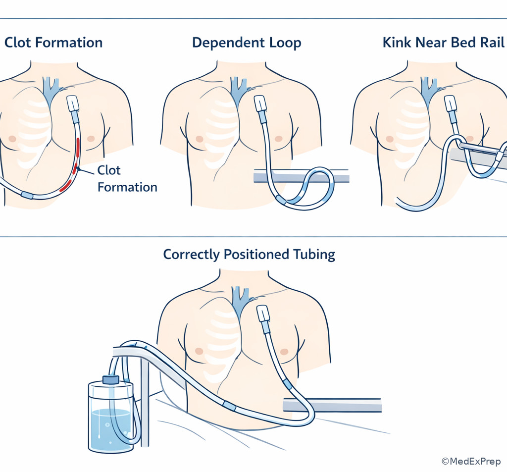
B Worked example
A 66-year-old man 5 hours after CABG x2 has peripheral vascular disease and poorly controlled diabetes. He was initially stable on minimal vasoactive support. Over 30 minutes, MAP falls from 72 to 58 mm Hg, HR rises from 88 to 112/min, CVP increases from 9 to 16 mm Hg, pulse pressure narrows, urine output drops, and the mediastinal chest tube that had drained 80–100 mL/hr is now nearly absent. The stem adds that the patient is “less agitated now” and oxygen saturation remains 95% on the ventilator.
Identify task: Highest-priority interpretation and action.
Extract key facts: Sudden drainage drop, hypotension, tachycardia, rising CVP, narrow pulse pressure, oliguria. Reduced agitation and preserved oxygen saturation are distractions.
Apply rule: This is tamponade/retained blood physiology until proven otherwise.
Eliminate distractors: Reassuring yourself because drainage has stopped is dangerous. Treating with additional sedation does not address the cause. Waiting for marked desaturation misses a circulation emergency.
Verify: Inspect system patency, support the patient, and urgently escalate suspected tamponade.
C Exam trap
D Checkpoint
Question: Which combination most strongly suggests tamponade after CABG?
- Increasing chest tube output, falling hemoglobin, tachycardia, and low CVP
- Suddenly decreased chest tube output, rising CVP, narrowing pulse pressure, and falling urine output
- Mild fever, diffuse crackles, stable chest tube output, and elevated white blood cell count
- New atrial fibrillation with adequate MAP, warm skin, and preserved urine output
Answer: B. A is tempting because it is unstable and dangerous, but it fits active bleeding more than tamponade. B is right because it combines obstructed drainage and impaired cardiac filling. C is tempting because crackles and leukocytosis sound urgent, but the pattern is not tamponade. D is tempting because new atrial fibrillation matters after CABG, but preserved perfusion argues against tamponade.
1.1.1.6 Respiratory priorities after CABG: common hypoxemia versus dangerous deterioration
A Explanation
The decision conflict is whether postoperative hypoxemia is expected atelectasis or a more urgent cardiopulmonary problem. The single rule that resolves it: mild oxygenation impairment with otherwise stable hemodynamics is common; hypoxemia plus rising work of breathing, hemodynamic decline, or pulmonary edema clues is not. See Fig 5.
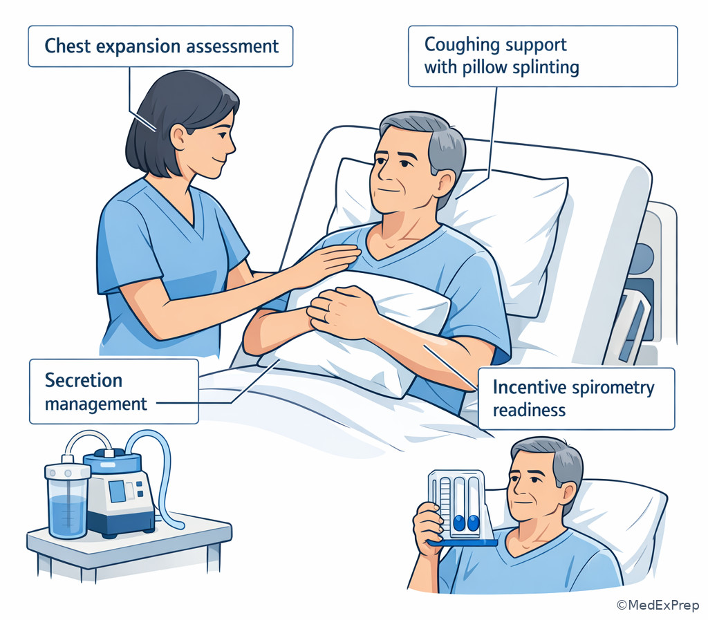
CABG patients commonly have atelectasis, pain-limited deep breathing, residual sedation, and transient secretion burden. These produce mild hypoxemia, bibasilar crackles or diminished bases, and the need for recruitment, pulmonary hygiene, early mobility when appropriate, and careful ventilator weaning. However, the exam may pivot to more dangerous causes: cardiogenic pulmonary edema from LV dysfunction, bleeding-related shock with poor oxygen delivery, pneumothorax, or less commonly severe postoperative respiratory failure. What changes the answer is the associated circulation pattern.
When oxygen saturation slips but MAP is stable, chest tube trends are acceptable, urine output is preserved, and breath sounds fit basilar atelectasis, the priority may be pulmonary recruitment and reassessment. When hypoxemia is accompanied by frothy secretions, new diffuse crackles, rising filling pressures, worsening output, or sudden unilateral absence of breath sounds, the priority escalates. The exam loves the tempting but wrong answer of “just encourage incentive spirometry” in a patient who is still intubated or is actually becoming hemodynamically unstable.
WHY THIS IS TESTED: Learners must separate a common postoperative pulmonary issue from respiratory findings that are really secondary to a cardiac problem.
Exam takeaway: The discriminator is not the oxygen saturation alone but whether respiratory findings occur with stable recovery or with worsening cardiac performance.
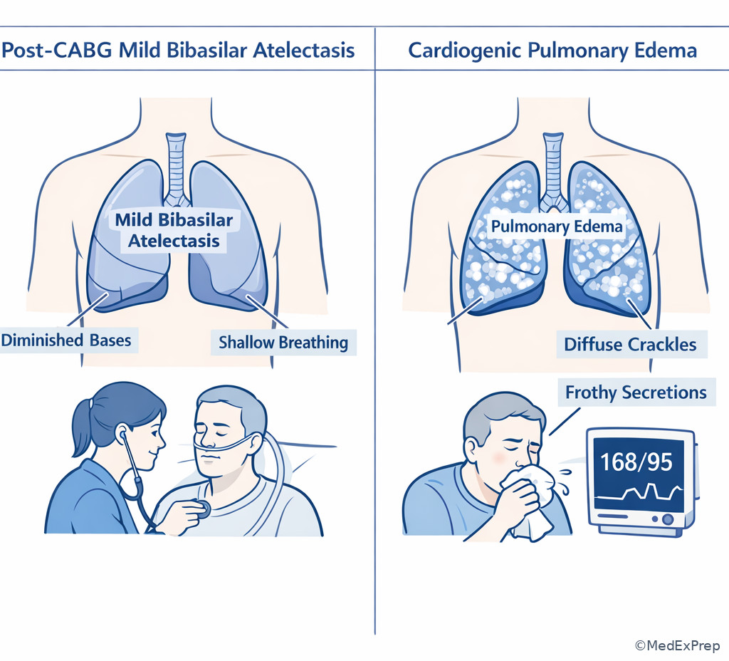
Exam takeaway: Use position, secretion clearance, and breathing pattern cues to identify common reversible respiratory causes before assuming a catastrophic complication.
B Worked example
A 71-year-old woman 8 hours after CABG x3 has obesity, obstructive sleep apnea, and hypertension. She was extubated 90 minutes ago. Oxygen saturation falls from 97% to 91% on supplemental oxygen. She has shallow respirations, diminished bibasilar breath sounds, pain with coughing, MAP 74 mm Hg, HR 92/min, stable chest tube output, warm skin, and urine output 45 mL/hr. The stem adds a mildly elevated white count and a history of heart failure with preserved EF.
Identify task: Best initial nursing action.
Extract key facts: Stable hemodynamics, preserved urine output, diminished bases, pain-limited breathing, early post-extubation timing. HF history and leukocytosis are distractors.
Apply rule: This most likely reflects atelectatic hypoventilation/pulmonary hygiene deficit rather than cardiogenic shock.
Eliminate distractors: Urgent tamponade or bleeding evaluation is less likely with stable circulatory data. Assuming infection is premature. The best step is oxygenation optimization, pulmonary hygiene, positioning, pain support, and reassessment.
Verify: Improvement in respiratory effort and oxygen saturation after these measures supports the interpretation.
C Exam trap
D Checkpoint
Question: Which finding most strongly shifts early post-CABG hypoxemia away from simple atelectasis and toward a cardiac cause?
- Diminished bibasilar breath sounds with pain-limited coughing and stable MAP
- Mild desaturation shortly after extubation with preserved urine output
- Diffuse crackles, worsening oxygenation, rising filling pressures, and declining perfusion
- Low-grade fever and shallow respirations on postoperative day 0
Answer: C. A is tempting because it is common and clinically relevant, but it supports atelectasis. B is tempting because it requires attention, but it does not by itself indicate a cardiac cause. C is right because pulmonary findings plus hemodynamic deterioration point toward cardiogenic pulmonary edema or another major cardiac problem. D is tempting because fever increases concern, but low-grade early fever is nonspecific and not the discriminator.
- The most common early pulmonary cause of mild post-CABG hypoxemia is atelectasis.
- True or False: Stable oxygen saturation excludes tamponade. False.
- Name 3 clues that favor a cardiac cause of respiratory decline: diffuse crackles, rising filling pressures, worsening perfusion.
1.1.1.7 Integration: choosing the best next action, delegation, and escalation
A Explanation
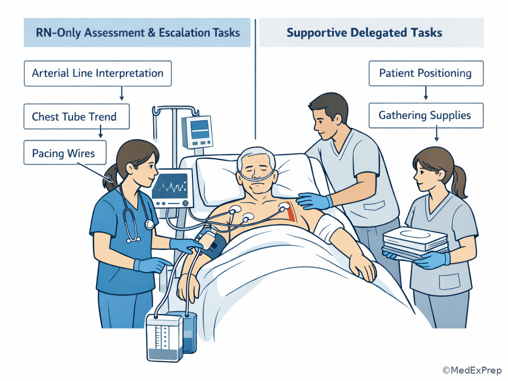
The final exam-level decision conflict is not just identifying the problem, but selecting the highest-priority nursing action within scope. The single rule that resolves it: the correct action is the one that immediately addresses ABCs or circulation, yields clarifying bedside data, and does not delay needed escalation. See Fig 6.
In CABG questions, all answer choices may be reasonable nursing actions: obtain labs, administer pain medication, reposition, notify the provider, reassess lines, or prepare blood products if ordered. Your job is to rank them. First comes direct bedside reassessment tied to the instability. Second comes immediate supportive intervention within protocol. Third comes closed-loop escalation. An action can be correct but still wrong because it is too late in the sequence. For example, notifying the provider without first confirming whether the chest tube is kinked or whether the arterial waveform has dampened may omit crucial bedside information. Conversely, collecting endless data without notifying anyone during a tamponade pattern is also wrong.
Delegation is also testable. Tasks requiring interpretation of hemodynamics, recognition of postoperative deterioration, management of vasoactives, and urgent reassessment remain with the RN. Routine comfort support, supply gathering, and non-interpretive tasks may be delegated according to local policy. The nurse must also know when to escalate beyond a routine page and activate higher-level resources.
WHY THIS IS TESTED: AACN CSC items emphasize professional judgment in a high-acuity monitored setting, not independent prescribing or surgical decision-making.
Exam takeaway: Pick the option that combines immediate RN-level reassessment with timely escalation when postoperative trends suggest instability.
flowchart TD
A[Concerning post-CABG change] --> B[ABCs and circulation check]
B --> C[Focused device and line reassessment]
C --> D{Threat persists?}
D -->|Yes| E[Implement protocol support and notify surgeon/intensivist now]
D -->|No| F[Continue frequent reassessment and document trend]
E --> G[Report key trend: rhythm, MAP, drainage, urine output, perfusion]
F --> G Exam takeaway: Distinguish bedside reassessment and interpretation tasks that must stay with the RN from supportive tasks that may be delegated.
B Worked example
A 63-year-old man 6 hours after CABG x4 has obesity, OSA, and insulin-treated diabetes. He is extubated, drowsy but arousable. HR 118/min, MAP 57 mm Hg, chest tube output has fallen from 90 mL/hr to 5 mL in the last hour, CVP has risen, extremities are cool, urine output is 10 mL in the last hour, and the monitor shows sinus tachycardia. A UAP reports that the patient “just looks tired,” and the family asks whether pain medication can be increased.
Identify task: Best next action and what can be delegated.
Extract key facts: Possible tamponade pattern, oliguria, hypotension, rising CVP, sudden drainage decrease. Family concern and drowsiness are distracting.
Apply rule: RN must remain at bedside, reassess chest tube/monitoring system and perfusion, initiate support per protocol, and urgently notify the provider.
Eliminate distractors: Delegating hemodynamic reassessment is unsafe. Addressing family concerns or pain first is lower priority. Simply calling the provider without checking for a mechanical drain issue omits important bedside data.
Verify: Appropriate care includes closed-loop communication of exact trend changes and continued reassessment.
C Exam trap
D Checkpoint
Question: Which action is most appropriate for the RN to delegate in a worsening post-CABG patient while the RN performs focused reassessment?
- Interpret the arterial pressure trend and determine whether tamponade is likely
- Inspect the chest tube system and decide whether the surgeon should be notified now
- Gather emergency supplies and assist with patient positioning while the RN evaluates hemodynamics
- Titrate vasoactive infusions independently according to clinical judgment without protocol
Answer: C. A is tempting because teamwork matters, but interpretation of invasive hemodynamics stays with the RN. B is tempting because tubing inspection sounds mechanical, yet deciding its significance in instability is an RN responsibility. C is right because supportive tasks that do not require clinical judgment can be delegated while the RN performs the critical assessment. D is tempting because urgent response is needed, but independent titration outside protocol is outside scope.
Exam Traps & Differentiators
Most common wrong answer and why: Choosing the most familiar postoperative task rather than the highest-priority one. Pain control, warming, pulmonary hygiene, lab collection, and routine documentation are all reasonable after CABG, but they become wrong when they delay circulation-focused reassessment in an unstable trend.
| Look-alike pattern | Why it is tempting | Single clue that separates it | Think |
|---|---|---|---|
| Bleeding vs low cardiac output | Both cause hypotension, tachycardia, oliguria | Increasing chest tube output favors bleeding | Volume loss first |
| Tamponade vs recovery | Lower drainage can look reassuring | Sudden drainage drop plus rising CVP or narrow pulse pressure | Obstructive physiology |
| Atelectasis vs pulmonary edema | Both can lower oxygen saturation | Diffuse edema findings with worsening perfusion favor cardiac cause | Cardiopulmonary deterioration |
| Pain tachycardia vs shock tachycardia | Incisional pain is common | Cool skin and low urine output point to perfusion problem | Compensation, not comfort issue |
| Routine page vs urgent escalation | Many changes can be “monitored” early after surgery | Trend worsens across multiple perfusion markers | Escalate now |
If the stem says ... think ...
- If the stem says “chest tube output suddenly decreased”, think tamponade or obstructed drainage if perfusion worsened.
- If the stem says “hourly drainage keeps rising”, think bleeding until proven otherwise.
- If the stem says “oxygenation worsened but hemodynamics are stable and bases are diminished”, think atelectasis/pulmonary hygiene.
- If the stem says “cool extremities, oliguria, rising lactate, pressor need”, think low cardiac output or another perfusion failure pattern.
- If the stem says “new dysrhythmia with ST changes or low output”, think ischemia/electrolytes/perfusion, not rhythm in isolation.
Forward connection: in Valve Surgery Bedside Patterns, rhythm and conduction issues become even more prominent; in Aortic And Combined Procedures, neurologic change and malperfusion cues compete more strongly with bleeding concerns. The trap is carrying a CABG-only mental model into those topics.
Tables
| Reference area | Expected early CABG finding | Concerning shift | Bedside meaning |
|---|---|---|---|
| Chest tube output | Serosanguineous drainage that trends toward reduction | Rapidly increasing output or sudden cessation with instability | Bleeding or tamponade/obstruction concern |
| MAP | May need support early but should stabilize | Persistent or worsening hypotension | Perfusion problem needing reassessment |
| Urine output | Adequate output with recovery | Falling output hour to hour | Early hypoperfusion clue |
| Skin/peripheral perfusion | Improving warmth and pulses | Cool, clammy, mottled, weak pulses | Low output or shock pattern |
| Breath sounds/oxygenation | Mild bibasilar atelectatic change common | Diffuse edema signs or worsening work of breathing | Cardiopulmonary deterioration |
| CVP/filling trend | Interpretable only in context | Rising CVP with shock and low drainage | Tamponade concern |
| Comparison point | Bleeding | Tamponade/retained blood | Low cardiac output |
|---|---|---|---|
| Chest tube output | Usually increasing | Often suddenly decreased or absent | May be modest or unchanged |
| CVP/filling trend | May be low or variable | Often rising | May be high if ventricular failure, variable otherwise |
| Pulse pressure | May narrow with shock | Often narrows prominently | Can narrow with poor stroke volume |
| Urine output | Falls with hypoperfusion | Falls with obstructive shock | Falls with poor forward flow |
| Key discriminator | Rising visible drainage | Worsening perfusion despite less drainage | Poor perfusion without dominant drain pattern |
| Priority nursing frame | Support and urgent bleeding escalation | Support, check patency, urgent escalation | Hemodynamic reassessment and support/escalation |
| Differential/classification | Typical bedside clues | Tempting wrong interpretation | Discriminator |
|---|---|---|---|
| Expected post-extubation atelectasis | Mild desaturation, diminished bases, pain-limited breathing, stable hemodynamics | Heart failure decompensation | Perfusion remains stable |
| Cardiogenic pulmonary edema | Diffuse crackles, worsening oxygenation, circulatory decline | Simple secretion burden | Hemodynamic worsening accompanies respiratory findings |
| Pain/agitation response | Tachycardia, restlessness, otherwise adequate perfusion | Shock compensation | Urine output and skin perfusion remain acceptable |
| Shock compensation | Tachycardia with cool skin and oliguria | Incisional pain only | End-organ perfusion is impaired |
| Routine drainage reduction | Gradual decline with stable hemodynamics | Tamponade | Perfusion improves rather than worsens |
Algorithm / Approach
Use this order on exam day: identify the unstable system → confirm the bedside trend → choose the action that addresses the highest threat first → escalate with concise, pattern-based reporting → reassess response.
Rapid Review
- Sudden low chest tube output + shock → tamponade/obstructed drainage until proven otherwise
- Increasing hourly mediastinal drainage + hypotension → bleeding pattern
- Tachycardia + cool skin + oliguria → compensation for poor perfusion, not pain until proven otherwise
- Mild post-extubation desaturation + stable hemodynamics → atelectatic pattern more likely
- Diffuse crackles + worsening oxygenation + poor perfusion → cardiac cause over simple pulmonary hygiene issue
- Normal-looking CVP alone → does not rule out low output
- Best next action → focused reassessment that changes immediate management
- Best escalation report → trend of MAP, rhythm, drainage, urine output, and support need
- Delegation after CABG instability → support tasks can go; interpretation cannot
- Less drainage is not always safer → context decides
- Urine output trend → early bedside perfusion marker before creatinine change
- Stable oxygen saturation does not exclude tamponade → circulation clue wins
Self-check quiz
1. A patient is 2 hours post-CABG and has HR 102/min, MAP 64 mm Hg, urine output 18 mL in the last hour, cool hands, and chest tube output 60 mL in the last hour. What is the most appropriate next nursing action?
- Administer PRN analgesia because tachycardia after sternotomy is most likely pain
- Perform focused perfusion reassessment and correlate hemodynamics with chest tube and urine output trends
- Reassure the family that low urine output is expected after bypass and recheck later
- Encourage incentive spirometry immediately
2. A patient 4 hours after CABG has MAP 58 mm Hg, HR 114/min, CVP rising, narrowing pulse pressure, and chest tube drainage that was 90 mL/hr but is now almost absent. Which finding requires immediate clarification?
- Low-grade fever to 37.9°C
- Restlessness during repositioning
- Sudden near-cessation of chest tube drainage with worsening perfusion
- Mild bibasilar diminished breath sounds
3. A 72-year-old woman with diabetes and CKD is 6 hours post-CABG x3. She is extubated, oxygen saturation is 91% on supplemental oxygen, breath sounds are diminished at the bases, pain limits coughing, MAP is 76 mm Hg, skin is warm, urine output is 45 mL/hr, and chest tube output is steadily decreasing. What is the best initial nursing action?
- Optimize positioning, support pulmonary hygiene and pain control, and reassess oxygenation
- Notify the provider for emergent tamponade evaluation
- Prepare for transfusion because falling oxygen saturation indicates bleeding
- Document probable cardiogenic pulmonary edema and request diuretics
4. A 69-year-old man with COPD and hypertension is 3 hours post-CABG. His chest tube output has been 180 mL, then 210 mL, then 250 mL in successive hours. HR is 118/min, MAP is 59 mm Hg, urine output has dropped to 15 mL/hr, and his skin is cool. Which action is most appropriate?
- Focus first on breath sounds because COPD makes respiratory failure the top concern
- Perform bleeding-focused reassessment, support circulation per protocol, and urgently notify the provider
- Delay action until the next hemoglobin result confirms the trend
- Treat the tachycardia as postoperative pain and reassess in 1 hour
5. A 65-year-old patient 5 hours after CABG becomes hypotensive with MAP 57 mm Hg. HR rises from 88 to 110/min, CVP rises from 8 to 15 mm Hg, chest tube output drops abruptly, urine output falls, and oxygen saturation remains 96% on the ventilator. What is the most appropriate next nursing action?
- Continue observation because oxygenation is preserved
- Inspect the drainage system and hemodynamic trend for tamponade/obstruction pattern while initiating urgent escalation
- Administer additional sedation because the tachycardia may reflect anxiety
- Prioritize pulmonary toilet because ventilated patients commonly retain secretions
6. A 67-year-old man with obesity, peripheral vascular disease, and insulin-treated diabetes is 7 hours post-CABG. He has HR 116/min, MAP 60 mm Hg, cool extremities, urine output 12 mL in the last hour, chest tube output unchanged at 35 mL/hr, and no obvious drain obstruction. A UAP asks whether to call the provider while the RN is helping another patient. What is the best RN response?
- Ask the UAP to interpret the chest tube trend and call the surgeon if it seems abnormal
- Have the UAP stay with the patient and obtain supplies while the RN immediately performs focused reassessment and then escalates with trend data
- Tell the UAP to increase the vasoactive infusion according to the bedside monitor
- Delegate a repeat hemodynamic interpretation to another unlicensed team member because the chest tube output is not high
7. A 73-year-old woman with heart failure, CKD, and atrial enlargement is 10 hours post-CABG and newly extubated. She develops oxygen saturation 89%, diffuse crackles, rising oxygen requirement, MAP 61 mm Hg, cool skin, and falling urine output. Chest tube output is low but has been gradually declining without an abrupt stop. Which action is the best priority?
- Assume simple atelectasis and focus only on coughing and deep breathing
- Recognize cardiopulmonary deterioration, support oxygenation, reassess perfusion, and escalate urgently
- Delay provider notification because low chest tube output excludes a major complication
- Administer routine analgesia first because shallow breathing is the likely driver
8. A 64-year-old man with CKD stage 3 and chronic anemia is 4 hours post-CABG. HR is 112/min, MAP is 62 mm Hg, urine output is falling, chest tube output has increased from 80 to 160 to 240 mL/hr, and he is drowsy but arousable. The family asks whether the drowsiness means he needs less monitoring. Which action requires immediate clarification as the best next step?
- Decrease bedside surveillance because drowsiness is expected after anesthesia
- Report the worsening drain-hemodynamic pattern, continue focused reassessment, and prepare for ordered interventions
- Assess current perfusion and line/drain function before assuming the cause is sedation
- Use the trend rather than one lab value to frame the urgency of provider communication
Answer key
1. Correct answer: B.
- A: Tempting because sternotomy pain commonly causes tachycardia. Incorrect for this scenario because ABC/circulation comes before comfort; cool hands and oliguria point to possible hypoperfusion. Single clue: low urine output with cool extremities. Sources: NCSBN clinical judgment prioritization principles; Lewis Medical-Surgical Nursing.
- B: Tempting because it requires more work than giving a medication, but it is correct because nursing priority is assessment of circulation before lower-level interventions. Correlating MAP, urine output, and chest tube trend follows the nursing process and addresses the highest risk. Single clue: multiple perfusion indicators are abnormal. Sources: NCSBN; AACN cardiac surgery critical care principles.
- C: Tempting because post-bypass oliguria can occur transiently. Incorrect because reassurance without reassessment ignores an active perfusion concern. Single clue: cool extremities make benign observation unsafe. Sources: ANA Standards of Practice; Lewis.
- D: Tempting because pulmonary care matters after CABG. Incorrect because incentive spirometry is not the top priority when circulation may be compromised. Single clue: the dominant abnormality is perfusion, not oxygenation. Sources: NCSBN; Lewis.
2. Correct answer: C.
- A: Tempting because fever after surgery prompts concern. Incorrect because low-grade early fever is common and does not outrank circulatory collapse clues. Single clue: fever is nonspecific here. Sources: Lewis; NCSBN prioritization.
- B: Tempting because restlessness may reflect hypoxia or pain. Incorrect because it is less specific than the dangerous chest tube-hemodynamic pattern. Single clue: the drainage change plus worsening perfusion is the emergency signal. Sources: AACN cardiac postoperative care principles.
- C: Correct because sudden near-cessation of drainage with rising filling pressure signs suggests tamponade or drain obstruction and requires immediate clarification. Single clue: sudden drainage stop with worsening perfusion. Sources: Lewis; AACN essentials for postoperative cardiac surgery monitoring.
- D: Tempting because diminished bases are common after cardiac surgery. Incorrect because this is usually expected and lower priority than suspected obstructive shock. Single clue: no major respiratory instability described. Sources: NCSBN; Lewis.
3. Correct answer: A.
- A: Correct because the stem fits common post-extubation atelectatic hypoxemia with stable perfusion; ABCs still guide care, but the best initial action is pulmonary support and reassessment. Single clue: stable MAP, warm skin, and adequate urine output. Sources: Lewis; NCSBN airway/breathing prioritization.
- B: Tempting because CABG patients can tamponade. Incorrect because the hemodynamic pattern is stable and chest tube output is steadily, not abruptly, decreasing. Single clue: no circulatory collapse pattern. Sources: AACN postoperative cardiac monitoring principles.
- C: Tempting because desaturation can feel ominous. Incorrect because there is no bleeding trend in chest tubes or perfusion. Single clue: chest tube output is not rising. Sources: Lewis.
- D: Tempting because HF history suggests edema risk. Incorrect because the respiratory pattern and preserved perfusion favor atelectasis rather than cardiogenic pulmonary edema. Single clue: diminished bases with stable circulation. Sources: Lewis; NCSBN.
4. Correct answer: B.
- A: Tempting because COPD is a real comorbidity and breath sounds matter. Incorrect because circulation takes priority when drainage is increasing and perfusion is worsening. Single clue: rising hourly chest tube output. Sources: NCSBN prioritization; Lewis.
- B: Correct because increasing drainage plus hypotension and oliguria strongly suggests bleeding, requiring focused reassessment, protocol support, and urgent escalation. Single clue: drain trend tracks with shock pattern. Sources: AACN cardiac surgery critical care; Lewis.
- C: Tempting because lab confirmation feels definitive. Incorrect because waiting delays response to active deterioration. Single clue: the trend is already sufficient for urgent concern. Sources: ANA Standards; NCSBN clinical judgment.
- D: Tempting because tachycardia can follow pain. Incorrect because cool skin and falling urine output indicate a perfusion problem. Single clue: end-organ hypoperfusion. Sources: Lewis; NCSBN.
5. Correct answer: B.
- A: Tempting because preserved oxygenation can falsely reassure. Incorrect because tamponade is primarily a circulation problem and may occur before oxygenation worsens. Single clue: rising CVP with abrupt drainage drop. Sources: AACN postoperative monitoring; Lewis.
- B: Correct because the combination suggests tamponade or tube obstruction; the nursing priority is immediate system inspection, perfusion reassessment, and urgent escalation. Single clue: sudden low drainage plus worsening hemodynamics. Sources: NCSBN prioritization; Lewis.
- C: Tempting because anxiety and discomfort can raise heart rate. Incorrect because sedation does not address obstructive physiology. Single clue: urine output and CVP trend indicate circulatory failure, not anxiety alone. Sources: ANA Standards; Lewis.
- D: Tempting because ventilated patients often need pulmonary care. Incorrect because it is the correct action at the wrong priority. Single clue: circulation is unstable now. Sources: NCSBN airway/breathing/circulation framework.
6. Correct answer: B.
- A: Tempting because the UAP wants to help and rapid communication matters. Incorrect because interpretation of trends and deciding to call based on clinical judgment is not delegated to unlicensed personnel. Single clue: interpretation is the task, not simple observation. Sources: NCSBN delegation principles; ANA Standards.
- B: Correct because the RN must perform focused reassessment and escalation while support tasks such as staying with the patient and bringing supplies may be assigned appropriately. Single clue: unstable hemodynamic pattern needs RN-level judgment. Sources: NCSBN delegation and prioritization guidance.
- C: Tempting because the patient is unstable and vasoactives may be used in ICUs. Incorrect because independent titration without protocol is outside the stated boundaries. Single clue: “without protocol.” Sources: ANA scope and standards; exam scope guardrails.
- D: Tempting because another person might speed care. Incorrect because unlicensed team members cannot perform hemodynamic interpretation. Single clue: clinical judgment remains with the RN. Sources: NCSBN delegation.
7. Correct answer: B.
- A: Tempting because atelectasis is common after extubation. Incorrect because diffuse crackles, rising oxygen need, low MAP, cool skin, and oliguria indicate broader cardiopulmonary deterioration. Single clue: hemodynamic decline accompanies respiratory findings. Sources: Lewis; NCSBN ABCs.
- B: Correct because airway/breathing support and immediate perfusion reassessment must occur together when respiratory decline coexists with shock features. Single clue: diffuse crackles plus poor perfusion. Sources: NCSBN prioritization; AACN critical care postoperative principles.
- C: Tempting because low chest tube output may seem reassuring. Incorrect because low output that declines gradually does not exclude a major cardiac cause, especially when other deterioration signs are present. Single clue: perfusion markers are worsening regardless of the drain number. Sources: Lewis.
- D: Tempting because shallow breathing and pain are common. Incorrect because analgesia may help later, but not before addressing combined breathing and circulation threats. Single clue: shock signs override comfort care. Sources: NCSBN; ANA.
8. Correct answer: A.
- A: Correct as the action that requires immediate clarification because decreasing surveillance is unsafe in a patient with increasing chest tube output and falling perfusion. Drowsiness may be tempting to attribute to anesthesia, but the circulation pattern is worsening. Single clue: chest tube output rises from 80 to 160 to 240 mL/hr. Sources: NCSBN prioritization; Lewis.
- B: Tempting because it sounds urgent, and it is actually appropriate. It is not the statement requiring clarification; it reflects correct nursing prioritization. Single clue: trend-based urgent communication is warranted. Sources: AACN cardiac surgery bedside principles.
- C: Tempting because it delays assumptions, and it is appropriate rather than problematic. The nurse should assess perfusion and drain function before attributing changes to sedation alone. Single clue: reassessment precedes benign attribution. Sources: ANA Standards; NCSBN.
- D: Tempting because some learners prefer lab confirmation, but this option correctly uses trend urgency rather than one data point. It does not require clarification. Single clue: the trend itself signals danger. Sources: NCSBN clinical judgment.
Keep going with the full CSC Prep Course (CSC)
You just studied the way members do, in the same lesson interface. Full access unlocks every lesson, the question bank and flashcards for a full year.
One payment · 1 year of access · No auto-renewal