1.1.1 - ABD Sonography Task Framework
ABD Sonography Task Framework
What you'll learn
- Map common ARDMS Abdomen tasks to learner-facing subskills: acquire, optimize, identify, measure, document, correlate, and communicate.
- Choose the most appropriate sonographer action when a stem asks for image acquisition, optimization, recognition, or escalation.
- Separate in-scope sonographer responsibilities from interpreting-physician or treatment decisions.
- Use a repeatable exam reasoning sequence for abdomen ultrasound questions.
- Recognize how protocol, anatomy, pathology pattern recognition, Doppler, and artifacts fit into one task framework.
- Abdominal sonography framework → Abdomen ultrasound task organization → exam-safe study structure
- Protocol and acquisition → Standard survey completion → view selection and documentation
- Optimization and artifacts → Image quality decisions → grayscale/Doppler adjustment
- Anatomy and pathology recognition → Normal vs abnormal pattern identification → correlate within scope
- Professional role → Significant finding communication → escalate appropriately without exceeding scope
Why it matters
- Many ARDMS stems are easier once you identify the task first: anatomy recognition, protocol completion, optimization, measurement, or urgent communication.
- High-scoring candidates do not just know organs; they know what the sonographer is expected to do with that knowledge.
- Common distractors are often good medical actions by the wrong provider or correct actions at the wrong step.
- This framework prevents overcalling pathology, under-documenting findings, and missing the safest scope-appropriate response.
Key Terms & Must-Know Facts
1.1.1.1 Key terms
- Acquire: obtain required images and cine clips
- Optimize: adjust technical settings to improve diagnostic quality
- Recognize: identify normal anatomy, variants, and abnormal patterns
- Measure: perform accepted sonographic measurements accurately
- Document: record views, measurements, limitations, and observations
- Correlate: connect sonographic findings with indication and provided clinical clues
- Escalate: notify per protocol when urgent/significant findings are seen
- Protocol: the standard required exam components for the organ/indication
- Scope boundary: limit between sonographer duties and physician interpretation/management
- Normal variant: benign anatomic pattern that can mimic pathology
- Artifact: image appearance produced by ultrasound physics, not anatomy alone
- Doppler assessment: evaluation of flow presence, direction, and pattern within exam scope
1.1.1.2 Must-know facts
- The ARDMS AB exam tests what the sonographer can acquire, optimize, identify, measure, document, and communicate.
- When a stem includes urgent findings, the expected action is usually document clearly and notify through protocol.
- A tempting wrong answer often asks you to diagnose definitively, prescribe treatment, or manage follow-up beyond scope.
- Image optimization is part of the answer when poor technique could explain the finding.
- Normal-versus-abnormal recognition is inseparable from anatomy knowledge.
- Measurement questions are often really landmark and protocol questions.
- Clinical clues matter only insofar as they help choose views, recognize pattern, or prioritize communication.
- Local protocols vary; use standard sonographic principles and safe escalation when details are not universal.
| Task category | What the stem is really asking | Best sonographer focus |
|---|---|---|
| Acquisition | Which view/window/position should be used? | Select transducer, position, breathing instruction, and scan plane |
| Optimization | Why does the image look poor or misleading? | Adjust depth, gain, focal zone, frequency, PRF, wall filter, angle |
| Recognition | What sonographic pattern is most likely present? | Match anatomy and echogenic pattern; compare with normal variant |
| Measurement | What should be measured and how? | Use accepted landmarks and caliper placement |
| Documentation | What images/observations must be recorded? | Capture required views, measurements, limitations, pertinent findings |
| Escalation | What is the safest next step for a significant finding? | Document and notify according to facility protocol |
Exam takeaway: Start by identifying the tested sonographer task before choosing an answer.
flowchart TD
A[Exam stem] --> B{What is being tested?}
B --> C[Acquire views]
B --> D[Optimize image]
B --> E[Recognize pattern]
B --> F[Measure/document]
B --> G[Communicate urgent finding]
C --> H[Choose window, plane, position]
D --> I[Adjust controls and Doppler]
E --> J[Normal vs variant vs pathology]
F --> K[Use landmarks and required images]
G --> L[Escalate per protocol] Exam takeaway: Visualize the sonographer role as a sequence from preparation to communication, not just image capture.
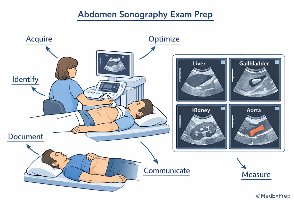
Core content
1.1.1.3 Foundational task categories: what the exam is actually testing
A Explanation
The first decision conflict in most ARDMS AB questions is not “What disease is this?” but “What task is being tested?” If you choose the wrong task, even strong anatomy knowledge will not rescue you. A stem about a poorly visualized common bile duct is often an optimization/acquisition question, not a pathology question. A stem about echogenic foci with posterior shadowing may be a recognition question. A stem asking what to do after seeing free fluid or absent flow is often a documentation-and-escalation question.
WHY THIS IS TESTED: exam writers reward candidates who can classify the problem before acting. Choose the answer that matches the sonographer’s task category; reject answers that jump ahead to physician-level interpretation or treatment. See Fig 1.
B Worked example
A 57-year-old woman with obesity, type 2 diabetes, and prior cholecystectomy presents for outpatient ultrasound for elevated liver enzymes. The sonographer notes that the porta hepatis is poorly seen due to body habitus and overlying bowel gas. The stem asks for the most appropriate next sonographer action. A distracting clue is that alkaline phosphatase is elevated, which can push the learner toward choosing a pathology interpretation.
Reasoning chain: Identify task: this is not yet a diagnosis question; it is an acquisition/optimization problem. Extract key facts: difficult habitus, bowel gas, poor visualization, post-cholecystectomy anatomy. Apply rule: when visualization is limited, first improve the examination with positioning, breathing, alternate windows, and technical optimization. Eliminate distractors: “document CBD obstruction,” “recommend MRCP,” and “diagnose choledocholithiasis” all exceed the role or skip the image-quality step. Verify: the best answer is to use alternate windows/positioning and optimize the exam, then document limitations if still incomplete.
C Exam trap
D Checkpoint
Question: During a limited abdominal study, a patient with cirrhosis, chronic kidney disease, and mild dyspnea has an indeterminate RUQ image because the diaphragm and posterior liver are obscured by shallow breathing. What is the most appropriate next step?
- A. Adjust patient breathing and obtain additional intercostal images
- B. Conclude the patient has a hepatic mass
- C. Recommend immediate contrast-enhanced CT to the patient
- D. Omit documentation because the study is technically limited
Answer: A
- A: Tempting because it sounds basic, but it is right because the primary problem is limited acquisition; breathing instruction and alternate windows are within scope and address the limitation.
- B: Tempting because posterior liver obscuration can mimic a lesion, but it is wrong because no adequate image supports that conclusion.
- C: Tempting because CT can evaluate the liver further, but it is wrong because recommending alternative physician-level management directly to the patient exceeds scope.
- D: Tempting because the exam is difficult, but it is wrong because technical limitations must still be documented.
1.1.1.4 Scope-safe thinking: choose sonographer action, reject physician action
A Explanation
The next major conflict is choosing between a clinically sensible action and a scope-appropriate action. ARDMS AB questions frequently include distractors that would be reasonable for a radiologist, surgeon, or treating physician but are not the best answer for a sonographer. Your job is to perform the exam, optimize images, recognize significant patterns, document them, and communicate through the appropriate chain. Your job is not to prescribe antibiotics for acute cholecystitis, schedule aneurysm repair, order biopsy, or provide definitive management counseling.
WHY THIS IS TESTED: scope boundaries protect patient safety and mirror real practice. The exam tests whether you can recognize what matters without stepping outside role. Choose actions such as “obtain Doppler,” “document measurements,” “notify interpreting physician per protocol,” or “record the limitation.” Reject answers that ask you to make the final interpretive report or direct treatment.
See Fig 2 for a quick role boundary map. This section builds directly toward the upcoming topic Sonographer Scope Boundaries, where the same principles will be tested in more detail.
B Worked example
A 71-year-old man with smoking history, hypertension, chronic obstructive pulmonary disease, and back pain is scanned for abdominal pulsation. A distracting detail is his recent constipation, which can mislead toward bowel pathology. The aorta appears enlarged on screening images. The stem asks for the most appropriate sonographer action.
Reasoning chain: Identify task: not treatment; this is documentation and escalation. Extract key facts: suspected enlarged aorta, symptomatic patient, risk factors. Apply rule: when a potentially significant vascular abnormality is seen, complete appropriate measurements and images and communicate per protocol. Eliminate distractors: “tell the patient to go directly to surgery” and “start blood pressure treatment” are outside scope; “ignore because bowel gas is present” misses a potentially important finding. Verify: document size and extent as able, note limitations, and notify appropriately.
C Exam trap
D Checkpoint
Question: A sonographer identifies marked biliary dilatation in a jaundiced inpatient with prior pancreatic surgery and atrial fibrillation. Which action is most appropriate within scope?
- A. Document the ductal findings and notify the interpreting physician according to protocol
- B. Inform the patient that malignant obstruction is present
- C. Order laboratory testing for bilirubin fractionation
- D. Prescribe fasting and repeat ultrasound in one week
Answer: A
- A: Tempting because it may seem too procedural, but it is right because recognition plus documentation and escalation is exactly the sonographer role.
- B: Tempting because the pattern suggests obstruction, but it is wrong because assigning a definitive cause and counseling exceed scope.
- C: Tempting because labs correlate with obstruction, but it is wrong because ordering tests is not the sonographer’s decision.
- D: Tempting because repeat imaging can be useful in some settings, but it is wrong because management planning is outside the sonographer’s role and urgent communication may be needed now.
- The sonographer’s first duty after recognizing a potentially urgent finding is to document and communicate per protocol.
- True or False: A technically limited study means you should omit images. False.
- Name the three role-safe verbs most commonly tested: acquire, optimize, document.
Exam takeaway: Prefer acquire-optimize-document-communicate answers over diagnose-prescribe-manage answers.
flowchart LR
A[Finding seen on ultrasound] --> B[In scope]
A --> C[Out of scope]
B --> D[Obtain additional views]
B --> E[Measure and document]
B --> F[Notify per protocol]
C --> G[Write final report]
C --> H[Prescribe treatment]
C --> I[Provide definitive diagnosis]
Exam takeaway: Distinguish technical and communication duties from physician interpretation and management.
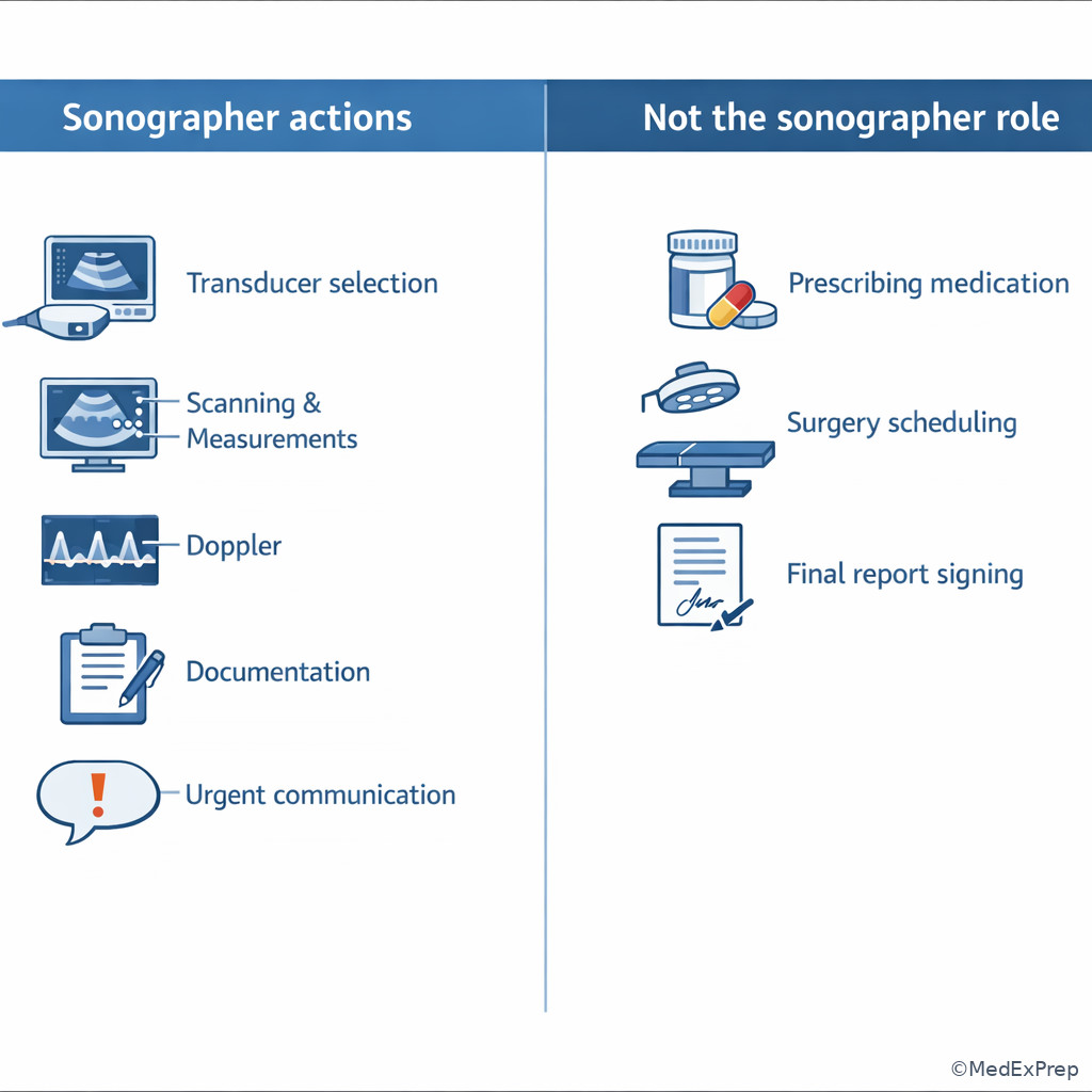
1.1.1.5 Image acquisition and optimization as task-based decisions
A Explanation
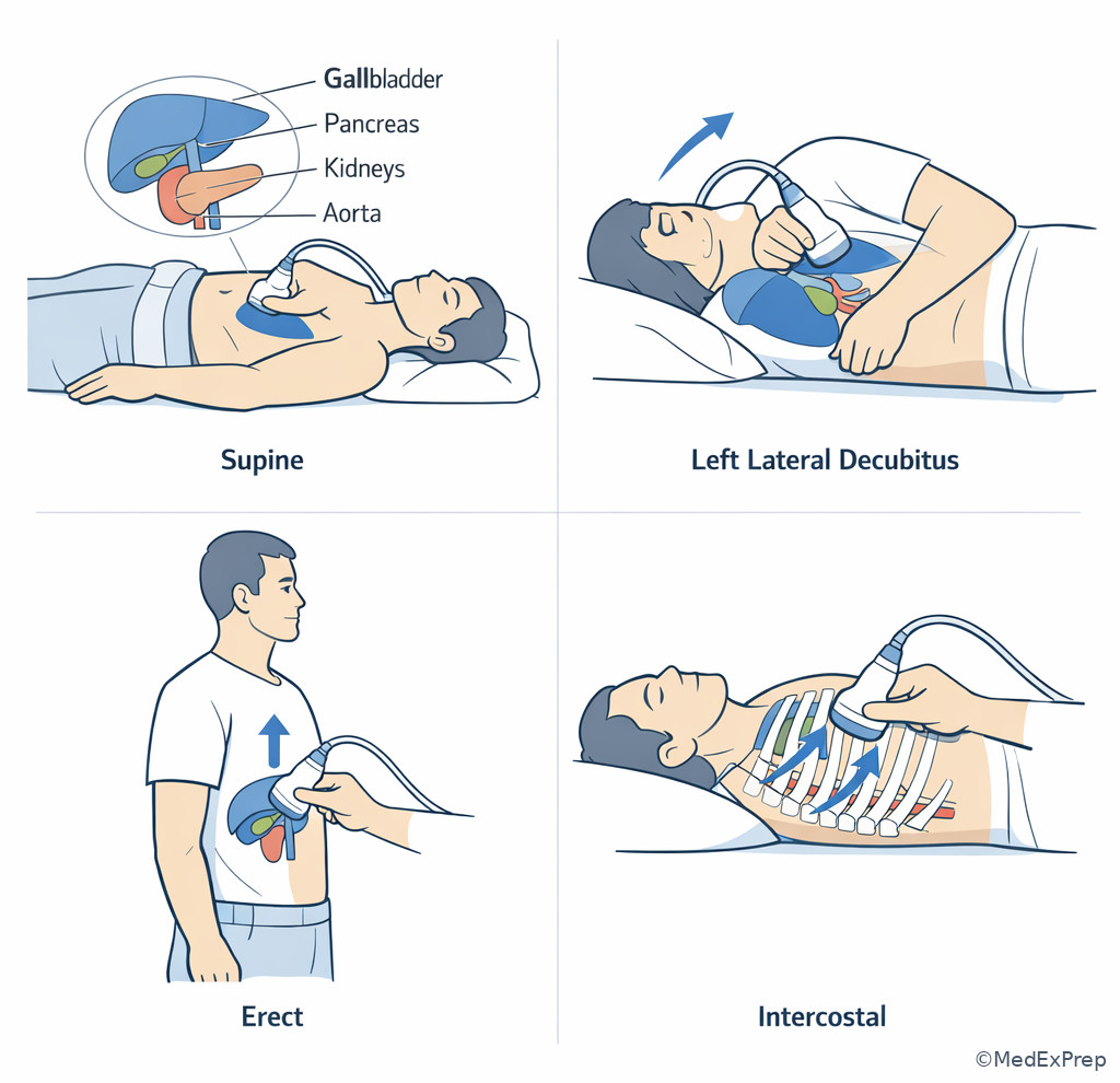
Foundational AB questions often hide the real issue inside a suboptimal image. The exam may show incomplete organ visualization, poor penetration in a large patient, color aliasing, absent flow due to excessive wall filter, or a false lesion caused by artifact. The decision conflict is whether to accept the image as truth or correct the technique first. The rule that resolves this: if the appearance could reasonably be explained by positioning, transducer choice, gain/depth/focus, Doppler settings, or artifact, optimize before concluding pathology.
WHY THIS IS TESTED: abdominal ultrasound is operator dependent, and exam writers expect you to know how technique changes what you see. Choose answers involving alternate windows, intercostal approach, left lateral decubitus or erect positioning when appropriate, breath control, fasting context, grayscale adjustments, and Doppler optimization. Reject answers that overinterpret a technically poor image. See Fig 3.
B Worked example
A 48-year-old man with obesity, recent heavy meal intake, and chronic reflux is scanned for RUQ pain. He also has mildly elevated lipase, a distracting clue that can pull attention toward the pancreas. The gallbladder is poorly distended and partly obscured by bowel gas; the stem asks for the best explanation for the limited evaluation.
Reasoning chain: Identify task: optimization and protocol. Extract key facts: nonfasting patient, poor distention, bowel gas, RUQ indication. Apply rule: an inadequately distended gallbladder in a nonfasting patient limits evaluation and can mimic wall thickening or obscure stones. Eliminate distractors: acute cholecystitis, porcelain gallbladder, and “normal study” all overcall the situation. Verify: the correct answer emphasizes limited evaluation from inadequate preparation and the need to document that limitation.
C Exam trap
D Checkpoint
Question: During evaluation of the portal vein in a patient with fatty liver, heart failure, and recent abdominal surgery, color fill-in is poor despite visible vessel lumen. Which next step is most appropriate before concluding thrombosis?
- A. Optimize color Doppler settings and angle, then reassess flow
- B. Immediately document complete portal vein occlusion
- C. Tell the patient anticoagulation is likely needed
- D. End the exam because surgery history explains absent flow
Answer: A
- A: Tempting because it feels technical, but it is right because inadequate Doppler parameters can create false absent flow.
- B: Tempting because poor color fill-in suggests thrombosis, but it is wrong until technical causes are addressed.
- C: Tempting because thrombosis may require treatment, but it is wrong because treatment counseling is outside scope and the finding is not yet confirmed.
- D: Tempting because recent surgery may complicate imaging, but it is wrong because history does not replace appropriate optimization.
Exam takeaway: If a finding could be created by technique or artifact, optimize first and only then interpret.
flowchart TD
A[Questionable image finding] --> B{Could technique explain it?}
B -->|Yes| C[Change window or position]
C --> D[Adjust grayscale or Doppler]
D --> E[Reassess appearance]
E --> F{Still abnormal?}
F -->|Yes| G[Document and communicate]
F -->|No| H[Artifact/technical limitation recognized]
B -->|No| I[Proceed with pattern recognition and documentation] Exam takeaway: Alternate windows and positioning often solve stems that appear pathologic at first glance.
1.1.1.6 Pattern recognition plus clinical correlation: enough context, not too much
A Explanation
After classifying the task and optimizing the image, the next decision is how much clinical context to use. On ARDMS AB, history matters, but only within sonographic scope. Use symptoms, surgery history, age, labs provided, and exam indication to choose likely views, prioritize structures, and interpret a sonographic pattern. Do not let the clinical story push you into physician-only conclusions. For example, jaundice plus intrahepatic ductal dilatation supports an obstructive pattern, but the sonographer still documents the pattern and communicates it rather than declaring the final etiology.
WHY THIS IS TESTED: stems intentionally add noise. A distracting lab, unrelated comorbidity, or prior surgery can tempt premature closure. The single rule: use clinical data to refine image acquisition and pattern recognition, not to exceed role or ignore discordant sonography. See Fig 4.
B Worked example
A 63-year-old woman with weight loss, prior hysterectomy, and chronic renal insufficiency presents with painless jaundice. Her AST is only mildly elevated, a distracting finding that might make diffuse liver disease seem less urgent. Ultrasound shows a distended gallbladder and dilated intrahepatic ducts; the pancreas is partly obscured by bowel gas. The stem asks for the most appropriate sonographer focus during the remainder of the exam.
Reasoning chain: Identify task: targeted acquisition and documentation guided by pattern recognition. Extract key facts: painless jaundice, ductal dilation, partially obscured pancreas. Apply rule: when obstructive biliary pattern is present, thoroughly assess and document the biliary tree, pancreatic region as visualized, associated measurements, and any limitations. Eliminate distractors: assigning pancreatic cancer, dismissing because liver enzymes are not extreme, or skipping the pancreas because it is difficult to see are all errors. Verify: the correct approach is complete targeted documentation and communication of the obstructive pattern with noted technical limitation.
C Exam trap
D Checkpoint
Question: A patient with fever, dialysis dependence, and recent abdominal surgery undergoes ultrasound for abdominal pain. Small-volume ascites is seen, but the stem’s question asks for the best explanation for poor pancreas visualization. Which clue should drive your answer most directly?
- A. Overlying bowel gas and postoperative anatomy
- B. Fever alone implies pancreatitis
- C. Dialysis dependence excludes pancreatic imaging abnormalities
- D. Ascites means the pancreas is necessarily enlarged
Answer: A
- A: Tempting because it seems too simple, but it is right because the question asks about visualization, and bowel gas/postoperative change directly affect the sonographic window.
- B: Tempting because fever suggests inflammation, but it is wrong because the ask is technical visualization, not diagnosis.
- C: Tempting because chronic disease can distract from the exam task, but it is wrong because dialysis status does not determine image quality that way.
- D: Tempting because ascites is abnormal, but it is wrong because it does not automatically imply pancreatic enlargement.
- The most useful clinical clue is the one that changes view selection, measurement, or urgency of communication.
- True or False: A strong clinical story can replace inadequate imaging. False.
- Name 3 helpful stem clues: indication, prior surgery, targeted labs.
Exam takeaway: Use history and lab clues to refine scanning and documentation, not to overstep the sonographer role.
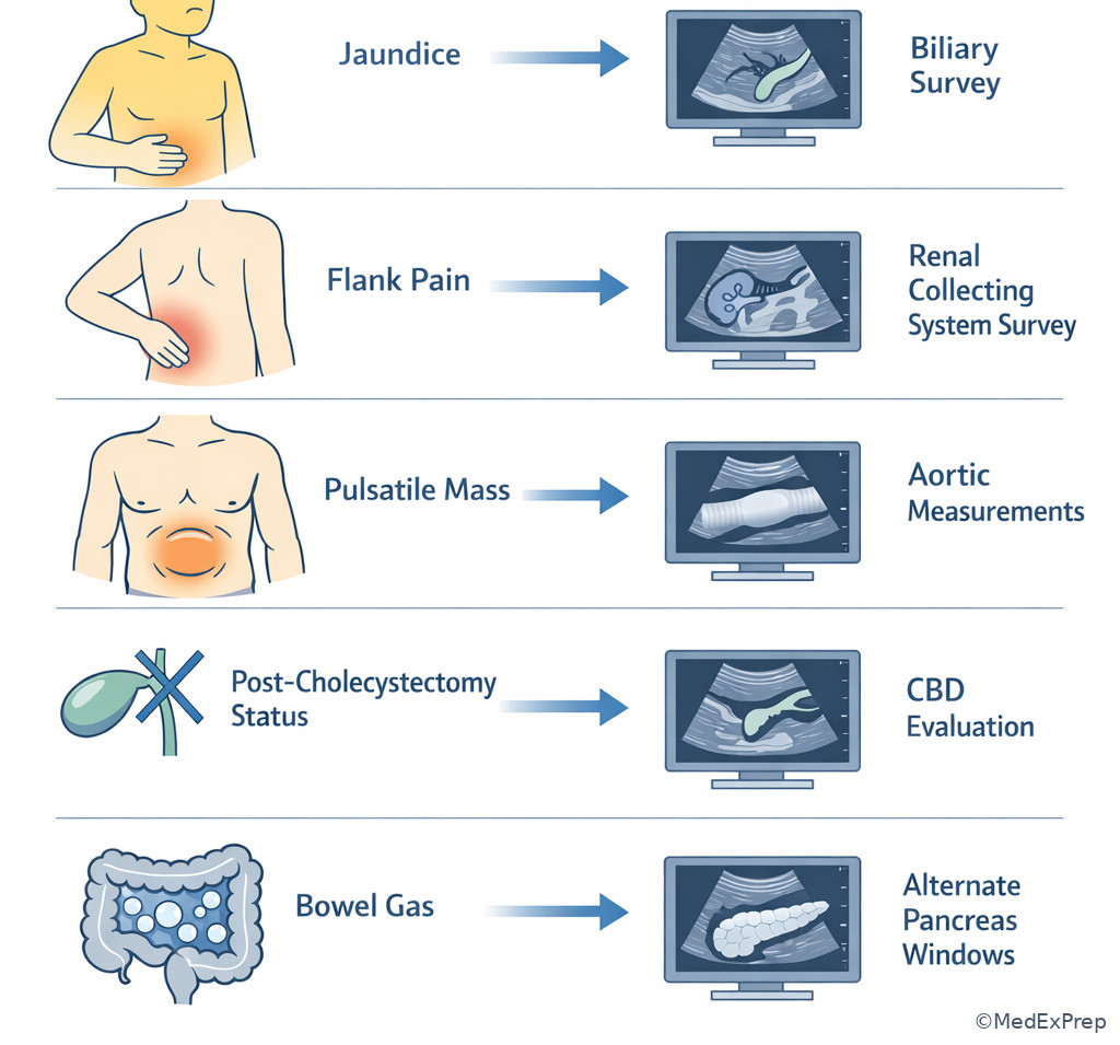
Exam takeaway: Distended ducts, shadowing stones, simple cysts, and hydronephrosis each point to different task priorities.
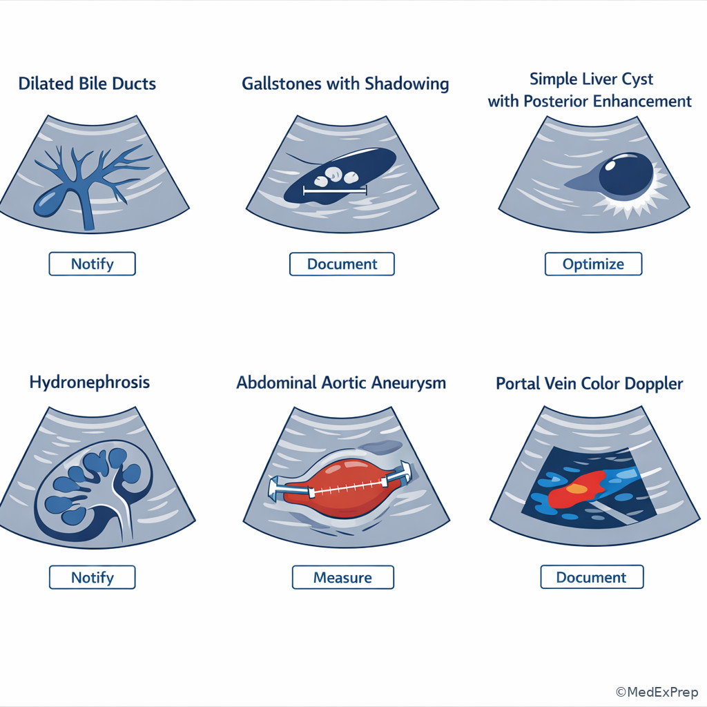
1.1.1.7 Documentation, communication, and test-day reasoning sequence
A Explanation
The final integration step is turning findings into a complete, exam-safe action sequence. Even when you recognize the right pattern, you can still miss the question by failing to document required views, note limitations, or communicate urgent findings appropriately. The board-style sequence is: identify the imaging task, extract key sonographic and clinical clues, apply the governing anatomy/criterion/artifact principle, eliminate distractors, and verify the best answer within scope. This topic previews the upcoming lesson Exam-Style Reasoning Sequence.
WHY THIS IS TESTED: ARDMS questions reward disciplined thinking, not impulsive recognition. Choose the answer that completes the workflow: obtain needed images, measure correctly, document what was seen and what limited the exam, then escalate if required. Reject answers that skip documentation or communicate uncertainty poorly. See Fig 5.
B Worked example
A 66-year-old woman with atrial fibrillation, hypertension, and prior nephrolithiasis undergoes abdomen ultrasound for elevated creatinine and abdominal pain. A distracting clue is a history of gallstones years earlier. The kidneys are partially visualized, mild right hydronephrosis is seen, and the bladder is underdistended. The stem asks for the best next sonographer action.
Reasoning chain: Identify task: complete and document the relevant urinary tract survey within scope. Extract key facts: renal collecting system finding, underdistended bladder, prior stones, nonspecific abdominal pain. Apply rule: when hydronephrosis is suspected, document renal appearance and, if protocol allows, evaluate the bladder and distal obstruction clues as feasible; note limitation if underdistended. Eliminate distractors: “diagnose ureteral calculus,” “advise urology intervention,” and “ignore bladder because kidneys already explain the pain” all skip workflow or exceed role. Verify: obtain/document the most complete urinary tract images feasible and note limitations.
C Exam trap
D Checkpoint
Question: A technically difficult abdominal exam reveals a possible splenic lesion in a patient with trauma history, obesity, and shallow respirations. What best completes the task framework?
- A. Obtain the best additional views possible, document limitations, and communicate significant concern per protocol
- B. Reassure the patient that the lesion is definitely benign
- C. Skip splenic imaging because the liver was already evaluated
- D. Delay all documentation until CT confirms the finding
Answer: A
- A: Tempting because it sounds procedural rather than diagnostic, but it is right because it follows the proper sonographer workflow from acquisition to communication.
- B: Tempting because many splenic findings are incidental, but it is wrong because definitive reassurance is outside scope and the exam is technically limited.
- C: Tempting because time pressure encourages early closure, but it is wrong because the spleen remains clinically relevant in trauma-related scanning.
- D: Tempting because confirmation imaging may occur, but it is wrong because ultrasound findings and limitations must be documented at the time of the exam.
Exam takeaway: Solve stems by task identification, clue extraction, rule application, elimination, and scope-safe verification.
flowchart LR
A[Identify the ask] --> B[Extract key clues]
B --> C[Apply anatomy criterion or artifact rule]
C --> D[Eliminate close distractors]
D --> E[Verify best answer within scope]
E --> F[Document or communicate if asked]
Exam takeaway: Required images, measurements, limitations, and communication steps are part of the answer, not afterthoughts.
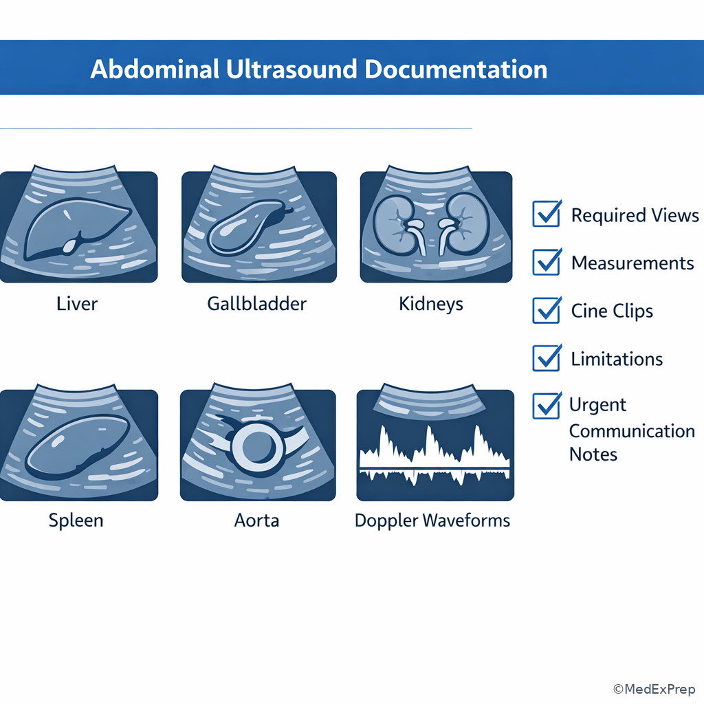
Exam takeaway: When important findings appear, document first and escalate through the proper chain rather than managing treatment.
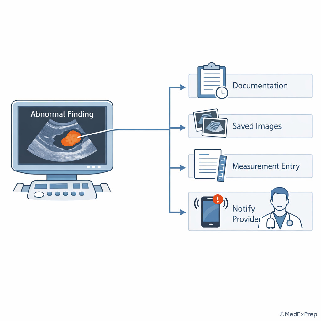
Exam Traps & Differentiators
Most common wrong answer and why: The single most common miss is choosing a medically reasonable answer that is outside the sonographer’s role. These options feel “advanced,” but ARDMS AB is usually asking what the sonographer should acquire, optimize, measure, document, or communicate.
| Looks similar | Actually tested task | Key differentiator |
|---|---|---|
| Diagnosis question | Optimization question | The image is incomplete or technically limited |
| Management question | Communication question | Urgent/significant finding within sonographer scope |
| Pathology recognition | Normal variant recognition | Stable benign pattern without aggressive features |
| True absent flow | Doppler optimization issue | Settings or angle could explain poor fill-in |
| Missed organ survey | Protocol completion problem | One finding does not end the required exam |
| If the stem says... | Think... |
|---|---|
| “Poorly visualized,” “limited by bowel gas,” or “suboptimal color fill-in” | Optimize first before naming pathology |
| “Most appropriate sonographer action” after a major abnormality | Document + notify per protocol |
| “Patient recently ate” with gallbladder question | Nondistention/limited evaluation is a strong possibility |
| “History of surgery” with altered anatomy | Expect different windows and careful documentation of limitations |
| “Best explanation for appearance” | Consider artifact or technique before disease |
| “Most important to monitor/communicate” | Prioritize significance and protocol-based escalation |
Related traps from upcoming neighboring topics matter here. In Sonographer Scope Boundaries, you will see more examples where the wrong answer is a correct physician action by the wrong provider. In Exam-Style Reasoning Sequence, you will refine the habit of identifying the ask before interpreting the image.
Tables
| High-yield framework summary | Key question to ask yourself | Common best answer style | Common wrong-answer style |
|---|---|---|---|
| Acquisition | What view or position is missing? | Additional planes, windows, breathing instructions | Premature diagnosis |
| Optimization | Could technique create this appearance? | Adjust controls or Doppler settings | Labeling artifact as disease |
| Recognition | What pattern best matches the image? | Normal, variant, or likely abnormal pattern | Using history alone to decide |
| Measurement | What landmark-based caliper placement is needed? | Measure according to accepted protocol | Ignoring landmark rules |
| Documentation | What still must be recorded? | Required images, cine, limitation statement | Stopping after one finding |
| Communication | Does this require prompt notification? | Escalate through protocol | Providing treatment plan |
| Comparison | Within sonographer scope | Outside sonographer scope |
|---|---|---|
| Image quality problem | Adjust depth/gain/focus/position | Declare final pathology without adequate images |
| Abnormal vascular finding | Obtain Doppler, measure, document, notify | Start anticoagulation plan |
| Biliary obstruction pattern | Survey ducts, measure, note limitations, communicate | Tell patient exact cause and treatment |
| Renal collecting system dilatation | Document kidneys/bladder as feasible | Schedule urologic intervention |
| Trauma-related abnormality | Capture images and escalate significant concern | Discharge or clear the patient independently |
| Differential/classification issue | Ask yourself | Discriminating clue |
|---|---|---|
| True lesion vs artifact | Does it persist in multiple planes and after optimization? | Change with technique suggests artifact |
| Poor visualization vs absent structure | Was the exam technically limited? | Bowel gas, fasting status, or habitus often explain nonvisualization |
| Urgent communication vs routine documentation | Would delay create safety risk? | Significant unexpected finding warrants escalation |
| Protocol complete vs incomplete | Were complementary views/measurements obtained? | One abnormality does not cancel the protocol |
| Clinical clue useful vs distracting | Does it change the scan plan? | Only use clues that affect acquisition, recognition, or urgency |
Algorithm / Approach
Rapid Review
- Task classification → identify what the question is asking before naming pathology
- Optimization issue → correct technique before concluding abnormality
- Scope boundary → document and notify, do not prescribe or definitively manage
- Clinical correlation → use clues that change scan strategy, not clues that tempt overreach
- Protocol completion → one abnormality does not end the survey
- Technical limitation → state it explicitly; never hide incomplete visualization
- Urgent finding → best next sonographer action usually includes prompt communication
- Doppler uncertainty → settings and angle can mimic absent flow
- Nonfasting gallbladder → limited evaluation can mimic wall abnormality or obscure stones
- Postoperative anatomy → adapt windows and document altered landmarks
- Artifact question → ask whether the finding persists after optimization and in multiple planes
- Good distractor → often the right medical action by the wrong provider
Self-check quiz
1.1.1.8 Question 1
A 52-year-old woman with obesity and gastroesophageal reflux presents for RUQ ultrasound after eating breakfast. The gallbladder is poorly distended, and the stem asks for the best explanation for limited evaluation. Which option is most appropriate?
- A. Nonfasting state causing poor gallbladder distention
- B. Definite acute cholecystitis
- C. Porcelain gallbladder
- D. Gallbladder agenesis
1.1.1.9 Question 2
A sonographer detects a suspiciously enlarged abdominal aorta in a 69-year-old smoker with hypertension and chronic back pain. Which response is the most appropriate within scope?
- A. Measure, document, and notify per protocol
- B. Tell the patient urgent surgery is required
- C. Start blood pressure management instructions
- D. Write the final diagnostic report
1.1.1.10 Question 3
A 61-year-old man with cirrhosis, diabetes, and recent abdominal surgery undergoes ultrasound for worsening ascites. Color Doppler shows poor fill-in within the portal vein, but the vessel lumen is visible and the angle is suboptimal. The stem asks for the most appropriate next sonographer action. Which is best?
- A. Optimize Doppler settings and angle, then reassess
- B. Document complete thrombosis immediately
- C. Advise the patient to begin anticoagulation
- D. Omit Doppler images because grayscale already shows the vein
1.1.1.11 Question 4
A 58-year-old woman with painless jaundice, prior cholecystectomy, and mild chronic kidney disease has dilated intrahepatic ducts on ultrasound. The pancreas is partly obscured by bowel gas. What is the best sonographer focus for the remainder of the exam?
- A. Document biliary dilatation thoroughly, assess the pancreatic region as visualized, and note limitations
- B. Conclude pancreatic malignancy is present
- C. Cancel the rest of the exam because bowel gas prevents certainty
- D. Ignore the ducts because creatinine is abnormal
1.1.1.12 Question 5
A 45-year-old man with obesity, recent abdominal pain, and prior appendectomy is scanned for pancreas evaluation. Overlying bowel gas obscures much of the gland, and the stem asks for the best next step. Which option is most appropriate?
- A. Use alternate windows and positioning, then document remaining limitations
- B. Diagnose pancreatitis based only on pain
- C. Send the patient directly for endoscopy
- D. Skip documentation because the pancreas was not fully seen
1.1.1.13 Question 6
A 74-year-old inpatient with atrial fibrillation, heart failure, and sepsis is scanned for elevated bilirubin. Marked biliary dilatation and a distended gallbladder are seen; the distal duct is partly obscured. The patient is uncomfortable and asks what treatment will be needed. What is the most appropriate sonographer response and action?
- A. Document the findings, state that the interpreting physician will review them, and notify per urgent protocol
- B. Explain that choledocholithiasis is definite and antibiotics are required
- C. Reassure the patient that obstruction is unlikely because the distal duct is not fully seen
- D. Delay all communication until a complete distal duct image is obtained
1.1.1.14 Question 7
A 67-year-old woman with obesity, prior gastric surgery, and shortness of breath undergoes ultrasound for RUQ pain. The liver dome is poorly seen on subcostal images, and a questionable echogenic focus appears in one plane only. Which is the best explanation and next step?
- A. Possible artifact or limited window; obtain intercostal views and reassess before documenting a focal lesion
- B. Hepatic calcification is confirmed because the focus is bright
- C. Immediate biopsy referral is indicated
- D. The liver can be excluded from the exam because the gallbladder is the pain source
1.1.1.15 Question 8
A 62-year-old man with chronic kidney disease, hypertension, and prior nephrolithiasis has mild unilateral hydronephrosis on renal images. The bladder is underdistended, and the stem asks for the best next sonographer action. Which choice is most appropriate?
- A. Document the renal finding, evaluate the bladder as feasible, and note underdistention as a limitation
- B. Conclude distal ureteral stone is definitely present
- C. Skip bladder imaging because the kidney abnormality already explains symptoms
- D. Recommend immediate urologic decompression directly to the patient
Answer key
1.1.1.16 Question 1 answer: A
A. Correct. This is tempting because it may seem too simple, but it is correct for this scenario because a recently fed patient often has a contracted or poorly distended gallbladder, which limits evaluation and can mimic pathology. The single stem clue is after eating breakfast. Authoritative abdomen sonography texts emphasize patient preparation and the effect of fasting on gallbladder assessment.
B. Incorrect. This is tempting because RUQ pain plus suboptimal gallbladder appearance can suggest inflammation. It is incorrect here because the described limitation is poor distention after eating, not a complete inflammatory sonographic pattern. The discriminating clue is the nonfasting state, not wall hyperemia, pericholecystic fluid, or a sonographic Murphy sign.
C. Incorrect. This is tempting because abnormal gallbladder wall appearance can trigger thoughts of chronic calcific disease. It is incorrect because porcelain gallbladder is not inferred from simple poor distention after a meal. The key clue is inadequate preparation, which provides a sufficient technical explanation.
D. Incorrect. This is tempting because a poorly seen gallbladder can make learners think it is absent. It is incorrect because the organ is present but poorly distended. The discriminating clue is that the gallbladder is poorly distended, not absent.
Reference: Rumack et al.; Hagen-Ansert.
1.1.1.17 Question 2 answer: A
A. Correct. This is tempting because it sounds procedural rather than dramatic, but it is the correct sonographer action: obtain appropriate measurements, document the finding, and notify according to protocol. The discriminating clue is within scope. Practice standards emphasize acquisition, documentation, and communication of significant findings.
B. Incorrect. This is tempting because an enlarged aorta may indeed be urgent. It is incorrect because telling the patient urgent surgery is required is a physician-level management statement. The single clue is the request for the most appropriate action within scope.
C. Incorrect. This is tempting because hypertension relates to aortic disease, but it is incorrect because medication or treatment counseling is outside the sonographer role. The key clue is that the sonographer is not the treating provider.
D. Incorrect. This is tempting because final reporting follows imaging, but it is incorrect because the sonographer does not write the official interpretive report as the radiologist. The discriminating clue is the role boundary built into the exam.
Reference: AIUM practice parameters; ARDMS role-focused exam scope.
1.1.1.18 Question 3 answer: A
A. Correct. This is tempting because it delays the certainty many learners want, but it is correct because suboptimal Doppler angle and settings can falsely reduce color fill-in. The single stem clue is angle is suboptimal. Doppler practice standards require optimization before concluding absent flow.
B. Incorrect. This is tempting because portal vein thrombosis is a recognized complication in cirrhosis. It is incorrect here because technical factors remain uncorrected. The discriminating clue is visible lumen with suboptimal angle, which makes immediate occlusion labeling premature.
C. Incorrect. This is tempting because thrombosis may need therapy, but it is incorrect because counseling on anticoagulation is outside scope and the finding is not yet technically confirmed. The single clue is that the task asks for the next sonographer action.
D. Incorrect. This is tempting because grayscale confirms vessel anatomy, but it is incorrect because Doppler assessment is central to evaluating flow questions. The key clue is the concern about color fill-in, which specifically requires Doppler optimization, not omission.
Reference: AIUM Doppler guidance; Zwiebel and Pellerito.
1.1.1.19 Question 4 answer: A
A. Correct. This is tempting because it sounds broad, but it is correct because the obstructive pattern requires thorough documentation of ducts, pancreatic region as visualized, and remaining limitations. The discriminating clue is dilated intrahepatic ducts with partial pancreatic obscuration. Sonographic protocol emphasizes targeted completion, not premature closure.
B. Incorrect. This is tempting because painless jaundice suggests pancreatic head obstruction. It is incorrect because the sonographer should document the pattern, not state a final cause. The single clue is that the pancreas is partly obscured, so certainty is unjustified.
C. Incorrect. This is tempting because bowel gas limits certainty, but it is incorrect because partial obscuration does not justify canceling the rest of a targeted survey. The stem clue is as visualized; you still document what can be seen.
D. Incorrect. This is tempting because renal dysfunction can distract attention, but it is incorrect because the dominant sonographic issue in the stem is biliary obstruction pattern. The discriminating clue is dilated intrahepatic ducts.
Reference: Rumack et al.; AIUM abdomen practice guidance.
1.1.1.20 Question 5 answer: A
A. Correct. This is tempting because it seems mundane, but it is correct because bowel gas commonly limits pancreatic imaging, and alternate windows/positions are the appropriate next sonographer step. The single stem clue is overlying bowel gas obscures much of the gland. Abdomen scanning technique texts emphasize optimization before interpretation.
B. Incorrect. This is tempting because abdominal pain raises concern for pancreatitis. It is incorrect because pain alone does not establish the sonographic finding, especially with incomplete visualization. The key clue is that the pancreas is obscured.
C. Incorrect. This is tempting because endoscopy may be useful in some clinical pathways, but it is incorrect because referring directly for another procedure is outside the sonographer’s task here. The discriminating clue is the request for the best next step during the ultrasound exam.
D. Incorrect. This is tempting because incomplete visualization feels unhelpful. It is incorrect because exam limitations must be documented. The single clue is the technically limited window, which increases—not removes—the need for documentation.
Reference: Hagen-Ansert; AIUM abdomen examination parameters.
1.1.1.21 Question 6 answer: A
A. Correct. This is tempting because it avoids answering the patient directly, but it is correct because the sonographer should document significant findings, avoid providing a treatment plan, and notify urgently per protocol when indicated. The single discriminating clue is marked biliary dilatation in a symptomatic inpatient plus the question asking for the most appropriate sonographer response and action.
B. Incorrect. This is tempting because the pattern may fit obstruction, and antibiotics sound appropriately urgent. It is incorrect because definitive cause and treatment recommendations exceed scope. The key clue is that the distal duct is partly obscured, and treatment selection is not the sonographer’s role.
C. Incorrect. This is tempting because incomplete visualization can create uncertainty. It is incorrect because significant proximal findings still warrant documentation and communication; uncertainty is not reassurance. The discriminating clue is marked biliary dilatation, which remains important even if the distal duct is incompletely seen.
D. Incorrect. This is tempting because complete imaging is desirable. It is incorrect because delaying communication of a significant pattern may be unsafe; partial limitation does not erase the importance of the abnormal findings already seen. The single clue is the combination of major abnormality plus urgent clinical context.
Reference: AIUM practice parameters; ARDMS candidate-role scope guidance.
1.1.1.22 Question 7 answer: A
A. Correct. This is tempting because it preserves uncertainty, but it is correct because a questionable focus seen in one plane only in a poorly visualized liver dome is more consistent with limited window or artifact until confirmed in additional planes. The discriminating clue is appears in one plane only with poor subcostal visualization. Sonographic lesion confirmation requires reproducibility and adequate technique.
B. Incorrect. This is tempting because echogenic foci can represent calcification. It is incorrect here because single-plane appearance in a poorly seen dome is insufficient confirmation. The key clue is one plane only, which strongly argues for further reassessment.
C. Incorrect. This is tempting because focal liver lesions can require biopsy in broader clinical care. It is incorrect because biopsy referral is outside the sonographer’s role and the imaging finding is not yet confirmed. The discriminating clue is both the scope boundary and the uncertain image quality.
D. Incorrect. This is tempting because RUQ pain often centers the gallbladder. It is incorrect because the liver remains part of the survey, especially when a possible abnormality is seen. The single clue is the presence of a questionable focus requiring reassessment, not omission.
Reference: Rumack et al.; standard abdominal sonography technique texts.
1.1.1.23 Question 8 answer: A
A. Correct. This is tempting because it seems incomplete, but it is correct because mild hydronephrosis should be documented, the bladder evaluated as feasible, and underdistention recorded as a limitation. The discriminating clue is bladder is underdistended. Renal/urinary tract assessment relies on as-complete-as-possible documentation with limitations stated.
B. Incorrect. This is tempting because prior nephrolithiasis and unilateral hydronephrosis suggest a stone. It is incorrect because mild hydronephrosis does not prove distal ureteral calculus on its own. The key clue is the limited bladder evaluation, meaning the survey is not complete enough for certainty.
C. Incorrect. This is tempting because the kidney finding already seems explanatory. It is incorrect because the urinary tract task often includes bladder evaluation, especially when obstruction is considered. The discriminating clue is that the stem asks for the best next sonographer action, not the most likely diagnosis.
D. Incorrect. This is tempting because obstruction can be clinically urgent. It is incorrect because recommending decompression directly to the patient exceeds the sonographer role. The single clue is the request for sonographer action within scope.
Reference: AIUM urinary tract sonography guidance; Hagen-Ansert.
Keep going with the full ARDMS Abdomen Prep Course (ARDMS AB)
You just studied the way members do, in the same lesson interface. Full access unlocks every lesson, the question bank and flashcards for a full year.
One payment · 1 year of access · No auto-renewal