1.1.1 - Cardiac Chambers And Valves
Cardiac Chambers And Valves
What you'll learn
- FOUNDATIONAL KNOWLEDGE: Anatomy -> Chamber-valve relationships: identify the internal anatomy of the atria, ventricles, annuli, papillary muscles, chordae, and outflow tracts that directly affect operative exposure and injury risk.
- ADULT CARDIAC SURGERY: Imaging/interpretation -> Valve lesion recognition: connect chamber remodeling patterns to aortic, mitral, tricuspid, and pulmonic valve pathology.
- ADULT CARDIAC SURGERY: Procedure selection -> Repair versus replacement framing: decide which anatomic features favor repair, replacement, or multidisciplinary reassessment.
- PERIOPERATIVE/CRITICAL CARE: Hemodynamics -> Chamber-pressure logic: interpret preload, afterload, ventricular interdependence, and valve competence in board-style scenarios.
- FOUNDATIONAL KNOWLEDGE: Safety -> Operative landmarks: anticipate conduction tissue, coronary ostial, subvalvular, and right-sided injury zones.
- Foundational Knowledge → Anatomy and physiology → chamber geometry, valve apparatus, fibrous skeleton
- Adult Cardiac Surgery → Valve evaluation → lesion pattern recognition from anatomic and hemodynamic clues
- Adult Cardiac Surgery → Operative planning → exposure, repair feasibility, prosthesis-related decision framing
- Perioperative/Critical Care → Hemodynamic interpretation → chamber-pressure consequences of stenosis versus regurgitation
Why it matters
- Thoracic surgery board stems often hide the answer in anatomy: chamber enlargement pattern, annular geometry, or subvalvular involvement may matter more than a memorized murmur.
- Operative judgment depends on precise landmarks: misunderstanding the aortic root, mitral subvalvular apparatus, or AV node neighborhood creates wrong-answer choices and real-world technical complications.
- Postoperative ICU decisions require chamber-valve logic: low output, pulmonary edema, RV failure, and unexplained shock are interpreted through valve competence and ventricular interaction.
- This topic is foundational for later lessons on echo interpretation, valve disease, aortic root surgery, congenital lesions, and postoperative complication recognition.
Key Terms & Must-Know Facts
1.1.1.1 Key terms
- Annulus: fibrous ring supporting a valve orifice
- Fibrous skeleton: dense connective tissue linking valve annuli and insulating atria from ventricles except through the conduction system
- Sinotubular junction: transition from aortic root sinuses to tubular ascending aorta
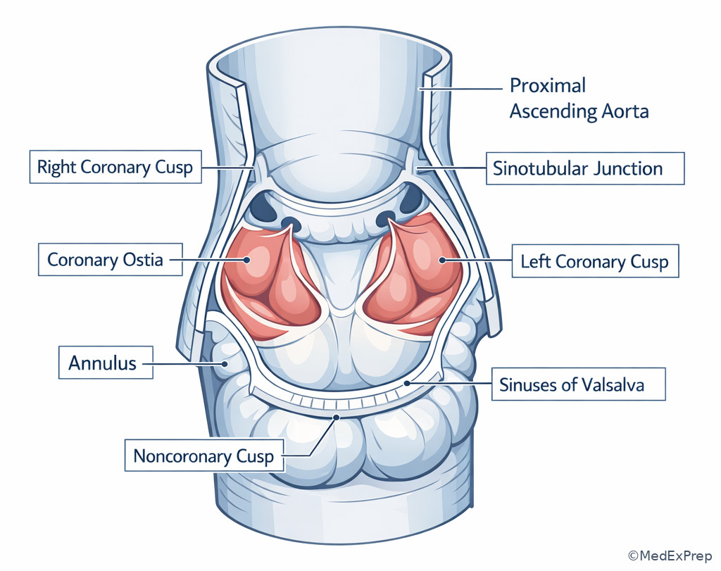
- Aortic root: annulus, cusps, sinuses of Valsalva, coronary ostia, sinotubular junction
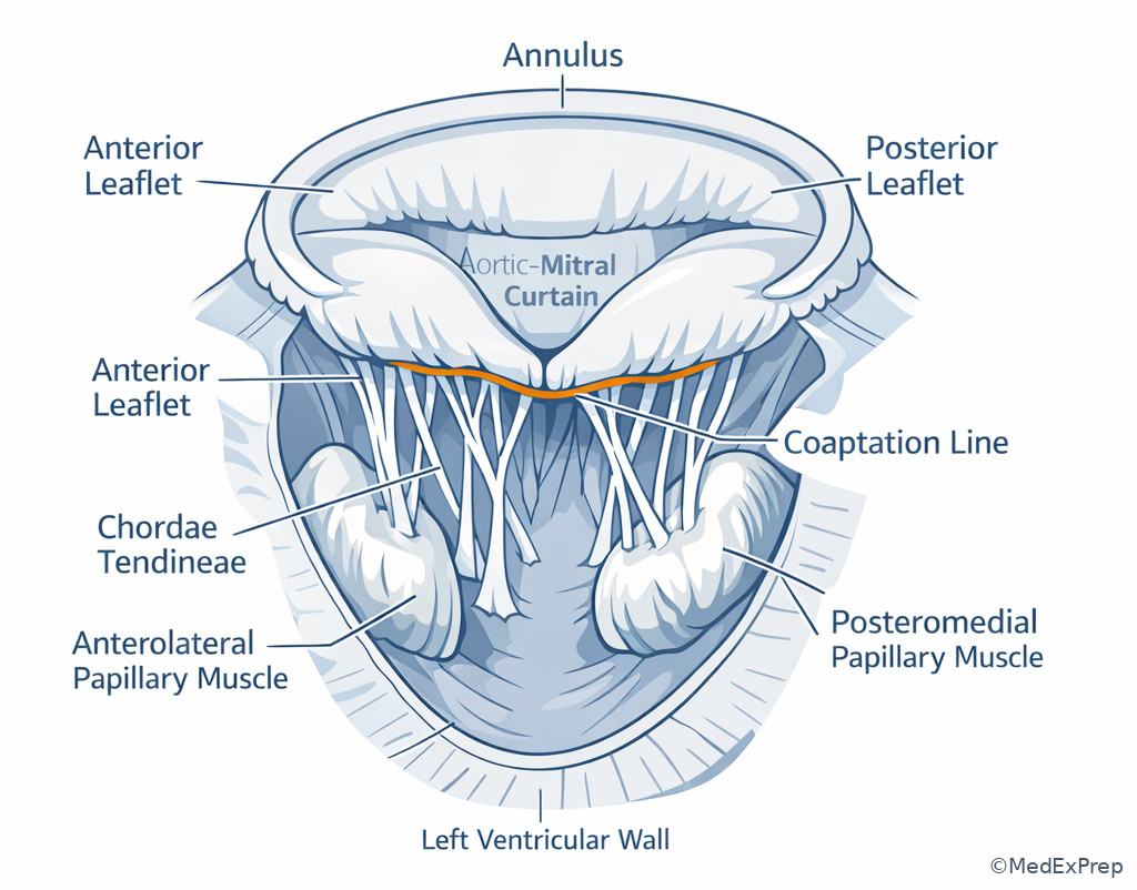
- Mitral apparatus: annulus, leaflets, chordae, papillary muscles, LV wall
- Tricuspid annular dilation: right-sided annular enlargement causing or worsening regurgitation
- Ventricular interdependence: one ventricle’s size/pressure affecting the other through the septum and pericardium
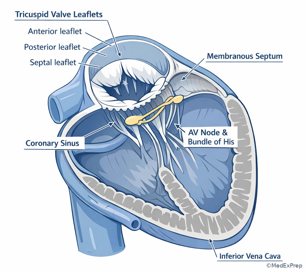
- Conduction tissue danger zone: membranous septum near the noncoronary-right coronary commissural region and septal tricuspid annulus
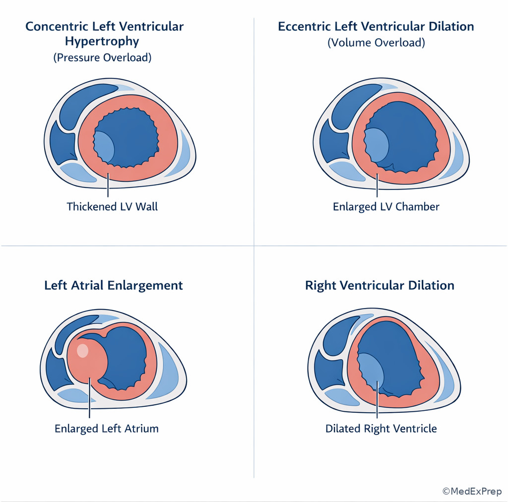
- Pressure overload: concentric hypertrophic response to stenotic lesions
- Volume overload: dilated eccentric remodeling from regurgitant lesions or shunts
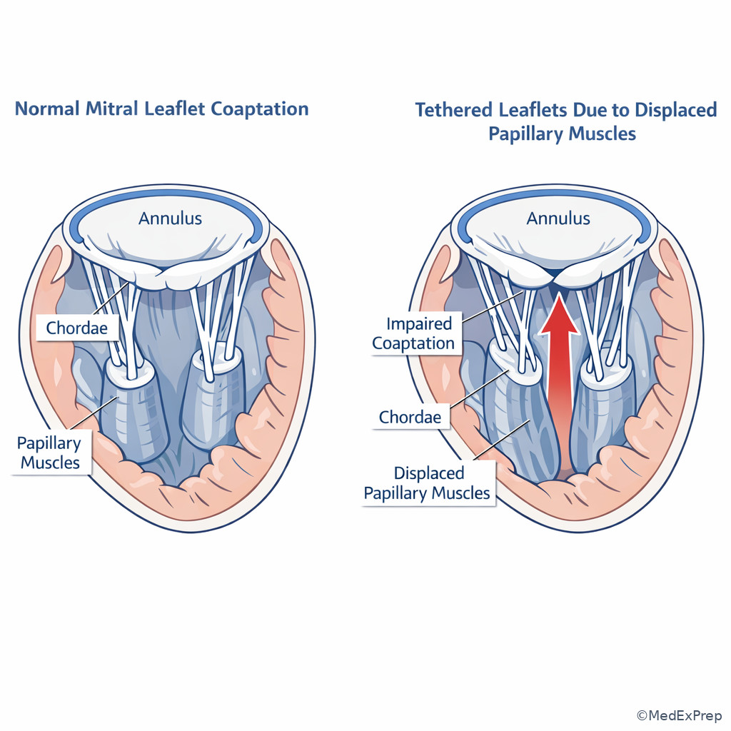
- Papillary muscle tethering: leaflet restriction due to ventricular remodeling
- Coaptation: leaflet meeting line that prevents regurgitation
- Afterload mismatch: LV dysfunction becoming apparent after relieving chronic regurgitation
1.1.1.2 Must-know facts
- The left atrium sits posteriorly and is the chamber most closely related to the esophagus.
- The right ventricle is the most anterior cardiac chamber behind the sternum.
- Aortic stenosis classically produces pressure overload and concentric LV hypertrophy.
- Aortic regurgitation classically produces LV volume overload and dilation.
- Primary mitral regurgitation is a valve/leaflet problem; secondary mitral regurgitation is usually a ventricular geometry problem.
- Functional tricuspid regurgitation is usually annular/RV dilation rather than leaflet destruction.
- The mitral valve has no true fibrous annulus circumferentially equivalent to the aortic annulus; posterior annular dilation matters in repair strategy.
- The AV node and bundle of His are vulnerable near the membranous septum during aortic and tricuspid surgery.
- Coronary ostia are part of aortic root anatomy, not just “adjacent structures.”
- Right-sided lesions frequently become hemodynamically important through pulmonary vascular resistance and RV reserve, not valve area alone.
| Structure | Key anatomic fact | Exam consequence |
|---|---|---|
| Right atrium | Receives SVC, IVC, coronary sinus | Dilation suggests chronic right-sided volume/pressure load |
| Left atrium | Posterior chamber, related to pulmonary veins and esophagus | Enlargement points toward chronic mitral disease or diastolic dysfunction |
| Right ventricle | Anterior chamber with crescent geometry | Most vulnerable in pulmonary hypertension and tricuspid pathology |
| Left ventricle | Elliptical pressure pump with thick wall | Concentric versus eccentric remodeling narrows lesion type |
| Aortic valve/root | Three cusps, sinuses, coronary ostia, sinotubular junction | Root disease changes operation beyond isolated valve replacement |
| Mitral valve | Leaflets plus subvalvular apparatus integrated with LV | Repair success depends on ventricle as much as leaflet |
| Tricuspid valve | Anterior, posterior, septal leaflets near conduction tissue | Septal work risks heart block |
| Pulmonic valve | Outflow valve of RV, lower-pressure system | Right-sided physiology and congenital context are common test settings |
Core content
Exam takeaway: Use chamber position plus adjacent landmarks to predict exposure, enlargement patterns, and injury risk.
flowchart LR
RA[Right atrium
venous inflow] --> TV[Tricuspid valve]
TV --> RV[Right ventricle
anterior]
RV --> PV[Pulmonic valve]
PV --> PA[Pulmonary artery]
LA[Left atrium
posterior] --> MV[Mitral valve]
MV --> LV[Left ventricle
systemic pump]
LV --> AV[Aortic valve/root]
AV --> AO[Ascending aorta]
ESO[Esophagus] --- LA
STERN[Sternum] --- RV
Exam takeaway: Chamber geometry often identifies the lesion class before you memorize any gradient or severity cutoff.
flowchart TD
Lesion[Valve lesion] --> Q{Primary hemodynamic burden}
Q -->|Pressure overload| P[Concentric hypertrophy
smaller cavity early]
Q -->|Volume overload| V[Eccentric dilation
larger cavity]
P --> ExamplesP[Aortic stenosis
Pulmonic stenosis]
V --> ExamplesV[Aortic regurgitation
Mitral regurgitation
Tricuspid regurgitation] Exam takeaway: Distinguish leaflet disease from ventricular or annular disease because the mechanism changes repair feasibility.
flowchart TD
MR[Regurgitant lesion] --> M{Where is the problem?}
M -->|Leaflet/chordal| Primary[Primary valve disease]
M -->|Annulus/ventricle| Secondary[Functional disease]
Primary --> RepairFavored[Often repair-favored if anatomy suitable]
Secondary --> VentricleDriven[Treat ventricle + geometry; repair durability varies]
TR[Tricuspid regurgitation] --> Annular[Often annular/RV dilation] Exam takeaway: The single best clue for postoperative heart block risk is work near the membranous septum or septal annulus.
flowchart LR
AV[Aortic valve] --> MS[Membranous septum]
TV[Septal tricuspid annulus] --> MS
MS --> AVN[AV node / His bundle vicinity]
AVN --> Risk[Heart block risk if injured]
Root[Aortic root procedures] --> Risk
SeptalRepair[Septal tricuspid repair] --> Risk
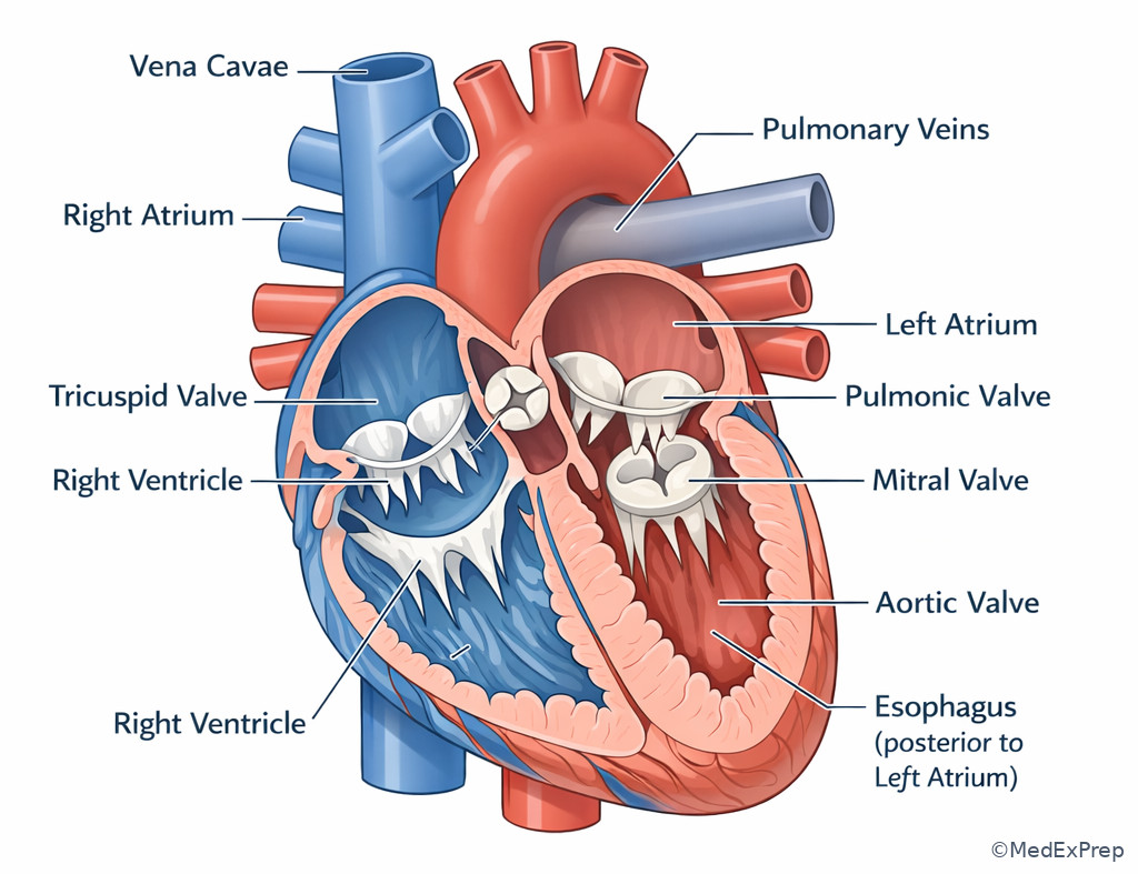
Exam takeaway: Visual recognition of chamber position and valve continuity helps solve exposure, pathology, and complication questions quickly.
Exam takeaway: Root operations require recognition that cusps, sinuses, commissures, and coronary ostia are one surgical unit.
Exam takeaway: Remember that leaflet competence depends on chordae, papillary muscles, annulus, and LV geometry, not leaflets alone.
Exam takeaway: Septal leaflet work is a safety question because conduction tissue lies nearby.
Exam takeaway: Concentric thickening suggests pressure load, while dilated cavities suggest chronic volume load.
Exam takeaway: Functional mitral regurgitation is often a ventricular geometry problem rather than a primary leaflet problem.
1.1.1.3 Chamber orientation and internal landmarks
A Explanation
On exam day, the decision conflict is often which chamber or landmark actually explains the finding, not which disease name sounds familiar. Choose chamber-position logic over murmur memorization when the stem gives you imaging, sternotomy risk, posterior compression, or intraoperative exposure clues. The right ventricle is most anterior; therefore it is the chamber most relevant to re-entry and anterior compression questions. The left atrium is posterior and closely related to the pulmonary veins and esophagus; therefore posterior mediastinal symptoms, dysphagia-related associations, and transesophageal imaging questions often center on the left atrium. The left ventricle is the main pressure chamber, so concentric thickening usually signals pressure overload, whereas dilation favors volume load. See Fig 1.
WHY THIS IS TESTED: board questions use spatial anatomy to force a choose-versus-reject decision between chambers that all sound plausible. The single resolving rule is position plus function: anterior chamber = RV, posterior chamber = LA, thick-walled systemic pressure chamber = LV, low-pressure venous receiving chamber = RA.
B Worked example
A 67-year-old man with hypertension, obesity, and chronic kidney disease is being evaluated before redo sternotomy. CT shows the most anterior cardiac chamber immediately behind the sternum. A trainee focuses on the patient’s mild left atrial enlargement and history of atrial fibrillation and argues the posterior chamber is highest risk during re-entry. A distracting note mentions dysphagia from reflux. What structure is most relevant to initial re-entry injury risk?
Reasoning chain: Identify the task: this is an operative exposure question, not a rhythm question. Extract key facts: redo sternotomy, most anterior chamber, immediately behind sternum. Apply the rule: the RV is the most anterior chamber. Eliminate distractors: LA enlargement and dysphagia point to posterior anatomy and are tempting because the LA is related to the esophagus, but that does not make it the re-entry danger structure. Verify: the chamber at greatest risk during sternal re-entry is the right ventricle.
C Exam trap
D Checkpoint
Question: A 58-year-old woman with long-standing pulmonary hypertension, obstructive sleep apnea, and prior pulmonary embolism undergoes TTE for progressive edema. Which chamber is most likely to be enlarged first as the direct consequence of sustained pulmonary vascular load?
- A. Right ventricle — Tempting because pulmonary vascular resistance primarily increases RV afterload; this is correct because the RV directly faces the pulmonary circuit.
- B. Left atrium — Tempting because dyspnea and atrial enlargement are common on echo; wrong because isolated pulmonary vascular disease first stresses the RV, not the LA.
- C. Left ventricle — Tempting because ventricular dysfunction causes edema; wrong because the load described is pulmonary, not systemic.
- D. Right atrium — Tempting because chronic right-sided pressure eventually dilates the RA; wrong as the earliest and most direct pressure-loaded chamber is the RV.
1.1.1.4 Valve anatomy and the fibrous skeleton
A Explanation
The decision conflict here is whether the valve can be thought of as an isolated flap or as part of a structural unit. On ABTS-style stems, reject the isolated-leaflet mindset when the root, annulus, subvalvular apparatus, or conduction tissue changes the operation. The aortic root is not just three cusps: it includes annulus, leaflets, sinuses of Valsalva, commissures, coronary ostia, and the sinotubular junction. If root geometry is distorted, the answer may shift from isolated AVR logic to root procedure logic. The mitral valve is even more integrated: annulus, anterior and posterior leaflets, chordae, papillary muscles, and LV wall form one apparatus. If the ventricle remodels, leaflet coaptation fails even when leaflets are not destroyed. See Fig 3 and Fig 5.
The fibrous skeleton matters because it supports the valves and electrically insulates atria from ventricles except through the conduction system. That is why septal tricuspid or membranous septal work risks heart block, and why aortic root procedures require careful respect for both coronary ostia and conduction pathways. See Fig 4.
WHY THIS IS TESTED: operative candidates must choose between repair, replacement, or more extensive reconstruction based on the full apparatus involved. The single rule is define the lesion by the structure driving incompetence.
B Worked example
A 61-year-old man with bicuspid aortic valve history, hypertension, and prior smoking presents with worsening exertional dyspnea. TTE shows severe aortic regurgitation. A distracting note says the leaflets appear thin and mobile. CT demonstrates root dilation with enlargement of the sinuses and sinotubular junction. A junior colleague suggests the lesion is “leaflet-only AR” and should be framed as isolated valve replacement without further anatomic concern. What is the correct operative-anatomic interpretation?
Reasoning chain: Identify the task: determine the structure driving incompetence. Extract key facts: severe AR, thin mobile cusps, root dilation, sinotubular junction enlargement. Apply the rule: aortic competence depends on root geometry as well as cusps. Eliminate the tempting distractor: mobile cusps make leaflet disease sound less severe, but that actually strengthens the case that root distortion is the mechanism. Verify: this is a root-driven regurgitant lesion, not simply a leaflet-only problem.
C Exam trap
D Checkpoint
Question: During tricuspid valve surgery, which anatomic neighborhood most strongly predicts risk of postoperative complete heart block?
- A. Anterior leaflet free edge — Tempting because it is prominent and commonly manipulated; wrong because conduction tissue is not primarily adjacent there.
- B. Septal annulus near the membranous septum — Tempting because septal work feels technically unforgiving; this is correct because the AV node/His bundle region lies nearby.
- C. Posterior annulus near the RV free wall — Tempting because annular sutures can be deep; wrong because conduction tissue risk is lower than at the septal portion.
- D. Pulmonic annulus — Tempting because it is another right-sided outflow structure; wrong because it is not the classic conduction danger zone in this setting.
- The chamber most closely related to the esophagus is the left atrium.
- True or False: The aortic root includes the coronary ostia. True.
- Name the 3 support elements beyond the mitral leaflets: annulus, chordae, papillary muscles.
1.1.1.5 Pressure versus volume lesions: how chambers remodel
A Explanation
Now the decision conflict becomes is the ventricle thick because it is pressure loaded, or dilated because it is volume loaded? This is one of the cleanest exam discriminators. Pressure overload lesions, especially aortic stenosis, drive concentric hypertrophy: increased wall thickness, smaller or normal cavity early, rising filling pressures, and later diastolic dysfunction. Volume overload lesions, especially aortic regurgitation and mitral regurgitation, drive eccentric dilation: larger chamber volume, chamber enlargement, and eventually systolic decompensation. See Fig 2.
Choose pressure-overload logic when the stem features severe stenosis, syncope/angina, a thick LV, or a high outflow gradient. Reject it when the main imaging clue is a dilated LV with preserved stroke volume but wide pulse pressure or regurgitant flow. For mitral disease, left atrial enlargement is a chronicity marker; for tricuspid disease, RA/RV enlargement suggests chronic right-sided volume or pressure burden. A common board move is to pair a regurgitant lesion with ventricular dysfunction and ask whether the lesion is primary or secondary. If the ventricle has remodeled first and papillary muscles are displaced, that supports secondary regurgitation rather than intrinsic leaflet failure.
WHY THIS IS TESTED: you must decide which lesion family is present before severity and procedure selection make sense. The single rule is pressure makes walls thick; volume makes chambers big, with some late overlap in advanced disease.
B Worked example
A 72-year-old woman with long-standing hypertension, type 2 diabetes, and peripheral arterial disease presents with progressive exertional dyspnea. TTE shows marked concentric LV hypertrophy, preserved cavity size, and elevated transvalvular aortic gradients. A distracting clue is a soft early diastolic murmur noted by an outside clinician after dehydration. Which hemodynamic pattern best explains the ventricular geometry?
Reasoning chain: Identify the task: determine the dominant chronic loading condition. Extract key facts: concentric LV hypertrophy, preserved cavity size, high aortic gradients. Apply the rule: pressure overload causes concentric hypertrophy. Eliminate distractors: the soft diastolic murmur could tempt you toward AR and volume load, but the geometry and gradients favor AS. Verify: chronic pressure overload from a stenotic outflow lesion is the best explanation.
C Exam trap
D Checkpoint
Question: A patient with chronic severe mitral regurgitation most appropriately would be expected to show which remodeling pattern before decompensation?
- A. Concentric LV hypertrophy with small cavity — Tempting because compensation often increases wall stress response; wrong because the dominant burden is volume, not pure pressure.
- B. LA enlargement with eccentric LV dilation — Tempting because chronic regurgitant backflow enlarges both receiving and ejecting chambers; this is correct.
- C. Isolated RV hypertrophy without LA change — Tempting because pulmonary hypertension can develop later; wrong because primary chronic MR first loads the LA and LV.
- D. Septal hypertrophy with normal LA size — Tempting because dyspnea may accompany hypertrophic physiology; wrong because it does not match chronic regurgitant remodeling.
1.1.1.6 Matching chamber findings to specific valve lesions
A Explanation
At this applied level, the decision conflict is which valve lesion best matches the chamber pattern. Do not choose based on a single buzzword if the chamber remodeling points elsewhere. Aortic stenosis usually produces LV pressure overload and later post-stenotic consequences such as elevated filling pressures and eventual LA enlargement. Aortic regurgitation produces LV dilation and widened stroke volume burden. Mitral stenosis produces LA enlargement and pulmonary venous congestion out of proportion to LV remodeling. Primary mitral regurgitation produces LA enlargement plus LV volume overload; secondary mitral regurgitation often appears in a dilated or ischemic LV with papillary tethering. Tricuspid regurgitation commonly shows RA/RV dilation, and severe right-sided disease should make you consider pulmonary hypertension, left-sided disease, or annular enlargement as the driver rather than isolated leaflet pathology.
Choose the lesion whose receiving chamber and pumping chamber both fit. For example, MR affects both LA and LV; MS affects LA much more than LV; AS affects LV first; TR affects RA and RV; pulmonic stenosis pressures the RV; pulmonic regurgitation volume-loads the RV. See Fig 3.
WHY THIS IS TESTED: exam writers present overlapping symptoms like dyspnea, edema, and atrial fibrillation, then force you to use chamber patterns as the tie-breaker. The single rule is ask where the abnormal blood volume or pressure is going.
B Worked example
A 54-year-old woman with prior mediastinal radiation, atrial fibrillation, and chronic anemia presents with progressive dyspnea. Echo shows severe left atrial enlargement, elevated transmitral gradient, preserved LV size, and no major LV dilation. A distracting clue is a holosystolic murmur at the apex described in an outside note. Which lesion most likely explains the chamber pattern?
Reasoning chain: Identify the task: choose the lesion that best matches chamber remodeling. Extract key facts: severe LA enlargement, elevated transmitral gradient, preserved LV size, no major LV dilation. Apply the rule: MS primarily enlarges the LA and raises pulmonary venous pressures without initial LV volume loading. Eliminate distractors: the outside note about a holosystolic murmur tempts MR, but MR usually produces LV volume overload in chronic severe disease. Verify: mitral stenosis best fits this pattern.
C Exam trap
D Checkpoint
Question: Which chamber pattern most strongly supports chronic severe tricuspid regurgitation rather than isolated pulmonic stenosis?
- A. RA and RV dilation with systemic venous congestion — Tempting because right-sided lesions often overlap; this is correct because chronic TR volume-loads the RA and RV.
- B. RV concentric hypertrophy with high RV outflow gradient — Tempting because both are right-sided valve lesions; wrong because this favors pulmonic stenosis pressure overload.
- C. LA enlargement with eccentric LV dilation — Tempting because edema and dyspnea may coexist; wrong because that is a left-sided regurgitant pattern.
- D. Isolated LV hypertrophy with small cavity — Tempting because fatigue may be nonspecific; wrong because it fits left-sided pressure overload instead.
- The lesion with LA enlargement out of proportion to LV remodeling is most often mitral stenosis.
- True or False: Chronic severe aortic regurgitation usually produces concentric LV hypertrophy as the dominant pattern. False.
- Name the 2 chambers typically enlarged in chronic severe MR: LA and LV.
1.1.1.7 Operative relevance: repair feasibility, replacement triggers, and safety zones
A Explanation
The final integration conflict is when anatomy supports repair-oriented thinking and when it should push you away from that answer. For primary degenerative mitral pathology with localized prolapse or flail and preserved ventricular geometry, repair is often favored because the mechanism is leaflet/chordal and potentially restorable. When MR is secondary to major LV dilation and papillary tethering, a pure leaflet repair answer is less attractive because the ventricle is the disease platform. For tricuspid disease, annular dilation accompanying left-sided disease or RV enlargement often favors annuloplasty-type thinking; for destructive endocarditic or severely distorted anatomy, replacement becomes more plausible. For the aortic valve, isolated cusp disease differs from root disease: once the annulus, sinuses, or sinotubular junction are fundamentally abnormal, broader reconstruction logic enters.
Safety zones matter just as much as lesion selection. The conduction system sits close to the membranous septum and septal tricuspid annulus; coronary ostia are inseparable from root procedures; papillary preservation influences LV function in mitral replacement strategy. On boards, the single clue that rejects an otherwise attractive repair option is often the disease lies outside the leaflet itself. See Fig 4.
WHY THIS IS TESTED: ABTS-level questions reward mechanism-based operations, not slogan-based ones. The rule is repair the anatomy you can restore; replace or broaden the operation when the supporting geometry is irreversibly diseased or safety is compromised.
B Worked example
A 70-year-old man with prior inferior MI, ischemic cardiomyopathy, diabetes, and CKD presents for evaluation of severe MR. Echo shows posteriorly displaced papillary muscles, leaflet tethering, annular dilation, and LV enlargement. A distracting clue notes a small flail-like segment suspected on one view. The most appealing answer choice is mitral leaflet repair because repair is generally preferred when possible. How should a board-focused surgeon frame this lesion?
Reasoning chain: Identify the task: determine whether anatomy truly favors repair. Extract key facts: prior MI, ischemic cardiomyopathy, LV enlargement, papillary displacement, tethering, annular dilation. Apply the rule: this is secondary/functional MR driven by ventricular geometry. Eliminate the tempting clue: a limited flail-like image on one view cannot outweigh the dominant ischemic tethering mechanism. Verify: the lesion must be framed as ventricle-driven MR, so any repair-versus-replacement decision must account for ventricular geometry and durability rather than reflexively choosing repair.
C Exam trap
D Checkpoint
Question: In a patient with severe aortic regurgitation and marked dilation of the sinuses of Valsalva and sinotubular junction, which statement best reflects the operative-anatomic issue?
- A. The lesion is purely cusp-based because regurgitation occurs at the leaflets — Tempting because cusps visibly fail to coapt; wrong because root geometry may be the primary cause.
- B. The operation must be framed with root anatomy in mind because coronary ostia and sinotubular geometry are involved — Tempting because root disease broadens the problem; this is correct.
- C. The main safety concern is LA perforation due to posterior chamber proximity — Tempting because posterior cardiac anatomy is clinically important; wrong because the root/coronary-conduction neighborhood is the key issue.
- D. The lesion primarily predicts isolated RA dilation — Tempting because chronic valve disease can eventually affect right-sided chambers; wrong because the direct anatomy is left-sided root incompetence.
Exam Traps & Differentiators
| Looks similar | Why it fools candidates | Single differentiator | Think instead |
|---|---|---|---|
| Mitral stenosis vs chronic mitral regurgitation | Both cause dyspnea, AF, and LA enlargement | Chronic severe MR also volume-loads and enlarges the LV | LA enlargement without LV dilation favors MS |
| Aortic stenosis vs chronic aortic regurgitation | Both may produce exertional symptoms and LV changes | Concentric hypertrophy favors AS; eccentric dilation favors AR | Use remodeling, not symptom overlap |
| Primary MR vs secondary MR | Both produce regurgitant jets and apical systolic findings | Papillary displacement/LV dilation identifies secondary MR | Mechanism determines repair durability |
| TR vs pulmonic stenosis | Both may present with right-sided failure signs | TR volume-loads RA/RV; pulmonic stenosis pressure-loads RV | Ask where the burden goes |
| Leaflet-only AR vs root-driven AR | Both have cusp malcoaptation | Sinus or sinotubular dilation signals root disease | Broaden the operation frame |
| Posterior cardiac structure vs anterior re-entry risk | LA is often emphasized in imaging and AF questions | Behind sternum = RV | Use spatial anatomy first |
If the stem says “posterior to the heart and near the esophagus,” think left atrium.
If the stem says “most anterior chamber at redo sternotomy,” think right ventricle.
If the stem says “thick LV with high outflow gradient,” think pressure overload, usually aortic stenosis.
If the stem says “dilated LV plus wide regurgitant lesion physiology,” think chronic volume overload such as AR or MR.
If the stem says “papillary displacement and tethering,” think secondary functional MR, not isolated degenerative leaflet disease.
If the stem says “membranous septum or septal tricuspid annulus,” think conduction injury risk.
Tables
| Valve | Core components | Receiving/pumping chamber consequence | High-yield operative relevance |
|---|---|---|---|
| Aortic | Annulus, cusps, sinuses, commissures, coronary ostia, sinotubular junction | LV pressure or volume burden depending on lesion | Root pathology can change operation beyond isolated AVR |
| Mitral | Annulus, anterior/posterior leaflets, chordae, papillary muscles, LV wall | LA and LV remodeling in regurgitation; LA-predominant consequences in stenosis | Repair durability depends on subvalvular and ventricular geometry |
| Tricuspid | Anterior/posterior/septal leaflets, annulus, chordae, RV | RA/RV dilation in chronic TR | Septal annulus lies near conduction tissue |
| Pulmonic | Semilunar cusps and RV outflow support | RV pressure or volume load | Often tested in congenital or pulmonary vascular contexts |
| Comparison | Pressure overload | Volume overload |
|---|---|---|
| Typical geometry | Concentric hypertrophy | Eccentric dilation |
| Classic left-sided example | Aortic stenosis | Aortic regurgitation or mitral regurgitation |
| Cavity size early | Normal to smaller | Enlarged |
| Wall thickness | Increased | May increase later but dilation predominates |
| Most useful discriminator | High resistance/outflow obstruction | Backward leak or excessive forward volume burden |
| Common exam error | Choosing AR because of a diastolic murmur clue | Choosing AS because the patient is symptomatic |
| Differential pattern | Chamber findings | Most likely lesion | Discriminator |
|---|---|---|---|
| Severe LA enlargement, preserved LV size | LA-predominant | Mitral stenosis | Transmitral obstruction without chronic LV volume load |
| LA enlargement plus eccentric LV dilation | LA + LV volume load | Chronic mitral regurgitation | Regurgitant volume returns to LA and cycles through LV |
| Concentric LV hypertrophy | Pressure-loaded LV | Aortic stenosis | Outflow obstruction pattern |
| Eccentric LV dilation with root abnormality | Volume-loaded LV | Aortic regurgitation | Backflow and/or root distortion |
| RA/RV dilation | Right-sided volume overload | Tricuspid regurgitation | Systemic venous congestion and annular dilation clues |
| RV hypertrophy with outflow gradient | Right-sided pressure overload | Pulmonic stenosis | Pressure, not volume, is dominant |
Algorithm / Approach
Exam takeaway: The fastest reliable route is chamber pattern first, lesion name second, and operation frame third.
flowchart TD
A[Identify chamber pattern] --> B{Pressure or volume?}
B -->|Pressure| C[Think stenosis / outflow obstruction]
B -->|Volume| D[Think regurgitation / shunt]
C --> E{Left or right?}
D --> F{Left or right?}
E -->|Left| AS[Aortic stenosis pattern]
E -->|Right| PS[Pulmonic stenosis pattern]
F -->|Left LA+LV| MR[Mitral regurgitation pattern]
F -->|Left LV/root| AR[Aortic regurgitation pattern]
F -->|Right RA+RV| TR[Tricuspid regurgitation pattern] Rapid Review
- Right ventricle → most anterior chamber behind sternum
- Left atrium → posterior chamber closest to esophagus
- Aortic root → annulus + cusps + sinuses + coronary ostia + sinotubular junction
- Mitral competence → leaflet problem alone is insufficient; subvalvular geometry matters
- Pressure overload → concentric hypertrophy, not primary cavity dilation
- Volume overload → eccentric dilation, not isolated wall thickening
- Mitral stenosis → LA-predominant enlargement with relatively spared LV size
- Chronic severe MR → LA enlargement plus LV volume-loading pattern
- Aortic stenosis → LV pressure chamber adaptation first
- Aortic regurgitation → LV dilation with regurgitant volume burden
- Functional TR → think annular/RV dilation before leaflet destruction
- Secondary MR → papillary tethering/LV remodeling is the discriminator
- Septal tricuspid work → conduction injury risk
- Root dilation with AR → reject cusp-only framing
Self-check quiz
1. A 63-year-old man with hypertension and prior smoking undergoes CT before redo sternotomy. The report states that the most anterior cardiac chamber abuts the posterior table of the sternum. Which chamber is most likely being described?
- Right ventricle
- Left atrium
- Left ventricle
- Right atrium
2. A 59-year-old woman with rheumatic heart disease, atrial fibrillation, and exertional dyspnea has severe left atrial enlargement on echocardiography with preserved LV size. Which valve lesion is most likely?
- Mitral stenosis
- Chronic severe mitral regurgitation
- Aortic regurgitation
- Aortic stenosis
3. A 71-year-old man with hypertension, diabetes, and exertional presyncope has concentric LV hypertrophy and a high transaortic gradient on TTE. An outside note mentions a faint diastolic murmur after dehydration. Which hemodynamic process best explains the ventricular geometry?
- Chronic pressure overload from aortic stenosis
- Chronic volume overload from aortic regurgitation
- Chronic volume overload from mitral regurgitation
- Right ventricular pressure overload from pulmonic stenosis
4. A 68-year-old man with ischemic cardiomyopathy, prior inferior MI, CKD, and severe symptomatic MR has LV dilation, papillary displacement, and leaflet tethering on TEE. Which interpretation is most appropriate for operative planning?
- Primary degenerative MR driven by isolated posterior leaflet disease
- Secondary functional MR driven by ventricular geometry
- Mitral stenosis with pseudoregurgitation from elevated LA pressure
- Isolated annular calcification without true valvular mechanism
5. A 64-year-old woman with bicuspid aortic valve history, hypertension, and progressive dyspnea has severe aortic regurgitation. CT shows enlarged sinuses of Valsalva and a dilated sinotubular junction; cusps appear mobile. Which finding requires immediate clarification before choosing an isolated valve-only operation?
- Root geometry as the driver of regurgitation
- Mild right atrial enlargement on echo
- Posterior left atrial location near the esophagus
- Presence of chronic atrial fibrillation alone
6. A 57-year-old woman with obesity, OSA, and chronic thromboembolic pulmonary hypertension presents with worsening edema and ascites. Echo shows severe RA/RV dilation, annular enlargement, and severe tricuspid regurgitation. RV free wall is moderately hypertrophied. Which lesion mechanism best explains the tricuspid incompetence?
- Functional tricuspid regurgitation from annular and RV dilation
- Primary pulmonic regurgitation causing isolated cusp prolapse
- Primary tricuspid leaflet perforation from endocarditis
- Mitral stenosis causing isolated right atrial enlargement without RV involvement
7. During tricuspid valve repair in a 66-year-old man with severe functional TR, cirrhosis, and renal dysfunction, the team reviews structures at highest risk for injury. Which structure most requires caution because it lies in the critical neighborhood for postoperative complete heart block?
- Membranous septum near the septal annulus
- Left atrial appendage near the mitral annulus
- Pulmonary vein ostia behind the left atrium
- RV outflow free wall near the pulmonic valve
8. A 74-year-old man with severe dyspnea, chronic hypertension, CAD, and prior chest radiation has TTE showing severe LA enlargement, pulmonary hypertension, preserved LV size, and elevated transmitral gradient. A soft apical systolic murmur and mild MR are also present. Which is the most likely dominant lesion?
- Mitral stenosis
- Primary degenerative mitral regurgitation
- Secondary mitral regurgitation from LV dysfunction
- Aortic stenosis with secondary LA enlargement
Answer key
1.1.1.8 1. Answer: A. Right ventricle
A. Right ventricle: Tempting because redo sternotomy questions often hinge on anterior anatomy; correct because the RV is the most anterior chamber and is classically at risk during sternal re-entry. Single clue: “abuts the posterior table of the sternum.” Sources: Otto and Bonow; Sellke, Del Nido, and Swanson.
B. Left atrium: Tempting because it is a high-yield chamber tied to AF and TEE imaging; incorrect because the LA is posterior, near the esophagus, not directly behind the sternum. Single clue: anterior relationship excludes LA. Sources: Sellke, Del Nido, and Swanson.
C. Left ventricle: Tempting because it is the dominant systemic chamber; incorrect because it is not the most anterior cardiac structure in standard orientation. Single clue: sternotomy adjacency favors RV. Sources: Otto and Bonow.
D. Right atrium: Tempting because it is right-sided and anterior relative to left atrium; incorrect because the RV is the key chamber directly behind the sternum in classic operative anatomy. Single clue: “most anterior cardiac chamber.” Sources: Sellke, Del Nido, and Swanson.
1.1.1.9 2. Answer: A. Mitral stenosis
A. Mitral stenosis: Tempting because rheumatic disease, AF, dyspnea, and marked LA enlargement fit tightly; correct because MS primarily obstructs LA emptying, producing LA enlargement and pulmonary venous hypertension without early LV dilation. Single clue: severe LA enlargement with preserved LV size. Sources: Otto and Bonow; AHA/ACC valve guideline.
B. Chronic severe mitral regurgitation: Tempting because MR also causes AF and LA enlargement; incorrect here because chronic severe MR usually also volume-loads and enlarges the LV. Single clue: preserved LV size argues against dominant severe chronic MR. Sources: AHA/ACC valve guideline.
C. Aortic regurgitation: Tempting because rheumatic disease can affect multiple valves; incorrect because AR classically causes LV volume overload rather than isolated LA enlargement. Single clue: no LV dilation. Sources: Otto and Bonow.
D. Aortic stenosis: Tempting because exertional dyspnea is nonspecific; incorrect because AS primarily produces LV pressure-overload remodeling. Single clue: LA-predominant enlargement with transmitral context. Sources: Otto and Bonow.
1.1.1.10 3. Answer: A. Chronic pressure overload from aortic stenosis
A. Chronic pressure overload from aortic stenosis: Tempting because the stem directly gives concentric hypertrophy and high transaortic gradient; correct because AS imposes pressure overload, driving concentric LV hypertrophy. Single clue: concentric LVH with high gradient. Sources: Otto and Bonow; AHA/ACC valve guideline.
B. Chronic volume overload from aortic regurgitation: Tempting because of the diastolic murmur mention; incorrect because chronic AR classically causes eccentric LV dilation rather than a preserved cavity with concentric hypertrophy. Single clue: chamber geometry overrides the murmur distraction. Sources: Otto and Bonow.
C. Chronic volume overload from mitral regurgitation: Tempting because dyspnea can occur in any left-sided lesion; incorrect because MR usually enlarges LA and LV with a regurgitant volume pattern, not isolated concentric LVH. Single clue: transaortic gradient indicates outflow obstruction. Sources: AHA/ACC valve guideline.
D. Right ventricular pressure overload from pulmonic stenosis: Tempting because pressure overload is the right concept family; incorrect because the affected chamber and valve in the stem are left-sided. Single clue: transaortic, not transpulmonic, gradient. Sources: Sellke, Del Nido, and Swanson.
1.1.1.11 4. Answer: B. Secondary functional MR driven by ventricular geometry
A. Primary degenerative MR driven by isolated posterior leaflet disease: Tempting because repair-favored MR often comes from localized leaflet pathology; incorrect because the dominant findings are LV dilation, papillary displacement, and tethering. Single clue: ventricular remodeling precedes leaflet malcoaptation. Sources: Otto and Bonow; AHA/ACC valve guideline.
B. Secondary functional MR driven by ventricular geometry: Tempting because ischemic cardiomyopathy commonly distorts the mitral apparatus; correct because papillary displacement and tethering identify ventricle-driven MR rather than primary leaflet failure. Single clue: leaflet tethering with ischemic LV dilation. Sources: AHA/ACC valve guideline.
C. Mitral stenosis with pseudoregurgitation from elevated LA pressure: Tempting because dyspnea and LA pressure can coexist in advanced disease; incorrect because the echo mechanism is classic for MR, not inflow obstruction. Single clue: papillary tethering and LV dilation. Sources: Otto and Bonow.
D. Isolated annular calcification without true valvular mechanism: Tempting because annular disease can contribute to MR; incorrect because the stem gives a complete functional ischemic MR mechanism beyond annular calcification alone. Single clue: papillary displacement. Sources: Sellke, Del Nido, and Swanson.
1.1.1.12 5. Answer: A. Root geometry as the driver of regurgitation
A. Root geometry as the driver of regurgitation: Tempting because mobile cusps can falsely reassure candidates that the valve itself is normal; correct because enlarged sinuses and a dilated sinotubular junction indicate root-based malcoaptation, which can change the operation. Single clue: root dilation with mobile cusps. Sources: Otto and Bonow; Sellke, Del Nido, and Swanson.
B. Mild right atrial enlargement on echo: Tempting because chronic valvular disease can secondarily affect right heart chambers; incorrect because it does not explain the primary aortic regurgitation mechanism. Single clue: direct root abnormality matters more. Sources: Otto and Bonow.
C. Posterior left atrial location near the esophagus: Tempting because anatomy facts are often testable; incorrect because it is unrelated to choosing a valve-only versus root-framed operation. Single clue: sinotubular junction dilation. Sources: Sellke, Del Nido, and Swanson.
D. Presence of chronic atrial fibrillation alone: Tempting because AF can increase operative complexity; incorrect because it does not define the structural mechanism of AR. Single clue: root geometry is the lesion driver. Sources: AHA/ACC valve guideline.
1.1.1.13 6. Answer: A. Functional tricuspid regurgitation from annular and RV dilation
A. Functional tricuspid regurgitation from annular and RV dilation: Tempting because pulmonary hypertension commonly produces RV remodeling and tricuspid annular dilation; correct because the echo shows severe RA/RV dilation and annular enlargement without primary leaflet destruction. Single clue: annular enlargement with pulmonary hypertension. Sources: Otto and Bonow; AHA/ACC valve guideline.
B. Primary pulmonic regurgitation causing isolated cusp prolapse: Tempting because both are right-sided regurgitant settings; incorrect because the stem localizes the mechanism to the tricuspid annulus/RV, not pulmonic cusps. Single clue: severe tricuspid annular enlargement. Sources: Sellke, Del Nido, and Swanson.
C. Primary tricuspid leaflet perforation from endocarditis: Tempting because severe TR can result from leaflet destruction; incorrect because there is no infectious history or leaflet destructive finding, and the anatomy instead shows functional dilation. Single clue: RV/annular dilation without leaflet injury. Sources: AHA/ACC valve guideline.
D. Mitral stenosis causing isolated right atrial enlargement without RV involvement: Tempting because left-sided lesions can eventually cause pulmonary hypertension; incorrect because the stem specifically shows RV involvement and tricuspid annular dilation as the mechanism. Single clue: severe RA/RV dilation. Sources: Otto and Bonow.
1.1.1.14 7. Answer: A. Membranous septum near the septal annulus
A. Membranous septum near the septal annulus: Tempting because septal anatomy is the classic safety zone in tricuspid operations; correct because the AV node and His bundle region lies near the membranous septum and septal tricuspid annulus. Single clue: septal annulus during tricuspid repair. Sources: Sellke, Del Nido, and Swanson; Otto and Bonow.
B. Left atrial appendage near the mitral annulus: Tempting because it is a known left-sided operative landmark; incorrect because it is not the principal heart block danger zone during tricuspid surgery. Single clue: right-sided septal work. Sources: Sellke, Del Nido, and Swanson.
C. Pulmonary vein ostia behind the left atrium: Tempting because posterior atrial anatomy matters in other procedures; incorrect because it does not explain AV block risk here. Single clue: conduction question, not posterior LA exposure. Sources: Otto and Bonow.
D. RV outflow free wall near the pulmonic valve: Tempting because it is right-sided and technically relevant; incorrect because the conduction system is not primarily threatened there in this context. Single clue: “complete heart block” points to septal conduction anatomy. Sources: Sellke, Del Nido, and Swanson.
1.1.1.15 8. Answer: A. Mitral stenosis
A. Mitral stenosis: Tempting because LA enlargement, pulmonary hypertension, preserved LV size, and elevated transmitral gradient all fit; correct because MS causes inflow obstruction with LA pressure overload and pulmonary venous consequences while sparing LV volume loading. Single clue: elevated transmitral gradient with preserved LV size. Sources: Otto and Bonow; AHA/ACC valve guideline.
B. Primary degenerative mitral regurgitation: Tempting because a systolic apical murmur and mild MR are present; incorrect because severe chronic primary MR would more strongly enlarge the LV as well as the LA. Single clue: preserved LV size despite severe symptoms. Sources: AHA/ACC valve guideline.
C. Secondary mitral regurgitation from LV dysfunction: Tempting because radiation and CAD can coexist with LV disease; incorrect because the stem lacks LV dilation or systolic dysfunction and instead gives a transmitral gradient. Single clue: transmitral obstruction, not ventricular tethering. Sources: Otto and Bonow.
D. Aortic stenosis with secondary LA enlargement: Tempting because elderly patients with dyspnea may have AS; incorrect because the dominant data point is the elevated transmitral gradient, not an aortic outflow lesion. Single clue: mitral inflow obstruction. Sources: Otto and Bonow.
Keep going with the full ABTS Thoracic Surgery Prep Course (CT Surg)
You just studied the way members do, in the same lesson interface. Full access unlocks every lesson, the question bank and flashcards for a full year.
One payment · 1 year of access · No auto-renewal