1.1.1 - Arterial Wall And Flow Principles
Arterial Wall And Flow Principles
What you’ll learn
- DOMAIN: Hemodynamics and vascular physiology → Core arterial wall structure — choose which wall layer explains dissection, aneurysm, plaque behavior, and restenosis patterns.
- DOMAIN: Vascular assessment → Flow physics — apply pressure, resistance, velocity, and shear principles to board-style interpretation of pulse exam, duplex, and runoff.
- DOMAIN: Imaging and vascular laboratory interpretation → Laminar versus disturbed flow — determine when turbulence, separation, and stenosis accelerate velocity and change waveform quality.
- DOMAIN: Operative and endovascular decision-making → Anatomy-to-procedure reasoning — connect vessel wall biology and flow patterns to open versus endovascular planning at a principle level.
- DOMAIN: Perioperative and longitudinal care → Graft/stent surveillance logic — recognize why intimal hyperplasia, compliance mismatch, and low-flow states create failure risk.
- DOMAIN: Foundations of vascular assessment → arterial anatomy/physiology → exam emphasis variable by form
- DOMAIN: Vascular lab and imaging → hemodynamic interpretation of pressure/velocity/waveforms
- DOMAIN: Arterial disease management → anatomy and flow concepts that govern intervention selection
Why it matters
- Board stems often hide the answer in a single hemodynamic clue: velocity increase, pulse dampening, runoff loss, or waveform change.
- Open versus endovascular decisions depend on more than anatomy; they depend on how wall biology and flow will affect durability.
- Many common vascular complications—thrombosis, dissection extension, graft failure, restenosis—are easiest to predict when you understand wall stress and disturbed flow.
- This topic is foundational for later sections on ABI/TBI/PVR, duplex criteria, carotid disease, aneurysms, dialysis access, and acute limb ischemia.
Exam takeaway: Match the diseased layer and the flow consequence to predict the tested complication or treatment choice.
flowchart TD
A[Normal artery] --> B[Intima]
A --> C[Media]
A --> D[Adventitia]
B --> E[Atherosclerotic plaque]
B --> F[Intimal hyperplasia]
C --> G[Aneurysm degeneration]
C --> H[Dissection plane]
D --> I[External support and vasa vasorum]
E --> J[Stenosis and embolization]
F --> K[Restenosis and graft failure]
G --> L[Dilation and rupture risk]
H --> M[True and false lumen physiology]
Use this map to organize later disease topics. See Fig 1.
Key Terms & Must-Know Facts
1.1.1.1 Key terms
- Intima — endothelial layer plus subendothelial region; site of plaque and intimal hyperplasia.
- Media — smooth muscle and elastic tissue; main determinant of strength and compliance.
- Adventitia — collagen-rich outer layer with vasa vasorum and nerves.
- Compliance — change in vessel volume/diameter per change in pressure.
- Resistance — opposition to flow, strongly affected by radius.
- Laminar flow — orderly layered flow with highest centerline velocity.
- Turbulent flow — disordered flow, often distal to high-grade stenosis or at abrupt caliber change.
- Shear stress — tangential force of blood on endothelium.
- Wall tension — circumferential stress related to pressure and radius.
- Pulse wave reflection — returning wave that shapes arterial waveform and systolic load.
- Compliance mismatch — different elasticity across an anastomosis or stented segment.
- Runoff — distal outflow bed available to accept flow.
- Pressure gradient — driving force for perfusion across a vascular bed.
- Peak systolic velocity — duplex-derived highest velocity during systole.
1.1.1.2 Must-know facts
- Small changes in vessel radius produce large changes in resistance.
- Velocity rises at a stenosis because area narrows, even if volume flow falls overall.
- Low or oscillatory shear promotes atherosclerosis-prone regions such as bifurcations and curvatures.
- Aneurysm behavior reflects wall weakness and increased wall stress with larger radius.
- Intimal hyperplasia is a healing response but becomes pathologic when flow disturbance and injury persist.
- Triphasic waveforms suggest normal high-resistance peripheral arterial beds at rest.
- Monophasic dampened waveforms suggest proximal disease or severe downstream hemodynamic compromise.
- Good inflow without runoff can still thrombose a reconstruction.
- Endothelial injury, low flow, and foreign surface exposure all matter in graft/stent failure.
- Stenosis severity cannot be judged by velocity alone without segment context and waveform interpretation.
| Core principle | High-yield rule | Why the exam tests it |
|---|---|---|
| Resistance | Radius dominates resistance more than modest length or viscosity changes | Explains why small stenoses can become hemodynamically major |
| Velocity | Increases through narrowed lumen | Basis of duplex stenosis grading |
| Shear | Low/oscillatory shear promotes plaque-prone zones | Explains branch-point disease distribution |
| Wall stress | Larger radius raises wall tension at the same pressure | Core aneurysm reasoning |
| Compliance | Stiffer vessels transmit pulse waves faster and alter wave reflection | Links calcification/aging to waveform behavior |
| Runoff | Poor distal bed lowers effective flow and jeopardizes patency | Graft target selection and limb salvage logic |
Core content
1.1.1.3 Foundational arterial wall structure and why layer identification changes the answer
A Explanation
The first decision conflict is simple but heavily tested: which wall layer is actually driving the pathology? Choose intima when the stem points to plaque, ulceration, thrombogenic surface, or restenosis. Choose media when the stem points to aneurysmal degeneration, loss of elastic recoil, or dissection propagation. Choose adventitia when the clue is external support, inflammatory extension, or vasa vasorum-related vulnerability. The single rule that resolves most questions is this: atherosclerosis and intimal hyperplasia are intimal processes; aneurysm and dissection are fundamentally medial failure problems.
WHY THIS IS TESTED: exam items often offer attractive but wrong answers because all arterial layers are present in every specimen. The board-level task is not naming layers in isolation; it is linking the diseased layer to clinical consequence. A bypass anastomosis that narrows late is usually not “recurrent plaque” first—it is commonly intimal hyperplasia. A chronic aneurysm is not primarily an endothelial problem—it reflects structural medial degeneration. An aortic dissection travels through a medial plane after an intimal tear creates entry.
In procedure planning, this matters because endovascular therapy can relaminate flow without removing diseased wall, whereas open surgery can excise or bypass the pathologic segment. In surveillance, you monitor different failure modes: recurrent intimal hyperplasia at graft edges, endoleak/continued sac pressurization in aneurysm treatment, and stent fracture or mismatch where stiffness alters stress transfer.
Exam takeaway: Distinguish intimal plaque/hyperplasia from medial aneurysm/dissection to choose the correct mechanism in board stems.
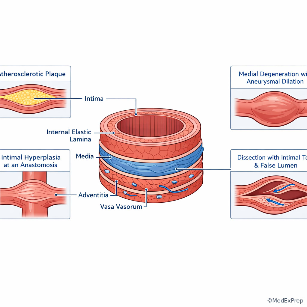
See Fig 2 for the layer-to-lesion mapping that underlies many later questions.
B Worked example
A 69-year-old man with hypertension, remote smoking history, chronic kidney disease stage 3, and prior femoral-popliteal bypass 14 months ago presents for surveillance. Duplex shows a focal high-velocity lesion at the distal anastomosis. He also has diffuse calcified native SFA disease on old imaging, and the report mentions “atherosclerotic burden.” The question asks for the most likely pathophysiologic process. Identify the task: mechanism of late focal graft-edge narrowing. Extract key facts: prior intervention, timing more than a year out but still in the restenosis window, focal anastomotic lesion, distracting clue of diffuse native plaque. Apply rule: graft-edge stenosis is classically driven by intimal hyperplasia related to injury and flow disturbance, not just progression of remote native plaque. Eliminate distractors: medial degeneration fits aneurysm, not focal stenosis; adventitial fibrosis is nonspecific; acute thrombosis is an event, not the primary remodeling mechanism. Verify: focal anastomotic acceleration on duplex is the classic setup for intimal hyperplasia.
C Exam trap
D Checkpoint
Question: A 74-year-old woman with hypertension and known infrarenal aneurysmal disease develops acute chest pain and is found to have an aortic dissection beginning with an intimal tear. Which arterial wall layer primarily allows propagation of the dissection plane?
- Intima — tempting because the initiating tear begins here, but propagation occurs through a deeper structural layer, so this is incomplete.
- Media — correct because the false lumen dissects through the medial lamellar structure after entry through the intima.
- Adventitia — tempting because rupture through the outer wall is catastrophic, but it is not the primary propagation plane.
- Endothelium alone — tempting because it is the luminal interface, but it lacks the structural substrate for dissection extension.
1.1.1.4 Pressure, resistance, and flow: the rules that explain pulse deficits and stenosis significance
A Explanation
The central decision conflict here is: is the lesion hemodynamically important or merely an anatomic narrowing? The resolving rule is that flow depends on a pressure gradient and resistance, and resistance is exquisitely sensitive to radius. For exam purposes, remember the spirit of Poiseuille’s relationship: \( Q \propto \frac{\Delta P \, r^4}{\eta L} \). You do not need to derive the equation on boards, but you must know what it means clinically. A modest decrease in radius can produce a major rise in resistance. That is why a lesion can look “moderate” on imaging yet produce substantial pressure drop, exercise limitation, or duplex acceleration.
Choose hemodynamic significance when you see pressure loss across a segment, dampened downstream waveform, collateral recruitment, exertional symptoms, or high focal velocity with post-stenotic disturbance. Reject “not significant” when the stem gives only plaque presence without pressure or waveform consequence. The board-tested insight is that anatomic disease burden and hemodynamic effect are related but not identical. Long diffuse disease, tandem lesions, poor cardiac output, and poor runoff can all modify presentation.
This principle also explains why pulse findings deteriorate downstream from inflow disease. If the pressure head delivered to distal beds falls, both pulsatility and tissue perfusion fall. The same logic underlies bypass planning: perfect proximal inflow is not enough if outflow resistance remains prohibitive. See Fig 3 for the decision logic that links radius change, velocity, and pressure drop.
Exam takeaway: A lesion becomes board-relevant when narrowing produces pressure loss and distal waveform change, not when plaque is merely present.
flowchart LR
A[Normal radius] --> B[Radius decreases]
B --> C[Resistance rises sharply]
C --> D[Pressure drop across lesion]
B --> E[Velocity rises at stenosis]
D --> F[Distal pulse and waveform dampen]
E --> G[Post-stenotic disturbance]
F --> H[Symptoms or abnormal vascular lab]
B Worked example
A 63-year-old woman with diabetes, hyperlipidemia, and prior coronary stenting reports exertional calf pain after one block. She has palpable femoral pulses but diminished pedal pulses. CTA from an outside hospital notes “multifocal femoropopliteal atherosclerosis,” and a resident focuses on the longest plaque segment. Duplex instead shows the highest velocity ratio at a short focal distal SFA stenosis with downstream waveform dampening. A mildly elevated creatinine is a distracting lab abnormality. Identify the task: find the lesion most responsible for symptoms. Extract key facts: exertional symptoms, focal velocity acceleration, distal waveform degradation. Apply rule: the culprit is the segment causing greatest hemodynamic consequence, not necessarily the longest plaque burden on anatomy alone. Eliminate distractors: diffuse noncritical plaque may coexist; neuropathy would not explain duplex changes; venous disease would not create focal PSV acceleration. Verify: pressure/flow consequence makes the short lesion clinically dominant.
C Exam trap
D Checkpoint
Question: In a patient with a focal arterial stenosis, which finding most strongly supports that the lesion is hemodynamically important?
- Calcification visible on plain radiograph — tempting because calcification implies disease, but it does not prove pressure loss or flow limitation.
- Increased peak systolic velocity at the lesion with dampened distal waveform — correct because it shows both local acceleration and downstream consequence.
- Nonobstructive mural irregularity on CTA — tempting because anatomic abnormality is present, but the lesion may not be flow-limiting.
- Presence of a bruit without imaging correlation — tempting because turbulence can generate bruit, but it is less specific than direct hemodynamic evidence.
- The vascular variable with the strongest effect on resistance is vessel radius.
- True or False: A lesion can have extensive plaque but limited hemodynamic effect at rest. True.
- Name 3 clues a lesion is flow-limiting: velocity rise, pressure drop, distal waveform dampening.
1.1.1.5 Laminar flow, shear stress, and why plaque and hyperplasia cluster in predictable locations
A Explanation
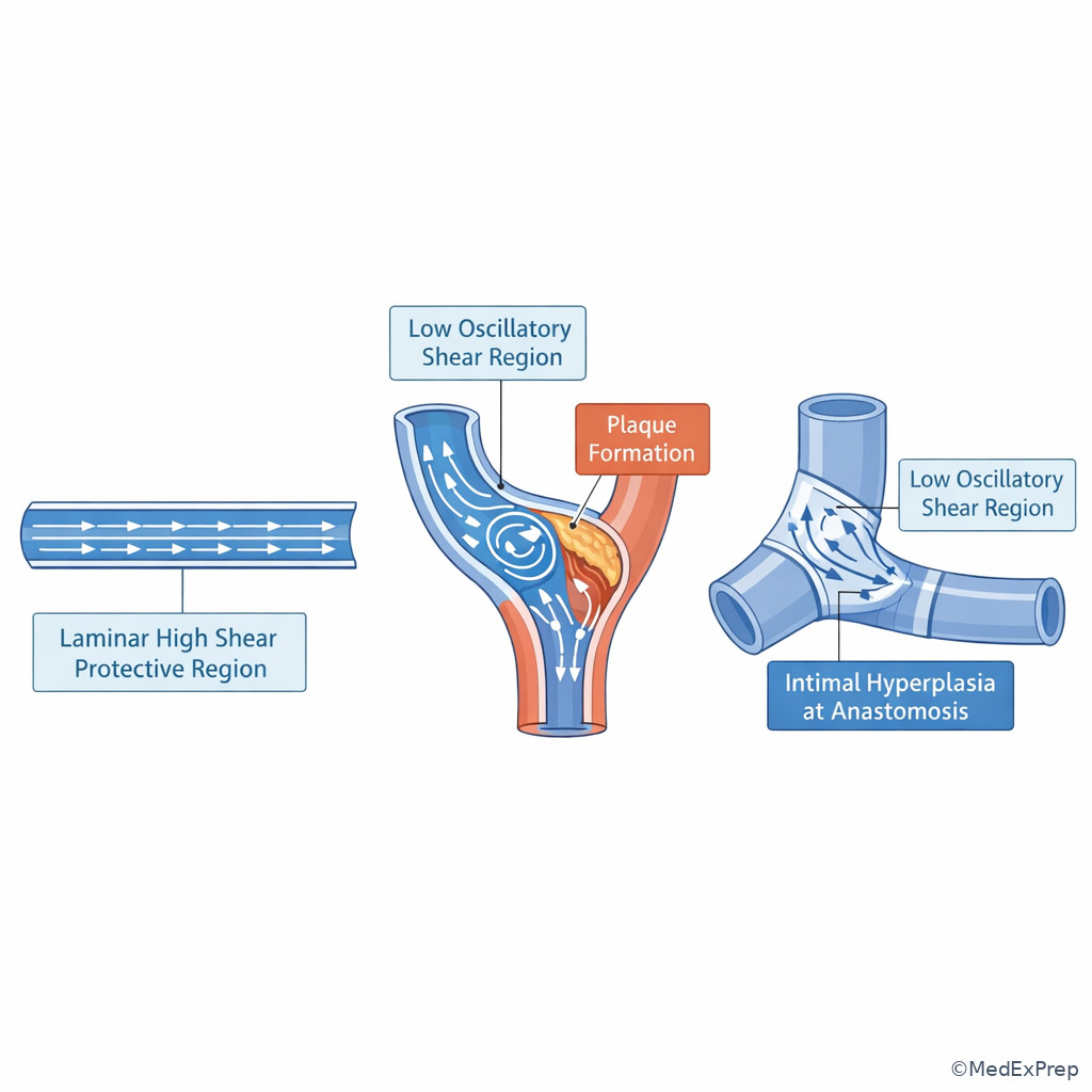
The decision conflict in this section is: why do some arterial regions repeatedly fail while others remain relatively protected? The single rule is that endothelial biology depends on the character of flow. Laminar high-shear flow tends to be protective; low or oscillatory shear promotes endothelial dysfunction, inflammation, and atheroprone signaling. This is why disease clusters at branch points, curvatures, bifurcations, and anastomoses—locations where streamline separation and recirculation occur.
Choose disturbed-flow biology when the stem highlights carotid bifurcation plaque, femoral bifurcation disease, proximal or distal graft-anastomotic restenosis, or post-stenotic dilation. Reject simplistic “uniform pressure exposure” explanations when disease distribution is focal and geometry-dependent. This principle matters clinically because intervention itself can create new disturbed-flow zones: abrupt diameter shifts, stent edges, sharp angles, kinks, and compliance mismatch all alter local shear. That is why a technically successful revascularization still needs surveillance; the repair changes hemodynamics.
WHY THIS IS TESTED: candidates often know that plaque favors branch points but cannot explain why. On the exam, that gap causes errors in graft surveillance questions, carotid disease interpretation, and questions about why a lesion recurs at one edge of a reconstruction. The vascular surgeon must think beyond “there is a narrowing” to “what local flow pattern produced it and will it recur?” See Fig 4 for atherosclerosis-prone zones and the flow logic behind them.
Exam takeaway: Low or oscillatory shear at bifurcations and anastomoses predicts plaque and intimal hyperplasia better than pressure alone.
B Worked example
A 71-year-old man with prior carotid endarterectomy, smoking history, COPD, and hyperlipidemia undergoes follow-up duplex. A focal recurrent lesion is seen near the endarterectomy endpoint. The trainee attributes this to “diffuse systemic atherosclerosis everywhere.” The stem, however, asks which local factor best explains recurrent narrowing. There is a distracting note about a mild troponin leak during a remote admission. Identify the task: mechanism of recurrence at a repair edge. Extract key facts: focal edge lesion, prior reconstruction, local geometry. Apply rule: disturbed flow and endothelial injury at transition zones promote intimal hyperplasia/restenosis. Eliminate distractors: systemic risk factors matter but do not explain why recurrence localizes to the endpoint; vasospasm is not the usual chronic mechanism; embolism would create downstream events, not fixed focal edge stenosis. Verify: location-specific recurrence is a geometry-plus-healing problem.
C Exam trap
D Checkpoint
Question: Which arterial location is most likely to develop plaque because of low or oscillatory shear?
- Long straight mid-segment of a healthy conduit with uniform caliber — tempting because disease can occur anywhere, but protective laminar flow makes it less likely.
- Outer wall of a bifurcation with flow separation — correct because disturbed flow and oscillatory shear favor endothelial dysfunction there.
- Uniformly pressurized proximal aorta without curvature effects — tempting because pressure is high, but focal plaque localization is better explained by flow pattern.
- Any artery with normal laminar centerline flow — tempting because all vessels age, but this does not identify the classic atheroprone zone.
1.1.1.6 Waveforms, velocity, and practical interpretation for vascular lab reasoning
A Explanation
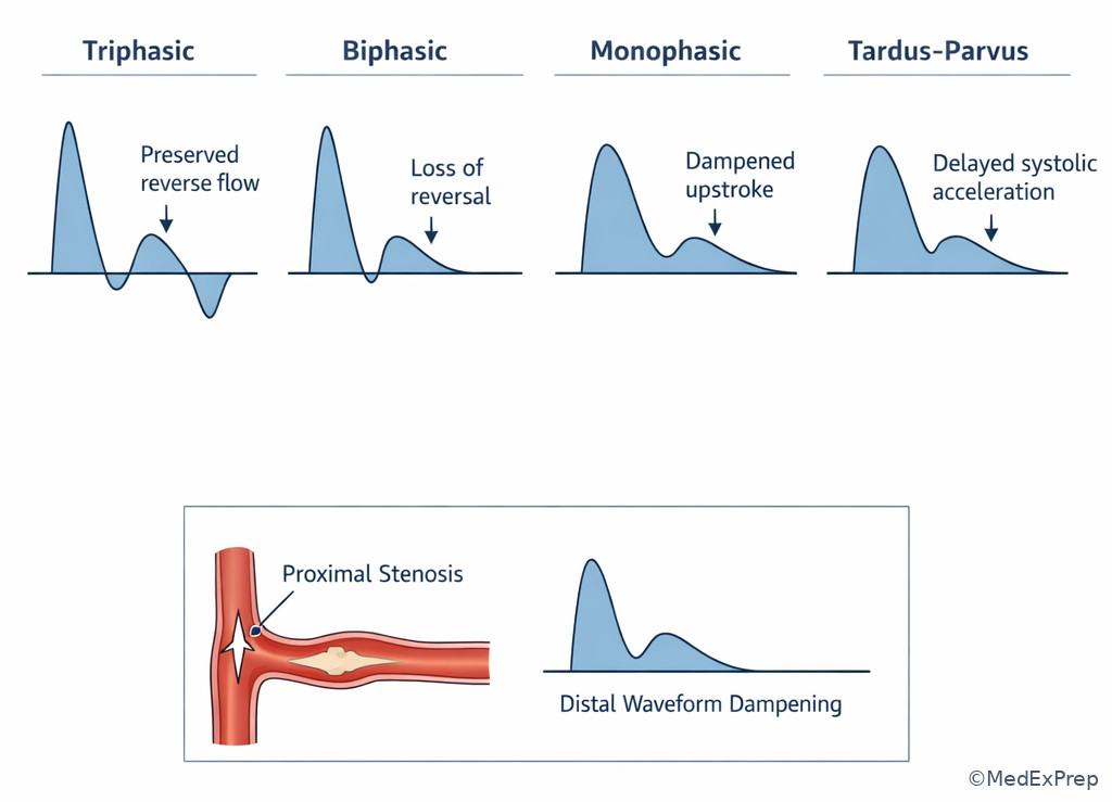
The decision conflict is: what does the waveform pattern actually mean? On board exams, learners overcall or undercall disease by reading velocity in isolation. The resolving rule is to integrate triphasic/biphasic/monophasic morphology, acceleration time, segment comparison, and clinical setting. Triphasic peripheral waveforms at rest imply preserved arterial compliance and unobstructed transmission. Biphasic may be age-related or mildly abnormal depending on territory and context. Monophasic, tardus-parvus, or markedly dampened waveforms suggest proximal inflow disease, severe stenosis, or major loss of pulsatile energy.
Choose a proximal lesion when distal waveforms are diffusely dampened across multiple segments. Choose a focal local stenosis when there is marked focal PSV increase with post-stenotic turbulence. Reject the simplistic idea that “higher velocity always means better flow.” In fact, a tight stenosis may have very high local velocity but poor net distal perfusion. Likewise, very low velocity may reflect poor cardiac output, severe inflow limitation, or impending graft thrombosis.
WHY THIS IS TESTED: vascular lab stems often provide one seductive number. The safer approach is pattern recognition. Ask: where is the transition from normal to abnormal waveform? Is there focal acceleration? Is the distal signal dampened? Does the waveform match the patient’s symptoms and exam? This topic prepares you for later duplex criteria and ABI/PVR interpretation. See Fig 5 for the waveform logic that prevents common exam errors.
Exam takeaway: Triphasic supports preserved pulsatility, whereas monophasic dampening localizes clinically important disease even before exact lesion grading.
B Worked example
A 58-year-old woman with diabetes, rheumatoid arthritis, and prior iliac stenting returns with recurrent buttock and thigh claudication. Resting ABI is borderline, creating a tempting impression of mild disease only. Duplex shows diffusely dampened monophasic waveforms from the common femoral distally on the symptomatic side, without a dramatic focal acceleration in the femoropopliteal segment. There is a distracting finding of chronic neuropathic foot numbness. Identify the task: localize the likely culprit territory. Extract key facts: widespread distal dampening beginning at the common femoral level, prior iliac intervention, no major local femoropopliteal acceleration. Apply rule: diffuse monophasic distal signals without focal downstream acceleration suggest inflow disease, likely proximal to the common femoral tracing point. Eliminate distractors: isolated tibial disease would not explain buttock symptoms; neuropathy does not create waveform abnormality; venous insufficiency does not explain monophasic arterial signals. Verify: recurrent iliac inflow compromise best fits the data.
C Exam trap
D Checkpoint
Question: A patient has a focal PSV increase in the superficial femoral artery and post-stenotic turbulence, but the distal pedal waveform remains relatively preserved. What is the best interpretation?
- No arterial disease is present because the distal waveform is preserved — tempting because runoff remains adequate, but focal hemodynamically relevant stenosis can still exist.
- A focal stenosis is present, but distal compensation currently preserves downstream pulsatility — correct because local acceleration identifies the lesion even before severe distal waveform collapse.
- The patient must have only venous disease — tempting if symptoms are nonspecific, but venous pathology does not explain focal arterial acceleration.
- The velocity rise proves globally increased limb perfusion — tempting because velocity sounds favorable, but stenotic jetting is not improved tissue flow.
- Most normal resting peripheral arterial waveform: triphasic.
- True or False: Higher velocity at a stenosis always means better perfusion. False.
- Tardus-parvus distal to a lesion suggests proximal inflow obstruction.
1.1.1.7 Integration: wall stress, compliance mismatch, runoff, and why repairs fail or succeed
A Explanation
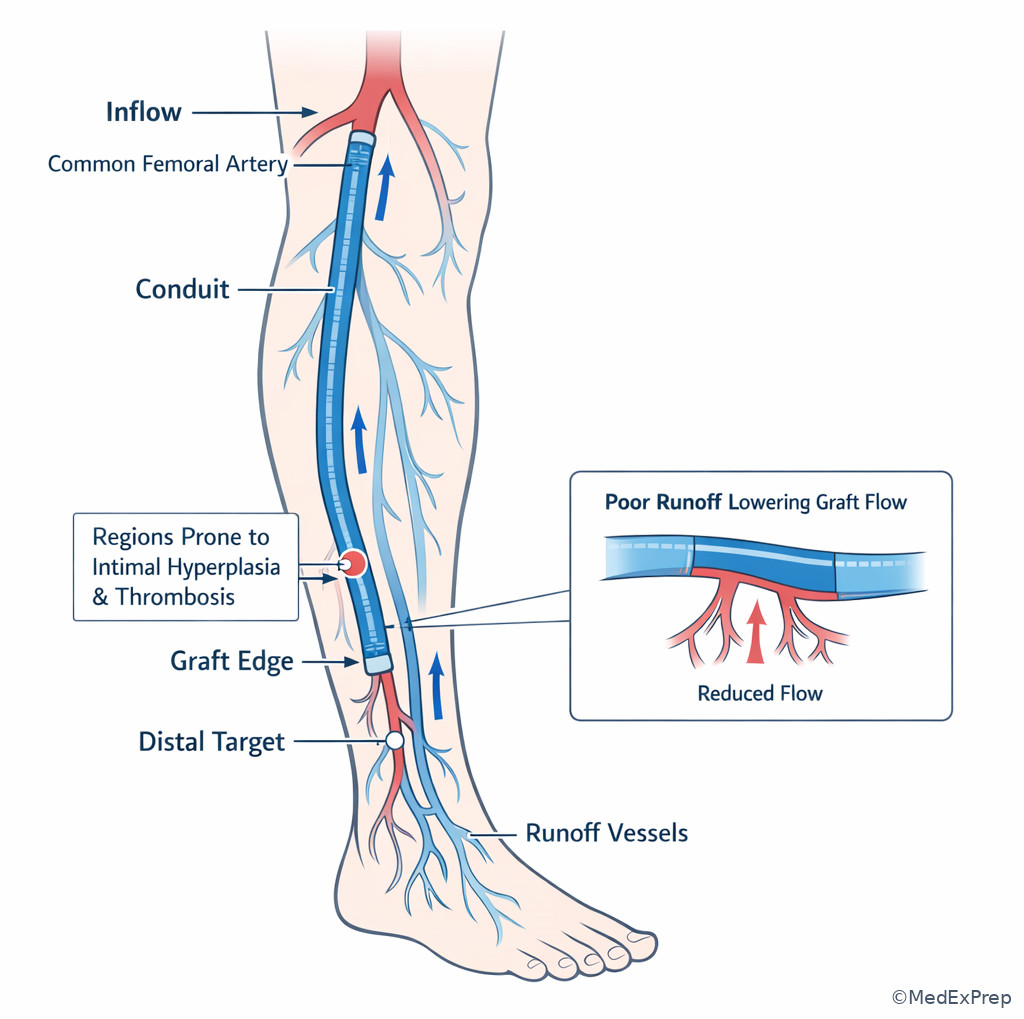
The final foundational integration conflict is: why does a technically successful repair later fail? The single resolving rule is that durability depends on the interaction of wall biology, conduit properties, and whole-circuit flow. A patent inflow segment can still fail if runoff is poor. A perfectly deployed stent can still create edge stenosis if geometry or compliance mismatch produces disturbed flow. A bypass can thrombose when low-flow states, small conduit, and poor distal target interact. An aneurysm can enlarge because sac pressure persists even when the repair initially appears satisfactory.
Wall stress helps explain aneurysm behavior. At a simplified level, circumferential tension rises with increasing vessel radius for a given pressure, often summarized conceptually by Laplace-type reasoning. As radius enlarges, further enlargement becomes mechanically easier, which is why aneurysms can progress. In reconstructive surgery, compliance mismatch matters because native artery, prosthetic graft, and stented artery do not deform identically with pulsation. The transition zone becomes a biologically active site for shear alteration and hyperplasia.
Choose poor runoff as the dominant explanation when distal targets are limited, tibial outflow is poor, and graft velocities are low without a focal inflow lesion. Choose compliance mismatch when the lesion localizes at graft or stent edges. Reject operator-centric overconfidence; on the boards, many “technical” failures are really circuit failures. See Fig 6 for how inflow, conduit, and runoff interact to determine patency.
Exam takeaway: A repair fails when the vascular circuit is mismatched, even if the treated segment itself initially appears satisfactory.
flowchart TD
A[Inflow] --> B[Conduit or stent]
B --> C[Runoff]
A --> D[If inflow poor: low pressure delivery]
B --> E[If edge mismatch: disturbed flow and hyperplasia]
C --> F[If runoff poor: low volume flow and thrombosis]
D --> G[Early failure risk]
E --> G
F --> G
G --> H[Need surveillance and reintervention logic]
Exam takeaway: Durable revascularization depends on the entire circuit rather than the treated lesion alone.
Exam takeaway: Larger radius increases wall tension and helps explain progressive aneurysm enlargement and rupture risk logic.
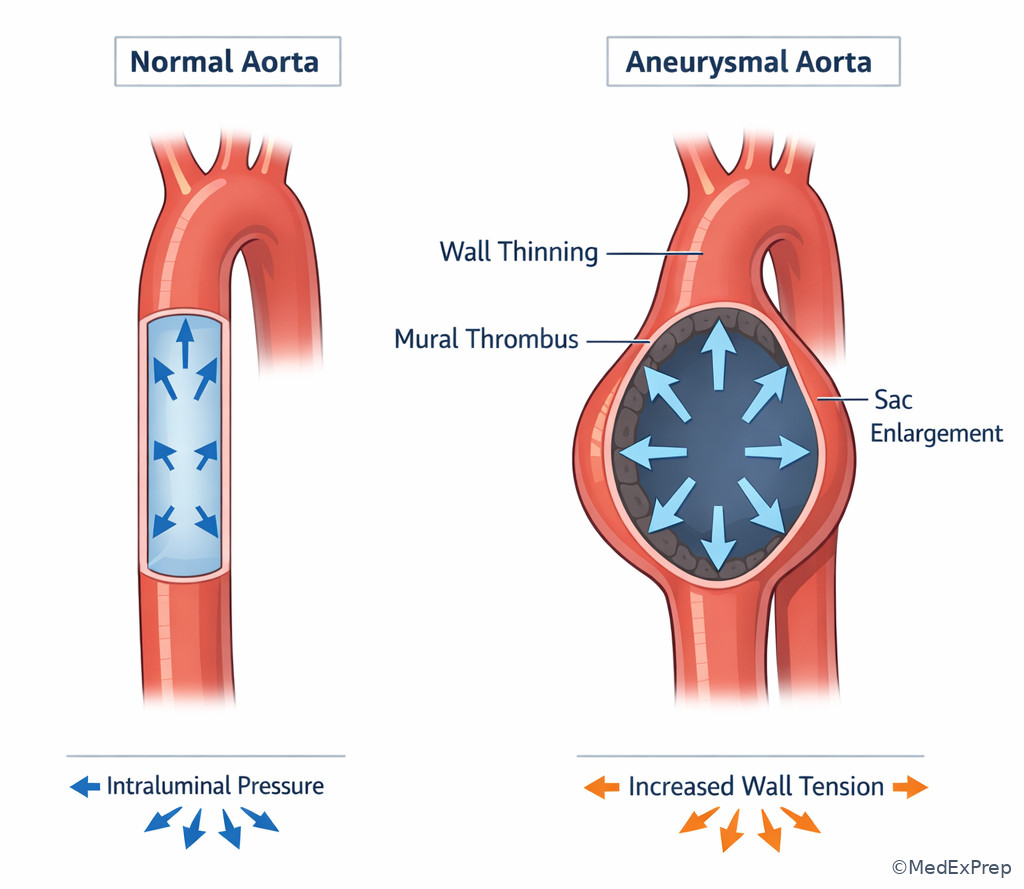
B Worked example
A 76-year-old man with diabetes, prior tibial angioplasty, congestive heart failure, and chronic limb-threatening ischemia undergoes femoral-to-distal bypass. Completion imaging is acceptable. Three months later, surveillance shows globally low graft velocities without a focal anastomotic jet. The distal target is a severely diseased single-vessel runoff bed. A tempting clue is mild dehydration at clinic, which could suggest transient low flow only. Identify the task: most likely reason for threatened patency. Extract key facts: globally low velocity, no focal edge lesion, poor distal bed. Apply rule: poor runoff can reduce circuit flow enough to threaten thrombosis even with acceptable inflow and no focal stenosis. Eliminate distractors: intimal hyperplasia usually causes focal acceleration; dehydration is too superficial to explain persistent pattern; cardiac output matters but the standout clue is the poor runoff target. Verify: whole-circuit analysis supports runoff-limited patency risk.
C Exam trap
D Checkpoint
Question: Which postoperative duplex pattern most strongly suggests poor runoff rather than focal anastomotic stenosis?
- Marked focal velocity acceleration at one graft edge — tempting because it indicates graft risk, but this pattern points to focal stenosis/hyperplasia instead.
- Globally low graft velocities throughout the conduit without a focal jet — correct because diffuse low-flow pattern fits poor runoff or low circuit flow.
- Normal triphasic signals throughout the graft — tempting because it sounds reassuring, but it does not suggest failure physiology.
- Isolated bruit over the incision with normal duplex — tempting because bruit raises concern, but it is less diagnostic than the diffuse low-flow duplex pattern.
Exam Traps & Differentiators
- Most common wrong answer: equating visible plaque with hemodynamic significance. Why it is tempting: anatomy is easier to see than physiology. What fixes it: require a pressure, velocity, waveform, symptom, or runoff consequence.
- Another common wrong answer: calling every late graft narrowing “progressive atherosclerosis.” Why it is tempting: the patient often has systemic plaque risk factors. What fixes it: focal anastomotic or stent-edge recurrence suggests intimal hyperplasia and disturbed flow.
- Looks similar but isn’t: high velocity at a stenosis versus good perfusion. Discriminator: stenotic jetting can coexist with low distal pressure and poor tissue delivery.
- Looks similar but isn’t: aneurysm enlargement versus simple ectasia. Discriminator: medial wall failure and rising wall stress make progressive dilation clinically important.
- Looks similar but isn’t: poor runoff versus focal stenosis. Discriminator: poor runoff gives diffuse low-flow patterns; focal stenosis gives a localized jet and post-stenotic change.
| Looks similar | Why it tempts you | Single clue that separates them | Think |
|---|---|---|---|
| Plaque burden vs flow-limiting stenosis | Both show narrowing on imaging | Pressure drop / PSV increase / distal dampening | Hemodynamic significance |
| Intimal hyperplasia vs recurrent diffuse atherosclerosis | Both can cause restenosis | Focal lesion at repair edge or anastomosis | Local healing + disturbed flow |
| High stenotic velocity vs robust perfusion | Velocity sounds favorable | Downstream waveform or pressure may still be poor | Jetting is not tissue delivery |
| Poor runoff vs inflow failure | Both can cause low graft flow | Inflow lesion shows proximal transition; runoff issue often diffuse low-flow with bad distal bed | Whole-circuit analysis |
| Dissection entry tear vs propagation plane | Both involve the same artery | Entry starts in intima; propagation tracks the media | Layer-specific mechanism |
If the stem says focal edge recurrence after prior repair, think intimal hyperplasia.
If the stem says branch point or bifurcation disease, think disturbed flow and low/oscillatory shear.
If the stem says marked PSV increase plus distal dampening, think hemodynamically important stenosis.
If the stem says globally low graft velocities without focal acceleration, think poor runoff or low-flow circuit.
If the stem says expanding dilation at constant pressure, think wall stress rising with radius and medial failure.
If the stem says diffuse distal monophasic signals from the groin downward, think proximal inflow disease.
Tables
| Wall layer / flow concept | Main structure | Common tested pathology | Board-use clue |
|---|---|---|---|
| Intima | Endothelium and subendothelial layer | Atherosclerosis, ulceration, intimal hyperplasia | Restenosis at repair edge, thrombogenic plaque surface |
| Media | Smooth muscle, elastic lamellae | Aneurysm degeneration, dissection propagation | Dilation, loss of wall strength, false lumen track |
| Adventitia | Collagen, nerves, vasa vasorum | External support failure, inflammatory extension | Outer-wall involvement, rupture containment question |
| Laminar flow | Ordered streamlines | Relative endothelial protection | Straight segments less plaque-prone than bifurcations |
| Disturbed flow | Separation/recirculation | Plaque, hyperplasia, post-stenotic change | Bifurcations and anastomoses recur |
| Poor runoff | Limited distal vascular bed | Low-flow graft failure | Diffuse low conduit velocities without focal jet |
| Comparison | Focal stenosis | Poor runoff | Proximal inflow disease |
|---|---|---|---|
| Typical duplex pattern | Localized high PSV and post-stenotic turbulence | Diffuse low velocities, no discrete jet | Dampened distal waveforms from a proximal level onward |
| Pressure consequence | Gradient across lesion | Low volume flow through whole circuit | Reduced pressure delivery to downstream bed |
| Exam clue | Short culprit lesion | Bad distal target/runoff bed | Buttock/thigh symptoms or proximal transition |
| Wrong answer trap | Assumed to be “good flow” because velocity is high | Missed because no dramatic focal number | Missed if only distal symptoms are considered |
| Planning implication | Targeted revision/intervention may help | Must address outflow strategy and expectations | Fix inflow before chasing distal lesions |
| Differential / classification issue | Correct interpretation | Common distractor | Discriminating clue |
|---|---|---|---|
| Late focal graft-edge narrowing | Intimal hyperplasia | Diffuse recurrent atherosclerosis | Edge/anastomotic localization |
| Aortic dissection plane | Media | Intima alone | Entry tear starts in intima but extension tracks medial layer |
| Branch-point plaque clustering | Disturbed low/oscillatory shear | Uniform systemic pressure alone | Focal geometry-specific distribution |
| Monophasic distal waveform | Loss of pulsatile energy from significant disease | Normal age-related variant | Dampened contour with clinical context |
| High local velocity | Stenotic acceleration | Improved tissue perfusion | Evaluate distal waveform and pressure, not velocity alone |
| Expanding aneurysm | Rising wall stress with radius and medial degeneration | Intimal plaque burden only | Dilation/rupture logic, not stenosis logic |
Algorithm / Approach
Exam takeaway: Localize the problem by deciding whether the key clue reflects wall pathology, focal hemodynamics, or whole-circuit failure.
flowchart TD
A[Board stem] --> B{What is being tested?}
B -->|Layer pathology| C[Intima vs media vs adventitia]
B -->|Significance of narrowing| D[PSV, gradient, distal waveform]
B -->|Failure of repair| E[Inflow, conduit, runoff]
C --> F[Plaque/hyperplasia vs aneurysm/dissection]
D --> G[Focal stenosis vs noncritical plaque]
E --> H[Edge lesion vs poor runoff vs inflow problem] Rapid Review
- Intimal hyperplasia → focal repair-edge restenosis, not diffuse native plaque progression
- Dissection → intimal entry tear with medial propagation
- Aneurysm progression → radius-driven wall stress increase, not stenotic jet physiology
- Bifurcation plaque → low/oscillatory shear clue
- Triphasic waveform → preserved pulsatile transmission at rest
- Monophasic dampening → significant loss of pulsatile energy, usually proximal or severe disease
- High PSV at lesion → stenotic acceleration, not proof of adequate tissue perfusion
- Diffuse low graft velocities → think poor runoff or low circuit flow before focal edge lesion
- Focal jet plus post-stenotic turbulence → localized hemodynamically important stenosis
- Longest plaque segment → not always culprit; hemodynamic consequence decides
- Compliance mismatch → recurrent trouble at transition zones
- Visible calcification → disease burden marker, not hemodynamic grading by itself
- Runoff failure → can defeat technically adequate revascularization
- Protective flow pattern → straight, laminar, nonseparating streamlines
Self-check quiz
1. A 72-year-old man with hypertension and smoking history is asked which arterial wall layer most directly accounts for aneurysmal degeneration and dissection propagation. Which is the most likely answer?
- Intima
- Media
- Adventitia
- Endothelium only
2. A 66-year-old woman with diabetes and hyperlipidemia has a focal femoral stenosis on duplex. Which finding most strongly indicates that the lesion is hemodynamically important?
- Visible calcification of the artery
- Localized PSV elevation with distal waveform dampening
- Any audible bruit over the groin
- Diffuse plaque on CTA without pressure change
3. A 61-year-old woman with prior iliac stenting, diabetes, and lumbar spinal stenosis reports recurrent buttock and thigh claudication. Resting ABI is borderline. Duplex shows monophasic dampened waveforms from the common femoral artery distally without a focal femoropopliteal jet. What is the most appropriate localization?
- Proximal inflow disease
- Isolated tibial runoff disease
- Pure neurogenic claudication
- Primary venous insufficiency
4. A 70-year-old man with CKD, prior femoral-popliteal bypass, and diffuse atherosclerosis has a new focal high-velocity lesion at the distal anastomosis 12 months after surgery. Which is the most appropriate mechanism?
- Intimal hyperplasia from local injury and disturbed flow
- Medial aneurysmal degeneration of the graft
- Uniform hypertension causing diffuse edge-independent narrowing
- Primary venous outflow obstruction
5. A 68-year-old man with tissue loss, heart failure, and severe tibial disease undergoes distal bypass. Surveillance at 3 months shows globally low graft velocities without a focal jet. Which is the most appropriate explanation?
- Poor runoff causing low circuit flow
- Focal anastomotic intimal hyperplasia
- Normal expected pattern after all distal bypasses
- Improved perfusion with reduced resistance
6. A 73-year-old woman with hypertension, COPD, and asymptomatic carotid disease has plaque concentrated at the carotid bifurcation. Which mechanism best explains this localization rather than uniform disease along the entire artery?
- High static pressure alone in all carotid segments
- Low or oscillatory shear caused by bifurcation flow separation
- Selective adventitial inflammation at every branch point
- Higher oxygen content in bifurcation blood
7. A 64-year-old man with diabetes, prior SFA intervention, and persistent calf claudication has a short focal distal SFA lesion with marked PSV elevation and post-stenotic turbulence. Longer proximal plaque segments are present but without major velocity change. Which is the most appropriate next interpretive conclusion?
- The longest plaque segment is the dominant culprit because plaque burden predicts symptoms best
- The short focal lesion is most likely the hemodynamically dominant culprit
- No lesion is significant because flow is fastest at the focal narrowing
- The findings are more consistent with isolated venous claudication
8. A 77-year-old man with an enlarging infrarenal aneurysm, hypertension, and coronary disease is asked why larger aneurysms are more concerning even at similar arterial pressure. Which explanation is most appropriate?
- Larger radius increases wall tension and promotes further expansion
- Higher centerline velocity in aneurysms protects against rupture
- Intimal plaque alone determines rupture behavior
- Adventitial nerves directly reduce wall stress as diameter enlarges
Answer key
1.1.1.8 1. Correct answer: B — Media
A. Intima: Tempting because dissections begin with an intimal tear. Incorrect for this scenario because the propagation plane and structural failure underlying aneurysm behavior are primarily medial. Single stem clue: the question asks what accounts for aneurysmal degeneration and dissection propagation, not the entry tear alone. Sources: Rutherford’s Vascular Surgery and Endovascular Therapy; Cronenwett and Johnston, Rutherford.
B. Media: Tempting because it is the structural layer, and it is correct because medial elastic/smooth muscle architecture determines wall strength and permits dissection tracking once blood enters the wall. Single stem clue: “aneurysmal degeneration and dissection propagation.” Sources: Rutherford’s Vascular Surgery and Endovascular Therapy; Harrison’s Principles of Internal Medicine.
C. Adventitia: Tempting because rupture through outer layers is clinically dramatic. Incorrect because adventitia is not the primary layer responsible for aneurysm formation or dissection propagation. Single stem clue: the question is about underlying mechanism, not final rupture containment. Sources: Rutherford; Robbins & Cotran.
D. Endothelium only: Tempting because it is the luminal interface and key to thrombosis signaling. Incorrect because it cannot account for the structural wall failure of aneurysm/dissection. Single stem clue: “wall layer” plus structural disease pattern. Sources: Robbins & Cotran; Rutherford.
1.1.1.9 2. Correct answer: B — Localized PSV elevation with distal waveform dampening
A. Visible calcification of the artery: Tempting because calcification signals arterial disease burden. Incorrect because it does not prove hemodynamic consequence. Single stem clue: the question asks what indicates significance, not mere presence of disease. Sources: Society for Vascular Surgery practice patterns in vascular lab interpretation; Rutherford.
B. Localized PSV elevation with distal waveform dampening: Tempting because it combines two independent clues, and it is correct because it shows both local stenotic acceleration and downstream energy loss. Single stem clue: combined focal and distal hemodynamic consequence. Sources: Rutherford; standard duplex interpretation principles in vascular laboratory texts.
C. Any audible bruit over the groin: Tempting because turbulence can generate bruit. Incorrect because bruit is less specific than direct duplex evidence and may not quantify significance. Single stem clue: “most strongly indicates.” Sources: Rutherford; Harrison’s.
D. Diffuse plaque on CTA without pressure change: Tempting because CTA looks anatomic and authoritative. Incorrect because anatomic plaque without hemodynamic consequence may not be flow-limiting. Single stem clue: “without pressure change.” Sources: Rutherford; AHA/ACC PAD guideline concepts.
1.1.1.10 3. Correct answer: A — Proximal inflow disease
A. Proximal inflow disease: Tempting because buttock/thigh symptoms and prior iliac intervention point proximally, and it is correct because diffuse dampened waveforms from the common femoral artery distally without a focal downstream jet suggest inflow compromise above that level. Single stem clue: monophasic signals begin at the common femoral level. Sources: Rutherford; AHA/ACC lower extremity PAD guideline framework.
B. Isolated tibial runoff disease: Tempting because distal disease is common in diabetes. Incorrect because isolated tibial disease would not usually create diffuse waveform dampening starting proximally or buttock symptoms. Single stem clue: common femoral-to-distal dampening. Sources: Rutherford.
C. Pure neurogenic claudication: Tempting because lumbar stenosis is present as noise. Incorrect because neurogenic disease does not explain objective monophasic arterial waveforms. Single stem clue: abnormal duplex hemodynamics. Sources: Harrison’s; Rutherford.
D. Primary venous insufficiency: Tempting because leg symptoms can overlap. Incorrect because venous insufficiency does not produce arterial monophasic inflow patterns. Single stem clue: arterial duplex abnormality. Sources: Rutherford.
1.1.1.11 4. Correct answer: A — Intimal hyperplasia from local injury and disturbed flow
A. Intimal hyperplasia from local injury and disturbed flow: Tempting because the lesion is focal at the distal anastomosis, and it is correct because repair-edge injury plus altered local shear are classic drivers of late focal restenosis. Single stem clue: new focal high-velocity distal anastomotic lesion 12 months after bypass. Sources: Rutherford; SVS surveillance concepts for infrainguinal bypass.
B. Medial aneurysmal degeneration of the graft: Tempting because graft complications can be structural. Incorrect because medial degeneration would not explain focal high-velocity edge stenosis. Single stem clue: stenotic duplex pattern rather than dilation. Sources: Rutherford.
C. Uniform hypertension causing diffuse edge-independent narrowing: Tempting because hypertension is a real risk factor. Incorrect because the localization to the anastomosis points to a local geometry/healing mechanism. Single stem clue: edge-specific focality. Sources: Rutherford; Robbins & Cotran.
D. Primary venous outflow obstruction: Tempting only if one mistakes graft failure for venous pathology. Incorrect because the scenario is an arterial bypass with duplex stenotic findings. Single stem clue: arterial distal anastomotic lesion. Sources: Rutherford.
1.1.1.12 5. Correct answer: A — Poor runoff causing low circuit flow
A. Poor runoff causing low circuit flow: Tempting because distal target quality is emphasized, and it is correct because globally low graft velocities without a focal jet point to a low-flow circuit, especially when runoff is severely limited. Single stem clue: severe tibial disease with diffuse low conduit velocities. Sources: Rutherford; SVS bypass surveillance principles.
B. Focal anastomotic intimal hyperplasia: Tempting because it is a common postoperative lesion. Incorrect because hyperplasia typically causes localized acceleration, not uniformly low velocities throughout the graft. Single stem clue: absence of focal jet. Sources: Rutherford.
C. Normal expected pattern after all distal bypasses: Tempting because distal targets can have lower flows than proximal bypasses. Incorrect because persistently globally low velocities are a patency warning, not a routine reassurance. Single stem clue: surveillance concern is built into the stem. Sources: Rutherford.
D. Improved perfusion with reduced resistance: Tempting because one may equate lower velocity with reduced resistance. Incorrect because in this scenario the bad runoff bed makes low flow pathologic, not favorable. Single stem clue: severe tibial disease and tissue loss context. Sources: Rutherford; AHA/ACC PAD framework.
1.1.1.13 6. Correct answer: B — Low or oscillatory shear caused by bifurcation flow separation
A. High static pressure alone in all carotid segments: Tempting because hypertension contributes to vascular disease. Incorrect because it does not explain why plaque localizes at the bifurcation rather than being uniform. Single stem clue: “localization.” Sources: Robbins & Cotran; Rutherford.
B. Low or oscillatory shear caused by bifurcation flow separation: Tempting because bifurcation geometry is specified, and it is correct because disturbed flow and endothelial dysfunction at branch points create atheroprone zones. Single stem clue: carotid bifurcation concentration of plaque. Sources: Rutherford; vascular biology literature summarized in major texts.
C. Selective adventitial inflammation at every branch point: Tempting because inflammation is involved in plaque biology. Incorrect because the tested mechanism for location specificity is hemodynamic shear pattern, not branch-point-selective adventitial inflammation. Single stem clue: flow localization concept. Sources: Robbins & Cotran.
D. Higher oxygen content in bifurcation blood: Tempting only superficially. Incorrect because oxygen content does not explain focal plaque distribution. Single stem clue: this is a hemodynamic location question. Sources: Rutherford.
1.1.1.14 7. Correct answer: B — The short focal lesion is most likely the hemodynamically dominant culprit
A. The longest plaque segment is the dominant culprit because plaque burden predicts symptoms best: Tempting because longer disease appears worse anatomically. Incorrect because symptoms correlate better with hemodynamic dominance than plaque length alone. Single stem clue: marked PSV elevation and post-stenotic turbulence at the short focal lesion. Sources: Rutherford; AHA/ACC PAD guideline concepts.
B. The short focal lesion is most likely the hemodynamically dominant culprit: Tempting because it has the physiologic signature, and it is correct because focal acceleration plus turbulence identify the segment producing the largest functional impact. Single stem clue: other longer segments lack major velocity change. Sources: Rutherford.
C. No lesion is significant because flow is fastest at the focal narrowing: Tempting because velocity sounds like stronger flow. Incorrect because stenotic jetting is a sign of narrowing and can coexist with impaired distal perfusion. Single stem clue: post-stenotic turbulence. Sources: Rutherford.
D. The findings are more consistent with isolated venous claudication: Tempting because leg discomfort has a differential. Incorrect because venous pathology does not create arterial focal PSV elevation with turbulence. Single stem clue: arterial duplex signature. Sources: Rutherford.
1.1.1.15 8. Correct answer: A — Larger radius increases wall tension and promotes further expansion
A. Larger radius increases wall tension and promotes further expansion: Tempting because aneurysm size is highlighted, and it is correct because simplified wall-stress reasoning links larger radius to greater tension at a given pressure, helping explain progressive enlargement and rupture risk logic. Single stem clue: “even at similar arterial pressure.” Sources: Rutherford; Harrison’s; major vascular surgery texts discussing Laplace-type principles.
B. Higher centerline velocity in aneurysms protects against rupture: Tempting because flow language is familiar. Incorrect because rupture concern is driven by wall strength and stress, not a protective effect of velocity. Single stem clue: aneurysm enlargement question, not stenotic flow question. Sources: Rutherford.
C. Intimal plaque alone determines rupture behavior: Tempting because atherosclerosis often coexists with aneurysm. Incorrect because aneurysm risk is not explained by plaque burden alone and centers on medial degeneration and wall stress. Single stem clue: asks why larger diameter matters. Sources: Robbins & Cotran; Rutherford.
D. Adventitial nerves directly reduce wall stress as diameter enlarges: Tempting only because adventitia is an outer wall structure. Incorrect because neural elements do not provide the tested mechanical explanation. Single stem clue: mechanical consequence of larger radius. Sources: Rutherford.
Keep going with the full ABS Vascular Surgery Prep Course (Vascular)
You just studied the way members do, in the same lesson interface. Full access unlocks every lesson, the question bank and flashcards for a full year.
One payment · 1 year of access · No auto-renewal