1.1.1 - Projectional Anatomy Landmarks
Projectional Anatomy Landmarks
What you’ll learn
- DOMAIN: Core Modalities → Radiography and Fluoroscopy → Projectional localization: Identify major chest, abdominal, and musculoskeletal landmarks on projection radiographs.
- DOMAIN: Integrated Image-Based Reasoning → Localization → Anatomic compartment assignment: Use silhouette, side, and level cues to localize structures on frontal and lateral projections.
- DOMAIN: Thoracic Radiology → Basic chest radiograph interpretation → Landmark recognition: Distinguish mediastinal, hilar, diaphragmatic, fissural, and pleural boundaries relevant to board-style questions.
- DOMAIN: Abdominal/GI Radiology → Plain-film anatomy → Gas pattern orientation: Recognize bowel distribution, solid-organ outlines, and key abdominal reference lines.
- DOMAIN: Physics/Safety → Modality-specific reasoning → Projection limitations: Explain how patient position and beam projection alter apparent anatomy and create common traps.
- Core Modalities → Radiography and fluoroscopy fundamentals → landmark-based interpretation
- Thoracic Radiology → chest radiograph anatomy → frontal/lateral localization
- Abdominal/GI Radiology → abdominal projectional anatomy → bowel and organ orientation
- Integrated Image-Based Reasoning → localization and pattern recognition → choose the correct compartment/structure
- Physics/Safety → image acquisition concepts → projection-dependent appearance
Why it matters
- Most “easy” radiology misses are not disease misses; they are anatomy-localization misses on plain films.
- Board stems often test whether you can decide where a finding is before asking what it is.
- Correct landmark recognition supports the next topic directly: line and tube position cannot be judged without a stable mental map of normal projectional anatomy.
- Many urgent chest findings are recognized through disturbed landmarks, silhouettes, or unexpected displacement rather than through a named diagnosis alone.
Key Terms & Must-Know Facts
1.1.1.1 Key terms
- PA view
- AP portable view
- Lateral view
- Silhouette sign
- Hilum overlay sign
- Cervicothoracic sign
- Cardiophrenic angle
- Costophrenic angle
- Aortic knob
- Paratracheal stripe
- Carina
- Major fissure
- Minor fissure
- Gastric bubble
- Psoas margin
1.1.1.2 Must-know facts
- On a standard PA chest radiograph, the left hemidiaphragm is usually slightly lower than the right.
- The right hilum is typically lower than the left hilum.
- The carina normally lies near the T5–T7 level and is a reliable central landmark for chest line/tube questions.
- AP portable films magnify the heart and can falsely suggest cardiomegaly.
- Loss of a normal border on frontal radiography localizes pathology to the structure touching that border: this is the silhouette sign.
- The gastric bubble usually sits beneath the left hemidiaphragm.
- Large bowel is peripheral; small bowel is more central, though overlap is common and absolute statements are dangerous.
- Psoas margins are best seen when there is adequate contrast and little overlying obscuration; loss is nonspecific.
| Landmark | Normal projection clue | High-yield use |
|---|---|---|
| Carina | Midline/slightly left of midline bifurcation level | Anchor for ET tube and central chest localization |
| Aortic knob | Left upper mediastinal contour | Orient left mediastinum; assess widened contour |
| Right paratracheal stripe | Thin vertical right upper mediastinal interface | Mass/adenopathy/interface abnormality questions |
| Right heart border | Created mainly by right atrium | Silhouette sign with right middle lobe disease |
| Left heart border | Created mainly by left ventricle | Silhouette sign with lingular disease |
| Right hemidiaphragm | Usually higher than left | Subpulmonic, pleural, and abdominal orientation |
| Gastric bubble | Subdiaphragmatic left lucency | Side confirmation; situs clues |
| Psoas margin | Soft-tissue edge in abdomen | Retroperitoneal process may obscure, but nonspecific |
Core content
Exam takeaway: Use side, height, and border cues together before naming a structure or abnormality.
flowchart TD
A[Start with projection] --> B[Check PA vs AP and rotation]
B --> C[Identify left side using gastric bubble and aortic knob]
C --> D[Locate central anchors: trachea, carina, hila]
D --> E[Trace borders: right heart, left heart, diaphragms]
E --> F[Localize any opacity by lost border or displaced line]
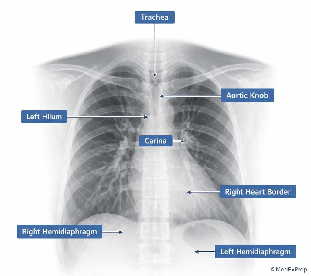
Exam takeaway: Memorize the normal contours of the hila, heart borders, diaphragms, and aortic arch because board stems often test disturbed landmarks rather than disease names.
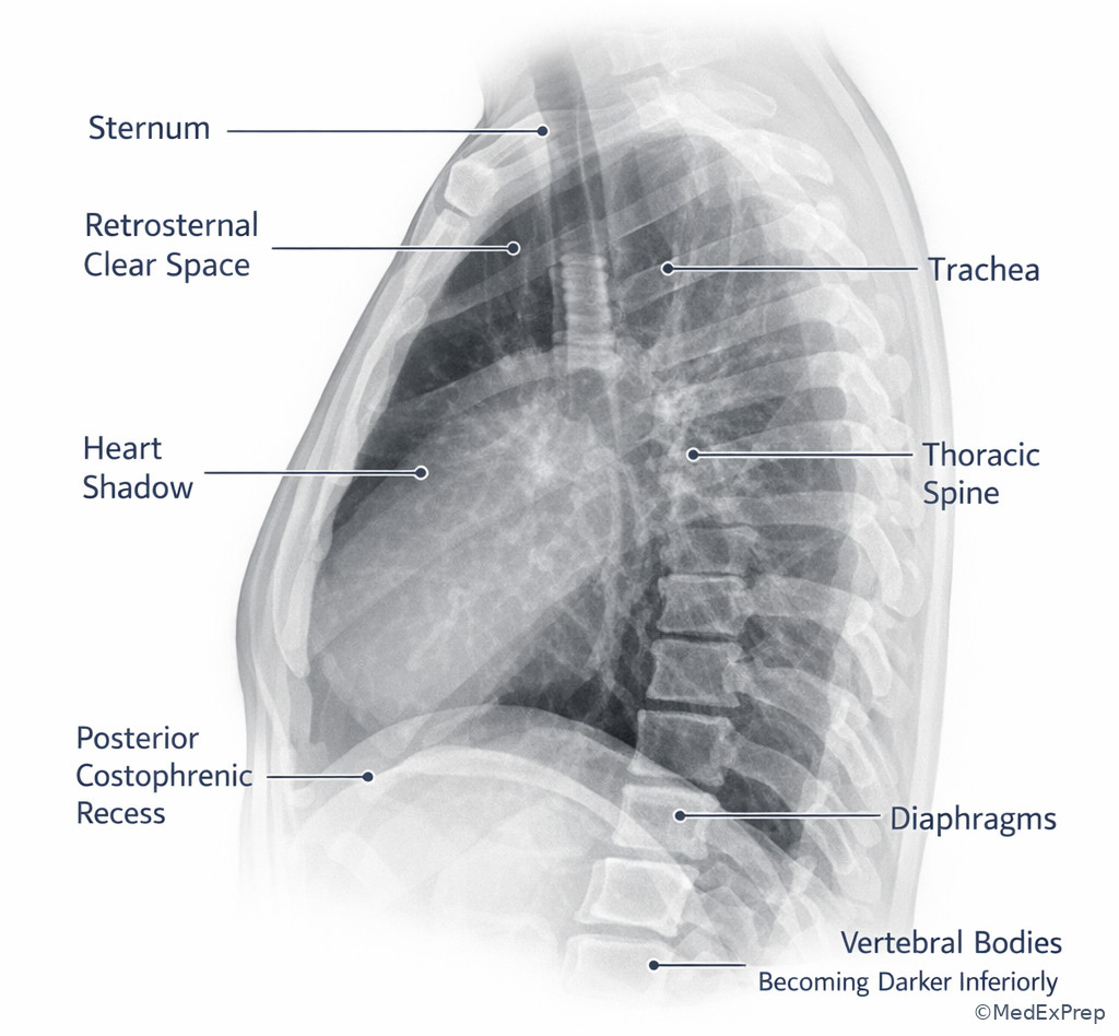
Exam takeaway: The lateral view is the fastest way to separate anterior, middle, and posterior structures when the frontal film is ambiguous.
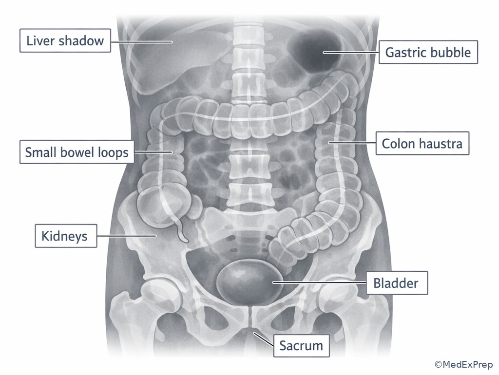
Exam takeaway: Peripheral colon, central small bowel, psoas margins, liver edge, splenic shadow, and gastric bubble form the basic abdominal orientation framework.
Exam takeaway: When a normal border disappears, localize the opacity to the lobe or segment touching that border.
flowchart LR
A[Lost right heart border] --> B[Right middle lobe]
C[Lost right hemidiaphragm] --> D[Right lower lobe]
E[Lost left heart border] --> F[Lingula]
G[Lost left hemidiaphragm] --> H[Left lower lobe]
I[Upper mediastinal contour lost] --> J[Adjacent upper lobe or mediastinal process]
Exam takeaway: Border loss is a localization tool, not a diagnosis; first identify the interface that should be visible.
flowchart TD
A[See an opacity on frontal chest radiograph] --> B{Which border is obscured?}
B -->|Right heart| C[Think right middle lobe contact]
B -->|Left heart| D[Think lingular contact]
B -->|Hemidiaphragm| E[Think lower lobe contact]
B -->|Aortic arch or upper mediastinum| F[Think upper lobe or mediastinal location]
C --> G[Confirm on lateral view if available]
D --> G
E --> G
F --> G
1.1.1.3 Foundational chest orientation: sides, heights, and central anchors
A Explanation
The first decision conflict on a chest radiograph is not “What disease is this?” but “Am I correctly oriented, and which structures are normal anchors?” The board-style rule is simple: choose landmark interpretation only after you decide projection, side, and whether magnification or rotation could distort appearance. A candidate who jumps directly to pathology will misread normal hila, overcall heart size on AP films, or mistake asymmetric diaphragms for disease. See Fig 1 and Fig 2.
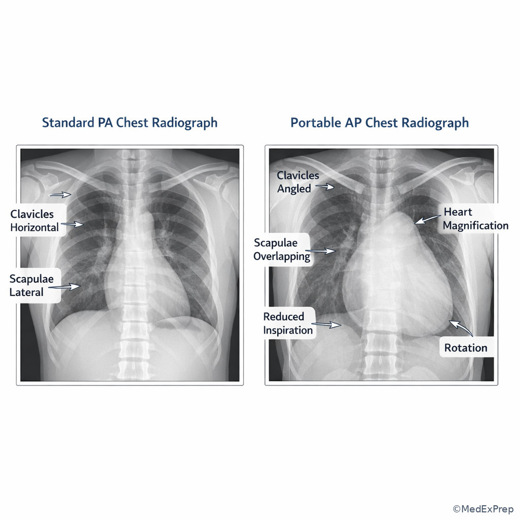
WHY THIS IS TESTED: ABR-style items often use normal or near-normal anatomy as the hidden discriminator. The single rule that resolves many stems is: establish left side and projection before assigning abnormality. Use the gastric bubble and aortic knob to orient left, then identify the trachea, carina, hila, and diaphragms. The right hemidiaphragm is usually higher; the left hilum is usually slightly higher than the right. On portable AP radiographs, the heart may appear enlarged because of magnification, so reject cardiomegaly as a conclusion if the stem emphasizes a supine or portable AP study without corroborating signs.
Choose PA when judging heart size and standard anatomy; reject strict size conclusions on AP portable films. Choose the carina as a robust central landmark; reject the clavicles or hilar density alone when rotation is present. Monitor rotation by checking whether the spinous processes are centered between the medial clavicles. If rotated, hilar asymmetry and mediastinal contour become less trustworthy.
Key rule: On a frontal chest radiograph, left side confirmation comes from the gastric bubble and aortic knob; normal hilar asymmetry is left higher, right lower.
Memory anchor: LAR = Left hilum Above, Right hemidiaphragm raised.
Recall: This is the first topic in the course, so build your base map here. In the upcoming topic on Line And Tube Position, the carina and diaphragms become operational landmarks for device safety questions.
B Worked example
A 67-year-old man with COPD, chronic kidney disease, and hypertension undergoes portable chest radiography for dyspnea after dialysis. The AP semiupright film shows a mildly enlarged cardiomediastinal silhouette, the left hilum slightly higher than the right, and a left subdiaphragmatic lucency. There is mild rotation, and the ordering team asks whether cardiomegaly explains the symptoms. A tempting clue is the “large heart,” but the exam task is to identify which statement is most accurate about the anatomy and projection.
Reasoning chain: Identify task: this is a projection/anatomy question, not a heart failure diagnosis question. Extract key facts: portable AP film, mild rotation, gastric bubble under the left hemidiaphragm, left hilum higher than right. Apply rule: AP magnification limits heart size assessment; hilar height relationship is normal; gastric bubble confirms the left side. Eliminate distractors: dextrocardia is unsupported because the aortic arch and gastric bubble are not reversed; hilar asymmetry is not necessarily adenopathy because left-higher-than-right is expected. Verify: the best answer is that the film is AP, left side is correctly identified by the gastric bubble, and cardiac enlargement cannot be confidently assessed from magnified projection alone.
C Exam trap
Common wrong answer: “Cardiomegaly on chest radiograph.”
Why it is tempting: The heart looks large on a portable film, and dyspnea creates a clinical frame that nudges you toward heart failure.
Single clue that eliminates it: The stem specifies a portable AP radiograph, where magnification alone can enlarge the cardiac silhouette.
D Checkpoint
Question: A frontal portable chest radiograph in a 59-year-old woman with asthma, obesity, and recent surgery shows a left subdiaphragmatic gastric bubble, a left hilar shadow slightly higher than the right, and apparent mild cardiomegaly. Which interpretation is most appropriate?
- The left hilum is abnormally elevated, suggesting left upper lobe collapse.
- The film confirms dextrocardia because the stomach bubble is visible below the diaphragm.
- The apparent cardiac enlargement may be projectional because this is an AP portable study.
- The right hilum should normally be higher than the left, so hilar inversion is present.
Answer: C
- A: Tempting because elevated hila can suggest volume loss, but the clue “left slightly higher than right” is normal rather than pathologic here.
- B: Tempting because side labels can be confusing, but a left-sided gastric bubble supports normal left-sided stomach position, not dextrocardia.
- C: Correct because AP portable acquisition magnifies the heart and weakens cardiothoracic ratio assessment.
- D: Tempting if you do not remember normal hilar asymmetry, but the left hilum is typically higher than the right.
Clinician Action: Diagnose by first confirming projection, side, and rotation; Order a standard PA/lateral chest radiograph if precise size or compartment localization matters and the patient can tolerate it; Treat none directly from anatomy alone; Refer/Escalate by promptly communicating if distorted landmarks suggest an urgent process, but do not overcall cardiomegaly on AP portable imaging.
1.1.1.4 Frontal-lateral chest localization: silhouette sign, fissures, and compartments
A Explanation
The next decision conflict is whether an opacity belongs to a lobe, mediastinal compartment, or pleural space. The single rule that resolves many questions is the silhouette sign: if two structures of the same radiographic density touch, their border disappears. If a border remains sharp, the lesion is not in direct contact with that border on that projection. See Fig 3 and Fig 4.
WHY THIS IS TESTED: Many board items disguise lobe localization as a “diagnosis” problem. Choose localization first. If the right heart border is obscured, choose right middle lobe contact. If the left heart border is obscured, choose lingular contact. If a hemidiaphragm is obscured, choose lower lobe contact. Reject unsupported mediastinal mass answers when a simple lobar airspace process explains the lost interface. The lateral view then resolves anterior versus posterior position. A lesion visible in the retrosternal clear space is anterior; one layering behind the heart or over the lower thoracic spine is posterior or lower lobe.
Use the major fissures on lateral radiographs conceptually even if not sharply visible. The right minor fissure helps identify right middle lobe localization on frontal and lateral correlation. The hilar overlay sign and cervicothoracic sign are advanced versions of the same localization logic: visible hilar vessels through a mass suggest the mass is not hilar; a superior mediastinal mass extending above the clavicles with sharp margins implies posterior location because posterior structures project above the clavicles against aerated lung.
Key rule: Lost border equals adjacent lobe or compartment contact; preserved border argues against direct contiguity on that projection.
High-Yield Connection: This concept is tested alongside Airspace And Interstitial Opacities later in thoracic imaging. Know both the localization mechanism here and the opacity pattern recognition there; exam stems often pair them.
B Worked example
A 48-year-old woman with diabetes, rheumatoid arthritis, and chronic steroid use presents with fever and cough. Frontal chest radiography shows an opacity adjacent to the right cardiac silhouette with partial loss of the right heart border. The right hemidiaphragm remains visible. On lateral view, the opacity is anterior and inferior. A tempting wrong clue is mild blunting of the posterior costophrenic angle, which may push you toward pleural effusion. The task asks for the most likely location of the opacity.
Reasoning chain: Identify task: localize the opacity, not determine the final organism or treatment. Extract key facts: right heart border obscured; right hemidiaphragm preserved; anterior-inferior opacity on lateral view. Apply rule: loss of the right heart border localizes contact to the right middle lobe. Eliminate distractors: right lower lobe would more often obscure the right hemidiaphragm; pleural effusion would blunt the angle and layer dependently rather than selectively erase the right heart border; anterior mediastinal mass usually would not create a classic lobar silhouette pattern. Verify: right middle lobe process is the best answer.
C Exam trap
Common wrong answer: “Right lower lobe pneumonia.”
Why it is tempting: Lower-lobe disease is common and the opacity is low in the chest, plus slight posterior angle blunting distracts toward basilar pathology.
Single clue that eliminates it: The right hemidiaphragm remains visible, but the right heart border is lost, pointing to right middle lobe contact instead.
D Checkpoint
Question: A chest radiograph in a 72-year-old man with coronary artery disease, emphysema, and chronic cough shows a focal left mid-lower lung opacity with obscuration of the left heart border. The left hemidiaphragm remains visible. Which localization is most appropriate?
- Left lower lobe
- Lingula
- Posterior pleural space
- Superior mediastinum
Answer: B
- A: Tempting because the opacity projects low, but lower-lobe contact more often obscures the hemidiaphragm rather than the left heart border.
- B: Correct because loss of the left heart border localizes the process to the lingula.
- C: Tempting because pleural fluid can project over the lower chest, but a focal silhouette sign with preserved diaphragm is more consistent with parenchymal lingular disease.
- D: Tempting if you anchor on left mediastinal contour, but the stem specifically describes left heart border loss, not upper mediastinal abnormality.
- Loss of the right heart border most strongly suggests ___ lobe contact.
- True or False: Preserved hemidiaphragm margin argues against lower-lobe contact on that side.
- Name the border classically obscured by lingular disease.
Clinician Action: Diagnose by pairing frontal border loss with lateral compartment location; Order lateral radiography or CT if the frontal view does not confidently localize; Treat none directly from the radiology seat, but frame the report around location and urgency; Refer/Escalate if the opacity suggests a mass, collapse, or urgent airspace complication requiring timely team communication.
Memory anchor: RML loves RA; Lingula loves LV — right middle lobe touches the right atrial border, lingula touches the left ventricular border.
1.1.1.5 Abdominal projectional landmarks: gas pattern, soft-tissue edges, and side recognition
A Explanation
In abdominal radiography, the decision conflict is usually whether you can trust a perceived bowel or soft-tissue pattern enough to localize the process. The resolving rule is: use distribution plus morphology plus landmark context, not one feature alone. Small bowel tends to be central and shows valvulae conniventes traversing the full lumen; colon tends to be peripheral and shows haustra that do not usually traverse the entire lumen. But overlap, postoperative change, and variable distention make single-clue interpretation dangerous. See the labeled abdominal figure above for the orientation framework.
WHY THIS IS TESTED: Plain-film abdominal questions often tempt overconfidence. Candidates may identify every dilated loop as small bowel or call every left subdiaphragmatic lucency free air. The better approach is to first orient sides using the gastric bubble, liver shadow, splenic shadow, kidneys, and psoas margins. Then ask whether lucencies are intraluminal and expected in location, or extra-luminal and abnormal. The liver usually occupies the right upper abdomen and may create a relatively homogeneous soft-tissue density. The spleen lies in the left upper quadrant, less conspicuous unless enlarged. Psoas margins, if visible, help orient retroperitoneal symmetry, but their loss is nonspecific and can reflect body habitus, technique, or overlying bowel.
Choose caution when interpreting bowel caliber on a single supine view. Reject strong statements about obstruction level without pattern support, decubitus/upright information, or cross-sectional correlation. For ABR-style reasoning, anatomy recognition comes before pathology narrowing. If the question is purely landmark based, the gastric bubble beneath the left hemidiaphragm and the liver density on the right are your fast orientation anchors.
Common pitfall: Loss of a psoas margin does not equal retroperitoneal hemorrhage by itself; it is a weak, nonspecific clue without corroborating findings.
High-Yield Connection: This plain-film orientation logic is tested later with Bowel Obstruction Patterns and Acute Abdomen Differential Logic. Know the normal gas map first, then layer in pathology.
B Worked example
A 61-year-old man with prior ventral hernia repair, obesity, chronic constipation, and diabetes has abdominal distention. Supine abdominal radiography shows multiple gas-filled loops centered in the mid-abdomen with folds traversing the full width of several loops. There is also peripheral colonic gas and a left subdiaphragmatic lucency. A tempting wrong clue is the left upper quadrant lucency, which may suggest free intraperitoneal air. The question asks which landmark interpretation is most appropriate.
Reasoning chain: Identify task: recognize normal projectional landmarks and bowel distribution. Extract key facts: central loops with full-width folds, peripheral colonic gas, left subdiaphragmatic lucency. Apply rule: central full-width folds favor small bowel; left subdiaphragmatic lucency in this setting most likely represents the gastric bubble rather than free air. Eliminate distractors: free air would often crescent under the right hemidiaphragm on upright imaging and requires context; peripheral gas alone does not exclude small-bowel dilation. Verify: the most appropriate statement is that the left upper quadrant lucency is the gastric bubble and the central loops are small bowel by fold pattern.
C Exam trap
Common wrong answer: “Pneumoperitoneum due to subdiaphragmatic lucency.”
Why it is tempting: Test-takers are trained to fear subdiaphragmatic air and may forget that the stomach normally creates a left-sided subdiaphragmatic lucency.
Single clue that eliminates it: The lucency is beneath the left hemidiaphragm in the expected gastric bubble location, not free air beneath the right hemidiaphragm.
D Checkpoint
Question: A supine abdominal radiograph in a 45-year-old woman with lupus, chronic opioid use, and prior C-section shows centrally distributed dilated loops with folds extending across the entire lumen. Which landmark-based interpretation is most appropriate?
- The loops are colon because they lie near the pelvis.
- The loops are small bowel because the folds traverse the full lumen.
- The finding localizes to the stomach because of left upper quadrant prominence.
- The finding most likely represents free intraperitoneal air.
Answer: B
- A: Tempting because pelvic loops can be colonic, but fold morphology is more decisive here than rough position alone.
- B: Correct because valvulae conniventes traverse the full bowel width and support small-bowel identification.
- C: Tempting if one loop is prominent in the upper abdomen, but the stem describes multiple central loops rather than a single gastric contour.
- D: Tempting because lucencies can raise concern, but the description is intraluminal bowel gas with traversing folds, not free extra-luminal air.
Clinician Action: Diagnose bowel location using gas distribution and fold pattern; Order upright/decubitus views or CT when free air or obstruction remains uncertain; Treat none directly from the radiology role; Refer/Escalate by promptly reporting signs of perforation, high-grade obstruction, or ischemic concern to the clinical team.
1.1.1.6 Skeletal and soft-tissue landmarks on projection radiography: alignment before lesion labeling
A Explanation
In MSK and trauma-style projection questions, the decision conflict is whether a density or lucency reflects true pathology versus summation, rotation, or normal overlap. The resolving rule is: name the alignment landmarks before naming the lesion. On chest radiographs, clavicles, ribs, scapulae, and vertebral bodies often create distracting summation. On extremity radiographs, joint congruity, cortical continuity, and expected ossified landmarks matter more than isolated density changes. Projectional anatomy is the grammar of fracture and dislocation questions.
WHY THIS IS TESTED: ABR-style items may show a “mass” that is actually a nipple shadow, skin fold, scapular overlap, or projection artifact. Or they may present shoulder trauma where recognizing the normal glenohumeral versus acromioclavicular relationship is the real test. Choose alignment-based interpretation first; reject isolated lucency/opacification as pathology if the expected anatomic lines remain intact and the clue suggests summation. On pediatric or variant anatomy questions later in the course, this same skill prevents overcalling normal ossification centers or unfused apophyses.
For foundational purposes, remember the simplest checklist: symmetry, continuity, joint relationship, and soft-tissue context. If a density is projected over lung but follows the contour of the scapula or rib, think overlap before nodule. If a line parallels the chest wall and lung markings extend beyond it, think skin fold rather than pneumothorax. Although that becomes more prominent in thoracic emergency topics, the anatomy logic starts here.
Recall: We established projection and side in 1.1.1.1. Here, we build on that by asking whether the apparent abnormality respects expected bone and soft-tissue landmarks or merely projects over them.
B Worked example
A 34-year-old woman with asthma, scoliosis, and prior breast reduction surgery undergoes chest radiography for cough. A curvilinear lucent line is seen along the right upper lateral chest wall, raising concern for pneumothorax. Lung markings are faintly visible peripheral to the line, and the line extends beyond the expected apex contour. A tempting wrong clue is the sharp pleural-like edge. The task asks for the best anatomic interpretation.
Reasoning chain: Identify task: distinguish a true pleural line from a projectional mimic. Extract key facts: curvilinear chest wall line, peripheral lung markings beyond it, line extends beyond expected pleural confines. Apply rule: skin folds can simulate pleural lines, but true pneumothorax should not have normal pulmonary markings peripheral to the visceral pleural edge. Eliminate distractors: rib edge and scapular border are denser and differently oriented; fissure would not create a lateral chest-wall pseudo-pleural line. Verify: the likely explanation is skin fold artifact rather than pneumothorax.
C Exam trap
Common wrong answer: “Small apical pneumothorax.”
Why it is tempting: A crisp line along the chest wall looks like a pleural edge, and test-takers often stop after spotting the line.
Single clue that eliminates it: Lung markings are visible peripheral to the line, which argues strongly against a true pneumothorax.
D Checkpoint
Question: A chest radiograph in a 70-year-old man with emphysema, kyphosis, and prior right shoulder surgery shows a dense triangular opacity over the right upper lung that aligns with the scapular border and is unchanged on prior films. Which interpretation is most appropriate?
- Scapular overlap causing summation opacity
- Right upper lobe collapse
- Pancoast tumor
- Loculated pleural effusion
Answer: A
- A: Correct because the opacity tracks the scapular contour and stability supports normal overlap rather than new intrathoracic disease.
- B: Tempting because upper lobe volume loss can create apical opacity, but the alignment with the scapular border and lack of displacement signs argue against collapse.
- C: Tempting because apical masses project in this region, but unchanged contour matching the scapula favors summation.
- D: Tempting because pleural processes can be peripheral, but a triangular density matching osseous contour is not the usual appearance.
- If lung markings extend beyond a suspected pleural line, think ___ rather than pneumothorax.
- True or False: A density that exactly follows scapular contour on chest radiography is more likely overlap than lung mass.
- Name the 3 quick checks before calling a projected opacity “real”: symmetry, continuity, and ___.
Clinician Action: Diagnose projected abnormalities by testing whether expected anatomic lines remain intact; Order repeat positioning, additional views, or CT only when the projectional mimic cannot be safely excluded; Treat none directly; Refer/Escalate urgently only when the finding remains convincing for true pneumothorax, fracture, or dislocation after landmark analysis.
1.1.1.7 Integration: choosing the next imaging step when projectional anatomy is insufficient
A Explanation
The final decision conflict is whether the projection radiograph answers the question or whether you should recommend a better view or cross-sectional study. The single rule is: when anatomy is ambiguous because of projection, choose the modality or projection that directly resolves the ambiguity. This is radiology-scope reasoning, not full clinical management. See Fig 5 and Fig 6.
WHY THIS IS TESTED: Board stems frequently ask for the “best next imaging step” after an equivocal radiograph. Choose a lateral chest radiograph when frontal localization is uncertain and the patient can cooperate. Choose decubitus or upright views when free air or layering effusion is the issue. Choose CT when overlap, patient habitus, trauma complexity, or urgent pathology makes projection insufficient. Reject routine repeat plain films if the unanswered question is inherently cross-sectional, such as subtle mediastinal mass characterization or retroperitoneal process.
This section also previews the next topic. Line and tube questions often rely on anatomy landmarks first, but if the film is rotated, clipped, or technically poor, the correct radiology answer may be to recommend repeat radiography with improved positioning. Likewise, in fluoroscopy contrast studies, projectional anatomy guides where the contrast should flow and which structure is being profiled.
High-Yield Connection: This concept overlaps with Best Initial Imaging Choices and with the upcoming Line And Tube Position lesson. On exam day, know when plain-film anatomy is enough and when a repeat view or CT is the safer radiology recommendation.
B Worked example
A 76-year-old woman with CHF, atrial fibrillation, chronic scoliosis, and recent fall has a frontal portable chest radiograph showing apparent left lower thoracic opacity. The left heart border and left hemidiaphragm are both partially obscured, but severe rotation and low lung volume limit confidence. A tempting wrong clue is to assign a lobe immediately. The task asks for the most appropriate next imaging step from a radiology perspective.
Reasoning chain: Identify task: select the next study, not the final clinical therapy. Extract key facts: portable AP film, severe rotation, low volume, ambiguous border loss. Apply rule: when projection and technique prevent confident localization, a standard PA/lateral chest radiograph is the best next step if feasible. Eliminate distractors: immediate chest CT may be appropriate in some unstable or complex cases, but here the uncertainty is largely projectional and may be solved with a lateral view; ultrasound is not first-line for this localization issue. Verify: recommend repeat chest radiography with standard views if the patient can tolerate them.
C Exam trap
Common wrong answer: “CT chest immediately.”
Why it is tempting: CT feels definitive and modern, and test-takers may prefer maximal information.
Single clue that eliminates it: The uncertainty here is mainly due to projection and rotation, which a standard PA/lateral chest radiograph can often resolve more appropriately.
D Checkpoint
Question: A 58-year-old man with obesity, chronic back pain, and limited mobility has a portable frontal chest radiograph with questionable retrocardiac opacity. The film is rotated and low volume. He is stable and can stand briefly. What is the most appropriate next imaging step?
- Repeat with standard PA and lateral chest radiographs
- Immediately recommend chest MRI
- Conclude left lower lobe pneumonia without further imaging
- Recommend abdominal radiography
Answer: A
- A: Correct because the unresolved issue is projectional localization, which is best addressed with standard orthogonal chest views.
- B: Tempting if one wants greater detail, but MRI is not the practical next study for basic chest opacity localization.
- C: Tempting because retrocardiac opacities often are pneumonia, but the technical limitations are the stem’s key discriminator.
- D: Tempting only if you mislocalize the problem below the diaphragm, but the stem is clearly chest-focused.
Clinician Action: Diagnose whether the remaining uncertainty is projectional or disease-intrinsic; Order repeat orthogonal views for localization problems and CT when overlap or urgency makes radiography insufficient; Treat none outside imaging scope; Refer/Escalate by directly communicating urgent findings or recommending immediate cross-sectional imaging when plain film cannot safely exclude a critical process.
Exam takeaway: Choose repeat orthogonal radiographs for projection-limited questions and CT when the unresolved issue is intrinsically cross-sectional or urgent.
flowchart TD
A[Equivocal radiograph] --> B{Why equivocal?}
B -->|Rotation or single-view limitation| C[Repeat or add orthogonal radiograph]
B -->|Need compartment detail| D[Lateral chest or targeted projection]
B -->|Overlap, trauma complexity, urgent structural concern| E[CT]
C --> F[Reassess landmarks]
D --> F
E --> G[Cross-sectional localization and characterization] Exam takeaway: Projection choice changes apparent heart size, clavicle position, and overall confidence in landmark interpretation.
Exam Traps & Differentiators
Most common wrong answer and why: Overcalling pathology from projection alone. Portable AP heart enlargement, left subdiaphragmatic gastric bubble, scapular overlap, and skin folds all create false positives when the candidate skips anatomy-first reasoning.
| Looks similar | Why it fools people | Single discriminator | Best radiology move |
|---|---|---|---|
| AP magnified heart vs true cardiomegaly | Large silhouette in dyspneic patient | Portable AP projection named in stem | Use caution; prefer PA if size matters |
| Gastric bubble vs free subdiaphragmatic air | Both are lucencies below diaphragm | Expected left-sided stomach location | Correlate with side and upright views |
| Skin fold vs pneumothorax | Sharp pleural-like line | Lung markings extend beyond line | Repeat view if uncertain |
| Scapular overlap vs lung mass | Peripheral upper lung opacity | Tracks osseous contour | Check positioning/prior films |
| Right middle lobe opacity vs right lower lobe opacity | Both may project low on frontal film | Lost right heart border points to RML | Use silhouette sign and lateral view |
| Lingular opacity vs left lower lobe opacity | Both can appear left basilar | Lost left heart border points to lingula | Use frontal-lateral correlation |
If the stem says portable AP, think projectional heart magnification before cardiomegaly.
If the stem says left subdiaphragmatic lucency, think gastric bubble before free intraperitoneal air.
If the stem says right heart border is obscured, think right middle lobe contact.
If the stem says left heart border is obscured, think lingular contact.
If the stem says lung markings are seen beyond a line, think skin fold or artifact rather than pneumothorax.
If the stem says rotated, low-volume, single-view film, think repeat orthogonal radiographs before overcommitting.
Related trap from neighboring topics: In the upcoming Line And Tube Position lesson, the same anatomy-first failure appears when candidates judge ET tube depth or enteric tube position without first locating the carina, diaphragm, and stomach.
Tables
| Reference item | Normal/expected projection pattern | Board relevance |
|---|---|---|
| Left hilum | Usually slightly higher than right | Prevents false hilar asymmetry calls |
| Right hemidiaphragm | Usually slightly higher than left | Side confirmation and lower-lobe localization |
| Gastric bubble | Usually under left hemidiaphragm | Left-right orientation |
| PA chest view | Preferred for heart size assessment | Avoid AP magnification trap |
| AP portable chest view | Heart often appears larger | Do not overcall cardiomegaly |
| Small bowel folds | Traverse full lumen | Differentiate from colon |
| Colonic haustra | Usually do not traverse full lumen | Differentiate from small bowel |
| Comparison | Feature 1 | Feature 2 | Best discriminator |
|---|---|---|---|
| PA vs AP chest | PA has less heart magnification | AP often portable, lower inspiration | Projection named in stem changes confidence in size calls |
| Right middle lobe vs right lower lobe | RML obscures right heart border | RLL obscures right hemidiaphragm | Which border is lost |
| Lingula vs left lower lobe | Lingula obscures left heart border | LLL obscures left hemidiaphragm | Which interface disappears |
| Skin fold vs pneumothorax | Skin fold may extend beyond thoracic boundaries | Pneumothorax lacks peripheral lung markings | Presence of lung markings beyond line |
| Gastric bubble vs free air | Bubble expected on left | Free air often seen under right hemidiaphragm upright | Expected location plus view type |
| Differential/classification problem | Key clue | Likely category | What to reject |
|---|---|---|---|
| Lost right heart border | Right hemidiaphragm preserved | Right middle lobe process | Right lower lobe by default |
| Lost left heart border | Left hemidiaphragm preserved | Lingular process | Posterior lower lobe by default |
| Peripheral chest-wall line with distal markings | Markings beyond line | Artifact/skin fold | Pneumothorax |
| Large heart on portable chest | AP projection | Projection effect until proven otherwise | True cardiomegaly from size alone |
| Left subdiaphragmatic lucency | Typical stomach location | Gastric bubble | Pneumoperitoneum from one clue alone |
| Central loops with full-width folds | Traversing folds | Small bowel | Colon based on location alone |
Algorithm / Approach
Rapid Review
- AP portable chest → heart size can be falsely enlarged
- Left hilum vs right hilum → left normally higher
- Right vs left hemidiaphragm → right normally higher
- Lost right heart border → right middle lobe contact
- Lost left heart border → lingular contact
- Lost hemidiaphragm border → ipsilateral lower lobe contact
- Left subdiaphragmatic lucency → gastric bubble until proven otherwise
- Lung markings beyond pleural-like line → skin fold/artifact, not pneumothorax
- Scapular contour-matching opacity → overlap before mass
- Central loops with traversing folds → small bowel
- Peripheral loops with nontransverse haustra → colon
- Rotated low-volume single-view chest → repeat orthogonal radiographs before overcommitting
Self-check quiz
1. A 63-year-old man with COPD and hypertension has a portable chest radiograph after overnight observation for wheezing. The film is AP and mildly rotated. Which finding is most appropriate to treat as potentially projectional rather than definitely pathologic?
- Apparent mild cardiomediastinal enlargement
- Left hilar position slightly above the right
- Left subdiaphragmatic gastric bubble
- Right hemidiaphragm slightly above the left
2. A 54-year-old woman with obesity and chronic reflux undergoes chest radiography for cough. The frontal image shows loss of the left heart border with preservation of the left hemidiaphragm. Which localization is most appropriate?
- Lingula
- Left lower lobe
- Posterior pleural space
- Superior mediastinum
3. A 69-year-old man with CHF, chronic kidney disease, and obesity has a rotated, low-volume portable AP chest radiograph showing a questionable retrocardiac opacity. He is stable and able to stand. What is the most appropriate next imaging step?
- Standard PA and lateral chest radiographs
- Chest MRI without contrast
- No further imaging because retrocardiac opacity confirms pneumonia
- Abdominal radiography to assess the left hemidiaphragm
4. A 46-year-old woman with rheumatoid arthritis, diabetes, and recent fever has a frontal chest radiograph with an opacity obscuring the right heart border while the right hemidiaphragm remains visible. What is the most likely localization?
- Right upper lobe
- Right middle lobe
- Right lower lobe
- Posterior mediastinum
5. A 58-year-old man with constipation, diabetes, and prior abdominal surgery undergoes supine abdominal radiography for distention. Multiple central loops show folds traversing the full lumen. What is the best landmark-based interpretation?
- Dilated colon
- Expected gastric contour
- Small-bowel loops
- Free intraperitoneal air
6. A 73-year-old woman with emphysema, scoliosis, and prior mastectomy has a chest radiograph showing a sharp curvilinear line along the right lateral hemithorax. The line extends beyond the expected thoracic apex contour, and faint lung markings are visible peripheral to it. What is the most likely explanation?
- Small right pneumothorax
- Skin fold artifact
- Loculated pleural effusion
- Major fissure seen en face
7. A 66-year-old man with coronary artery disease, smoking history, and chronic cough has a frontal chest radiograph with left basilar opacity. The left hemidiaphragm is obscured, but the left heart border remains distinct. Which localization is most appropriate?
- Lingula
- Left lower lobe
- Anterior mediastinum
- Pericardial fat pad
8. A 62-year-old woman with prior fundoplication, obesity, and chronic steroid use has upright chest radiography for acute abdominal pain. A lucency is seen beneath the left hemidiaphragm; the right subdiaphragmatic space is not lucent. Which interpretation is most appropriate?
- Pneumoperitoneum requiring no further distinction
- Normal gastric bubble in expected location
- Chilaiditi interposition under the left diaphragm
- Left subpulmonic pneumothorax
Answer key
1. Correct answer: A — Apparent mild cardiomediastinal enlargement.
A: Tempting because heart enlargement may be real in a patient with cardiopulmonary disease, but on an AP portable radiograph the heart is magnified and can look enlarged even without true cardiomegaly; the discriminating clue is the specified AP portable projection. Source: Brant and Helms; Felson’s Principles of Chest Roentgenology.
B: Tempting because hilar asymmetry can suggest pathology, but the left hilum normally sits slightly higher than the right; the discriminating clue is the normal directional relationship. Source: Felson’s Principles of Chest Roentgenology.
C: Tempting if side orientation is uncertain, but a left subdiaphragmatic gastric bubble is the expected normal landmark; the discriminating clue is its classic location. Source: Brant and Helms.
D: Tempting because diaphragmatic asymmetry can reflect subphrenic or thoracic pathology, but the right hemidiaphragm is normally slightly higher than the left; the discriminating clue is normal right-sided elevation. Source: Felson’s Principles of Chest Roentgenology.
2. Correct answer: A — Lingula.
A: Tempting and correct because obscuration of the left heart border classically localizes a process to the lingula; the discriminating clue is preservation of the left hemidiaphragm despite loss of the left cardiac silhouette. Source: Felson’s Principles of Chest Roentgenology; Webb, Higgins, and Jett.
B: Tempting because the opacity projects low in the chest, but left lower lobe disease more typically obscures the left hemidiaphragm; the discriminating clue is that the diaphragm remains visible. Source: Felson’s Principles of Chest Roentgenology.
C: Tempting because pleural processes can project along the lower thorax, but a selective silhouette sign with left heart border loss supports parenchymal lingular contact; the discriminating clue is the specific interface lost. Source: Brant and Helms.
D: Tempting if one broadly thinks “left mediastinal border,” but superior mediastinal disease would affect upper mediastinal contours, not the left heart border specifically; the discriminating clue is the named border. Source: Webb, Higgins, and Jett.
3. Correct answer: A — Standard PA and lateral chest radiographs.
A: Correct because the uncertainty is projectional—rotation and low lung volume on a portable AP film—and orthogonal standard views are the most appropriate next step if the patient can stand; the discriminating clue is that he is stable and able to cooperate. Source: ACR Appropriateness Criteria; Brant and Helms.
B: Tempting because MRI provides detail, but it is not the practical next study for basic chest opacity localization and adds complexity without solving the immediate projection problem; the discriminating clue is that a simpler orthogonal radiographic solution is available. Source: ACR Appropriateness Criteria.
C: Tempting because retrocardiac opacities often represent pneumonia, but technical limitation rather than diagnostic certainty is the stem’s main message; the discriminating clue is rotated low-volume AP imaging. Source: Felson’s Principles of Chest Roentgenology.
D: Tempting only if the opacity is misassigned below the diaphragm, but the question is explicitly a chest localization problem; the discriminating clue is retrocardiac chest opacity on chest radiography. Source: Brant and Helms.
4. Correct answer: B — Right middle lobe.
A: Tempting because upper-lobe processes can distort mediastinal contours, but isolated loss of the right heart border is not the classic upper-lobe silhouette pattern; the discriminating clue is preserved right hemidiaphragm with erased right cardiac border. Source: Felson’s Principles of Chest Roentgenology.
B: Correct because right middle lobe disease lies adjacent to the right atrial border and obscures it on frontal radiography; the discriminating clue is selective right heart border loss. Source: Felson’s Principles of Chest Roentgenology; Webb, Higgins, and Jett.
C: Tempting because lower-lobe pathology is common, but right lower lobe disease more often obscures the right hemidiaphragm instead; the discriminating clue is preserved diaphragmatic margin. Source: Brant and Helms.
D: Tempting because mediastinal abnormalities can alter central contours, but the silhouette sign here maps directly to a lobar process rather than posterior mediastinal disease; the discriminating clue is the specific cardiac border involved. Source: Webb, Higgins, and Jett.
5. Correct answer: C — Small-bowel loops.
A: Tempting because colon can dilate and occupy much of the abdomen, but haustra generally do not traverse the entire lumen; the discriminating clue is full-width folds. Source: Brant and Helms; Federle et al.
B: Tempting if one anchors on upper abdominal gas, but the stem describes multiple central loops rather than a single gastric bubble; the discriminating clue is plural centrally distributed loops with traversing folds. Source: Federle, Diagnostic Imaging: Abdomen.
C: Correct because valvulae conniventes traverse the entire lumen and support small-bowel identification; the discriminating clue is fold morphology rather than position alone. Source: Brant and Helms.
D: Tempting because lucency can imply extra-luminal air, but free air is not described within bowel loops with traversing folds; the discriminating clue is intraluminal bowel pattern. Source: Federle et al.
6. Correct answer: B — Skin fold artifact.
A: Tempting because a sharp curvilinear line near the chest wall mimics a visceral pleural edge, but true pneumothorax should not have pulmonary vascular markings peripheral to the pleural line; the discriminating clue is visible distal lung markings. Source: Webb, Higgins, and Jett; Felson’s Principles of Chest Roentgenology.
B: Correct because skin folds commonly simulate pleural lines and may extend beyond expected thoracic boundaries while preserving lung markings outside the line; the discriminating clue is peripheral markings beyond the line. Source: Brant and Helms.
C: Tempting because pleural disease can create peripheral curvilinear opacity, but effusion is fluid density rather than a thin line and does not preserve distal lung markings in this manner; the discriminating clue is the line-like morphology. Source: Webb, Higgins, and Jett.
D: Tempting because fissures can appear linear, but a major fissure seen en face would not typically extend to mimic a lateral pleural line with distal lung markings; the discriminating clue is chest-wall location and extension. Source: Felson’s Principles of Chest Roentgenology.
7. Correct answer: B — Left lower lobe.
A: Tempting because lingular disease can project over the left lower chest, but lingula classically obscures the left heart border; the discriminating clue is that the left heart border remains visible. Source: Felson’s Principles of Chest Roentgenology.
B: Correct because loss of the left hemidiaphragm with preserved left heart border localizes the process to the left lower lobe; the discriminating clue is the diaphragmatic interface loss. Source: Felson’s Principles of Chest Roentgenology; Webb, Higgins, and Jett.
C: Tempting because anterior mediastinal masses may project over the lower chest, but they do not typically produce isolated hemidiaphragm silhouette loss; the discriminating clue is the specific interface erased. Source: Webb, Higgins, and Jett.
D: Tempting because pericardial fat pads occur near the cardiophrenic angle, but they do not classically obscure the entire left hemidiaphragm in a basilar opacity pattern; the discriminating clue is broader diaphragmatic loss rather than focal cardiophrenic fullness. Source: Brant and Helms.
8. Correct answer: B — Normal gastric bubble in expected location.
A: Tempting because subdiaphragmatic lucency raises concern for free air, but isolated left-sided subdiaphragmatic lucency often reflects the normal gastric bubble; the discriminating clue is absence of right subdiaphragmatic free air and the expected left-sided location. Source: Brant and Helms; Federle et al.
B: Correct because the stomach normally creates a left subdiaphragmatic lucency on upright chest or abdominal radiography; the discriminating clue is the expected side and configuration. Source: Brant and Helms.
C: Tempting because colonic interposition can mimic subdiaphragmatic gas, but Chilaiditi syndrome/classic sign is more often right-sided and associated with visible bowel markings; the discriminating clue is expected simple gastric location under the left hemidiaphragm. Source: Federle et al.
D: Tempting because subpulmonic pleural collections can distort diaphragmatic contours, but a pure subdiaphragmatic left lucency without pleural signs is more consistent with gastric bubble; the discriminating clue is beneath rather than above the diaphragm. Source: Webb, Higgins, and Jett.
Keep going with the full ABR Diagnostic Radiology Prep Course (Rad)
You just studied the way members do, in the same lesson interface. Full access unlocks every lesson, the question bank and flashcards for a full year.
One payment · 1 year of access · No auto-renewal