1.1.1 - Cell Adaptation Patterns
Cell Adaptation Patterns
What you’ll learn
- AP: General Principles -> Cellular response patterns: Distinguish hypertrophy, hyperplasia, atrophy, and metaplasia by mechanism and morphology.
- CROSS-CUTTING: Diagnostic Reasoning -> Pattern recognition: Use a rule-based framework to choose the best adaptation diagnosis from a board-style stem.
- AP: General Principles -> Reversible versus pathologic change: Separate physiologic adaptation from injury, dysplasia, and neoplasia.
- CROSS-CUTTING: Specimen Adequacy and Test Selection -> Ancillary interpretation: Decide when morphology alone is enough and when clinical correlation or additional sampling is needed.
- CROSS-CUTTING: Error Recognition -> Overcall avoidance: Avoid common pathology board traps such as equating metaplasia with malignancy or atrophy with cell death.
- AP: General Principles -> Cell injury and adaptation -> Morphologic classification and mechanism
- CROSS-CUTTING: Diagnostic Reasoning -> Compare close morphologic differentials in tissue and cytology samples
- CROSS-CUTTING: Error Recognition -> Reject overcalling adaptation as necrosis, dysplasia, or carcinoma
- CROSS-CUTTING: Specimen Adequacy and Test Selection -> Recognize when limited sampling cannot exclude adjacent higher-grade pathology
Why it matters
- Cell adaptation patterns are the first gate in pathology reasoning: many later diagnoses depend on whether a tissue is adapting, injured, inflamed, dysplastic, or frankly neoplastic.
- Board questions often test one discriminating clue: increased cell size versus increased cell number, reduced cell size versus cell loss, or epithelial lineage switch versus malignant transformation.
- In surgical pathology, overcalling adaptation can trigger unnecessary ancillary studies or misleading reports; undercalling can miss a lesion that requires additional sampling.
- These patterns connect directly to the next lessons on cell death and inflammation, where the exam expects you to separate reversible adaptation from irreversible injury and inflammatory response.
Key Terms & Must-Know Facts
1.1.1.1 Key terms
- Hypertrophy: Increased cell size causing enlarged organ or tissue.
- Hyperplasia: Increased number of cells due to proliferative response.
- Atrophy: Decreased cell size and often cell number with reduced tissue mass.
- Metaplasia: Reversible replacement of one differentiated cell type by another better suited to stress.
- Physiologic adaptation: Normal response to hormonal or functional demand.
- Pathologic adaptation: Response to chronic stress, injury, or abnormal signaling.
- Autophagy: Lysosomal degradation pathway often increased in nutrient deprivation; linked more to atrophy and survival responses than to classic adaptation categories.
- Dysplasia: Disordered growth with cytologic atypia; not synonymous with metaplasia.
- Compensatory hyperplasia: Proliferation restoring tissue mass after loss.
- Hormonal hyperplasia: Growth driven by endocrine signals.
- Pressure atrophy: Tissue shrinkage from compression or reduced perfusion.
- Disuse atrophy: Shrinkage from reduced workload.
- SER expansion: Smooth endoplasmic reticulum induction in some adaptive metabolic states.
- Squamous metaplasia: Replacement by squamous epithelium, often in smokers or vitamin A deficiency contexts.
- Intestinal metaplasia: Replacement by intestinal-type epithelium, classically in Barrett esophagus or chronic gastritis settings.
1.1.1.2 Must-know facts
- \(Hypertrophy = cell size up\); \(hyperplasia = cell number up\).
- Permanent tissues such as cardiac myocytes adapt mainly by hypertrophy, not meaningful proliferative hyperplasia.
- Atrophy can occur through decreased protein synthesis and increased protein degradation.
- Metaplasia is adaptive and reversible, but persistent stress can create a setting for dysplasia and carcinoma.
- Metaplasia does not mean cancer, but it may mark an at-risk field.
- Atrophy is not the same as necrosis; viability may be preserved.
- Uterine enlargement in pregnancy combines hypertrophy and hyperplasia.
- Endometrial proliferation in response to estrogen is a classic hyperplasia setting.
- Bronchial squamous metaplasia in smoking is a classic lineage-switch example.
- Adaptation is identified by integrating stimulus, cell type, and morphology.
Exam takeaway: Start with the stressor and ask whether the tissue responded by bigger cells, more cells, smaller cells, or a lineage switch.
flowchart TD
A[Chronic stimulus or altered demand] --> B{What changes most?}
B --> C[Cell size increases
Hypertrophy]
B --> D[Cell number increases
Hyperplasia]
B --> E[Cell size and function decrease
Atrophy]
B --> F[One mature cell type replaced by another
Metaplasia]
C --> G[Think myocardium, skeletal muscle]
D --> H[Think endometrium, liver regeneration, BPH]
E --> I[Think disuse, denervation, ischemia, aging]
F --> J[Think smoker bronchus, Barrett esophagus] Exam takeaway: If the cells remain viable and the change improves survival under stress, favor adaptation rather than necrosis or apoptosis.
flowchart TD
A[Abnormal tissue appearance] --> B{Viable patterned response?}
B -->|Yes| C[Adaptation]
B -->|No| D[Consider cell injury or death]
C --> E{Key feature}
E --> F[Size up = hypertrophy]
E --> G[Number up = hyperplasia]
E --> H[Size down = atrophy]
E --> I[Type switch = metaplasia]
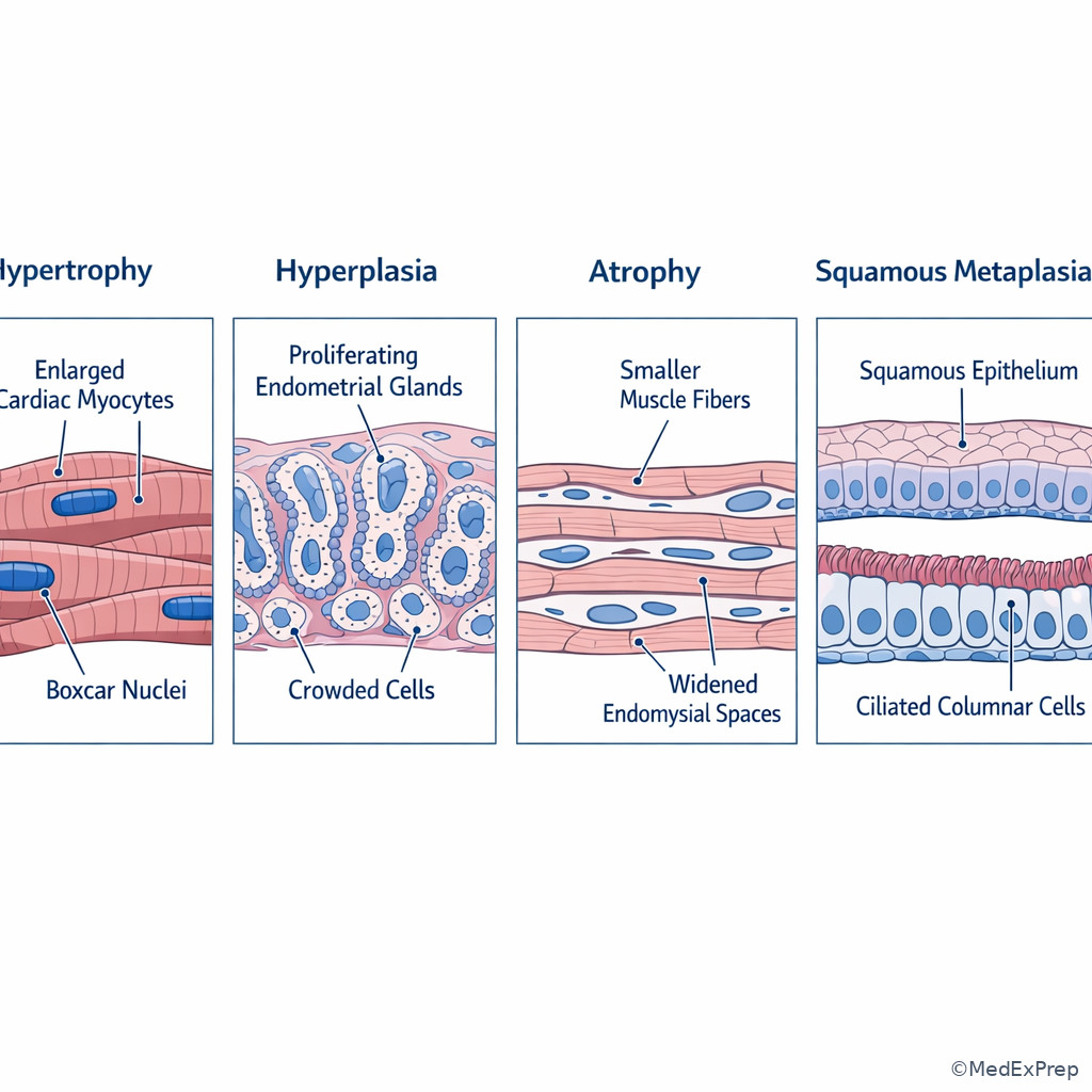
D --> J[Next lesson: necrosis, apoptosis, autophagy] Exam takeaway: Visual memory improves board performance when you can pair each adaptation with its signature microscopic pattern.
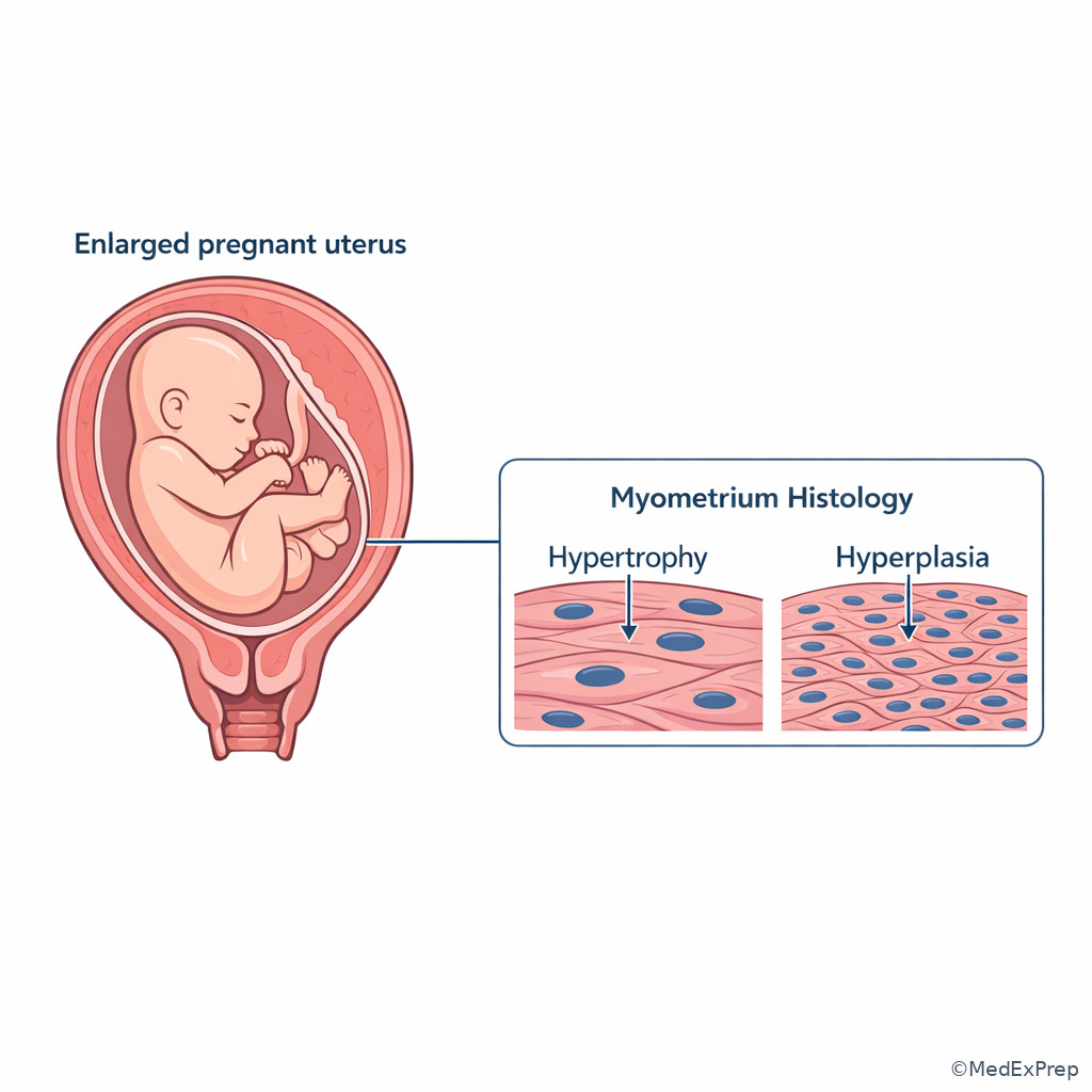
Exam takeaway: The gravid uterus is the classic physiologic example where hypertrophy and hyperplasia occur together.
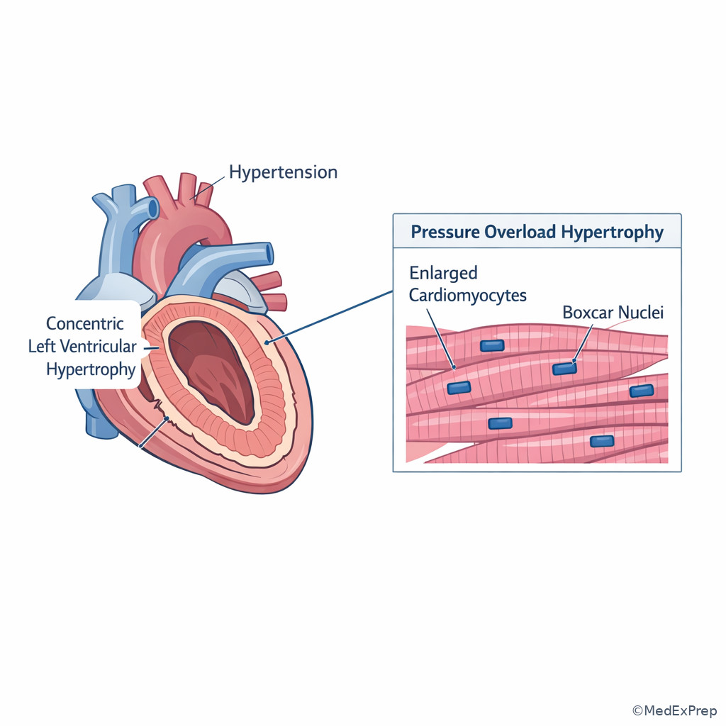
Exam takeaway: Myocardial adaptation to pressure overload is a size increase in existing myocytes, not true hyperplasia.
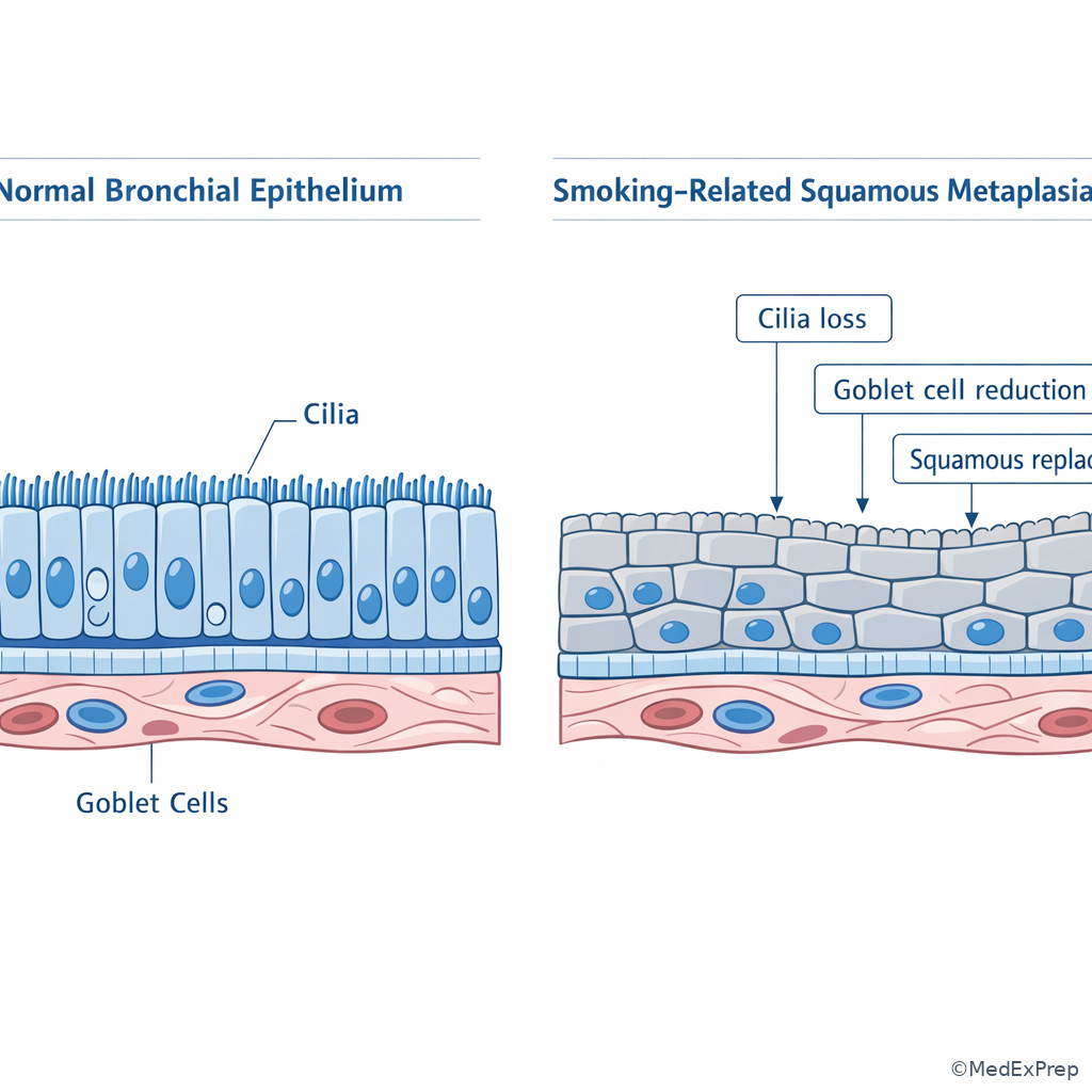
Exam takeaway: Loss of cilia and replacement by squamous epithelium signals metaplasia, a reversible adaptation that can precede dysplasia if the insult persists.
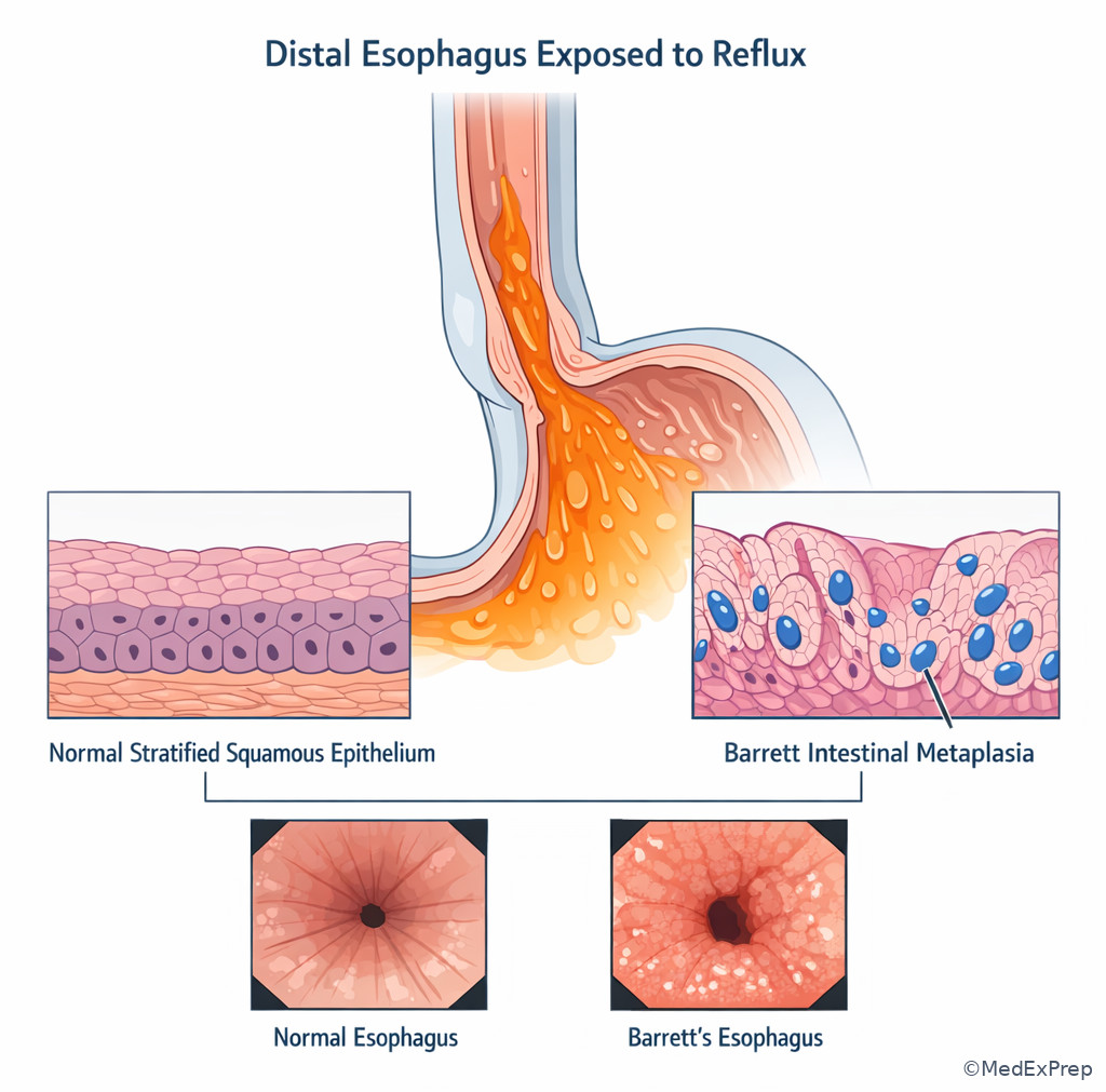
Exam takeaway: Columnar intestinal-type epithelium with goblet cells in distal esophagus is metaplasia, not simply hyperplasia.
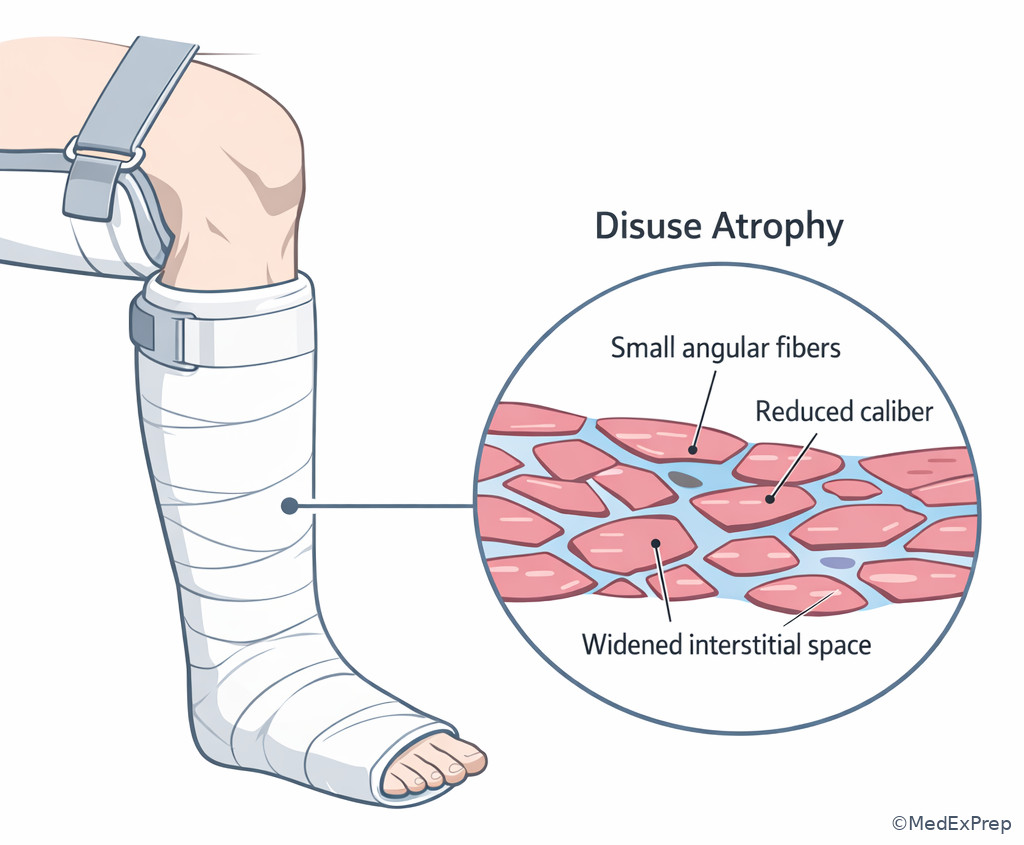
Exam takeaway: Reduced workload produces smaller fibers and decreased mass without the coagulative or inflammatory features of necrosis.
Core content
1.1.1.3 Foundational classification: which adaptation pattern fits the morphology?
A Explanation
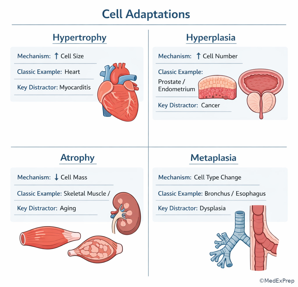
The first board-style decision conflict is simple but heavily tested: are you looking at bigger cells, more cells, smaller cells, or a changed cell type? That single rule resolves most stems. Hypertrophy means increased cell size. Hyperplasia means increased cell number. Atrophy means decreased cell size, often with reduced function and sometimes reduced cell number. Metaplasia means one mature differentiated cell type is replaced by another mature differentiated cell type that better tolerates the chronic stressor. See Fig 1 for the high-yield map.
WHY THIS IS TESTED: Many questions add distracting clinical context, but the answer still comes from identifying the dominant morphologic change. If the tissue is composed of nondividing permanent cells, reject hyperplasia first. If the epithelium changes lineage, reject hypertrophy and hyperplasia even if the tissue looks thicker. If the organ shrinks because individual cells are smaller and metabolically quieter, favor atrophy rather than cell death.
Choose hypertrophy when workload or hormonal stimulation enlarges existing cells, especially in cardiac or skeletal muscle. Choose hyperplasia when a cell population retains proliferative capacity and expands by mitosis, such as endometrium, breast epithelium, prostate stroma and glands, or regenerating liver. Choose atrophy when nutrient supply, use, innervation, or endocrine support falls. Choose metaplasia when chronic irritation or altered cytokine signaling reprograms stem cells or undifferentiated mesenchymal cells toward a new differentiation pathway.
Key rule: The best single discriminator is not the gross size of the organ; it is the microscopic basis of the size change.
Recall: This is the first topic, so build your baseline framework here. In the upcoming lesson on Cell Death Mechanisms, you will use this same framework to decide when adaptive change gives way to irreversible injury.
B Worked example
A 62-year-old man with long-standing hypertension, stage 3 chronic kidney disease, and obesity dies of unrelated trauma. At autopsy, the heart is enlarged with a thickened left ventricular wall. Microscopy shows enlarged myocytes with prominent rectangular nuclei. Serum troponin from admission is mildly elevated after chest wall injury, which is a distracting clue. The task is to identify the adaptation pattern. Key facts: pressure overload, permanent cells, enlarged individual myocytes. Apply the rule: myocardium adapts by hypertrophy, not meaningful hyperplasia. Eliminate hyperplasia because cardiomyocytes do not respond to chronic pressure primarily by increasing cell number. Eliminate atrophy because the cells are larger, not smaller. Eliminate metaplasia because cell lineage has not changed. Verify: the classic morphology is myocyte hypertrophy with boxcar nuclei. See Fig 3 for the visual pattern and the myocardium image panel.
C Exam trap
Common wrong answer: Hyperplasia.
Why it is tempting: The organ is larger, and many learners equate enlargement with increased cell number.
Single clue that eliminates it: Cardiac myocytes are permanent cells that respond to increased workload mainly by enlargement, not proliferative expansion.
D Checkpoint
Question: A 48-year-old woman with poorly controlled hypertension and obstructive sleep apnea undergoes autopsy after a motor vehicle collision. Cardiac histology shows enlarged myocytes with enlarged hyperchromatic rectangular nuclei and no increase in cellular crowding. Which adaptation pattern best explains this finding?
- Hypertrophy - tempting because the heart is enlarged; correct because the stem specifies increased cell size in nondividing myocytes.
- Hyperplasia - tempting because organ enlargement can result from proliferation; wrong because the key clue is enlargement of existing myocytes without increased cellular number.
- Atrophy - tempting if one overfocuses on chronic disease; wrong because the cells are enlarged rather than shrunken.
- Metaplasia - tempting if one thinks any chronic stress changes cell phenotype; wrong because no replacement by a different mature cell type is described.
Answer: A. Hypertrophy
If you missed this: Review 1.1.1.1 Foundational classification: which adaptation pattern fits the morphology? - focus on the discriminator of cell size versus cell number.
Clinician Action: Diagnose by matching morphology to the adaptive category. Order no ancillary study if classic morphology and context are present. Treat is outside the pathologist’s primary role; instead, report the adaptation and correlate with the chronic stressor. Refer/Escalate if findings suggest superimposed injury that requires clinicopathologic correlation.
1.1.1.4 Hypertrophy and hyperplasia: when enlarged tissue means size, number, or both
A Explanation
The second decision conflict is between hypertrophy and hyperplasia because both can enlarge a tissue. The resolving rule is whether the tissue can divide and whether the stimulus favors proliferation, increased synthetic capacity, or both. Hypertrophy reflects increased synthesis of structural proteins and organelles, often driven by mechanical stress and growth factor signaling. Hyperplasia reflects increased proliferative signaling in cells capable of division. Some organs show both. The gravid uterus is the board-classic example because estrogen stimulates smooth muscle cell enlargement and proliferation.
WHY THIS IS TESTED: Exam writers often pair a proliferative tissue with a nonproliferative tissue or present a hormonal context that can support mixed adaptation. If a stem gives pressure overload in the heart, choose hypertrophy. If it gives endometrial proliferation, benign prostatic enlargement, or liver regeneration, choose hyperplasia. If it gives pregnancy-related uterine enlargement, choose both.
Physiologic hyperplasia includes hormonal hyperplasia, such as glandular epithelium in the female breast at puberty or pregnancy, and compensatory hyperplasia, such as liver regeneration after partial hepatectomy. Pathologic hyperplasia often reflects excessive hormone or growth factor stimulation, as in endometrial hyperplasia from unopposed estrogen. Pathologic hyperplasia is still a controlled process and differs from neoplasia because the stimulus dependence persists, but it may create a fertile environment for malignant transformation in some settings. See Fig 2 and Fig 1.
Memory anchor: \(HYPErTROPHY = TALLER cells\); \(HYPErPLASIA = PLENTIER cells\). Think T in hypertrophy for thicker individual cells, and P in hyperplasia for proliferation.
High-Yield Connection: This concept is tested alongside neoplasia. Know both stimulus-dependent proliferative expansion in hyperplasia and autonomous growth in neoplasia for exam day; they are frequently paired in stems.
B Worked example
A 34-year-old pregnant woman with type 2 diabetes and chronic iron deficiency anemia undergoes hysterectomy for placenta accreta after delivery. Grossly, the uterus is enlarged. Histology shows thickened myometrium with enlarged smooth muscle cells and increased smooth muscle bundles. A distracting clue is postpartum hemorrhage, which does not determine the adaptation category. Identify task: classify the cellular response. Extract key facts: pregnancy, hormone-responsive smooth muscle, increased cell size plus increased cell number. Apply rule: pregnancy induces both hypertrophy and hyperplasia of the uterine smooth muscle. Eliminate isolated hypertrophy because proliferation is also present. Eliminate isolated hyperplasia because enlarged myocytes are specifically described. Eliminate metaplasia and atrophy because lineage and size direction do not fit.
C Exam trap
Common wrong answer: Hypertrophy alone in the gravid uterus.
Why it is tempting: Smooth muscle enlargement is memorable and true.
Single clue that eliminates it: Pregnancy also increases cell number, so the best answer is combined hypertrophy and hyperplasia when both are offered.
D Checkpoint
Question: A 57-year-old man with obesity, diabetes, and lower urinary tract symptoms has a transurethral resection specimen showing nodular enlargement with increased numbers of benign glands and fibromuscular stroma. Which adaptation pattern is most appropriate?
- Hypertrophy - tempting because the prostate is enlarged; wrong because the microscopic clue is increased numbers of glands and stromal cells.
- Hyperplasia - tempting because benign proliferative nodules are classic here; correct because the lesion reflects increased cell number in glandular and stromal compartments.
- Atrophy - tempting if one thinks aging always causes shrinkage; wrong because the tissue is expanded rather than reduced.
- Metaplasia - tempting if one assumes chronic irritation changes epithelial type; wrong because no lineage replacement is described.
Answer: B. Hyperplasia
If you missed this: Review 1.1.1.2 Hypertrophy and hyperplasia: when enlarged tissue means size, number, or both - focus on the discriminator of dividing tissue plus increased cell number.
Clinician Action: Diagnose enlargement by identifying whether the microscopic basis is size increase, proliferative increase, or both. Order clinicopathologic correlation if hormonal or regenerative context matters. Treat remains outside pathology scope; the pathologist clarifies the process and whether atypia is absent or present. Refer/Escalate if hyperplasia shows atypia or if sampling is limited and cannot exclude adjacent higher-grade disease.
- The classic organ that adapts to pressure overload by hypertrophy rather than hyperplasia is the heart.
- True or False: Pregnancy-related uterine enlargement is purely hypertrophy. False.
- Name the 2 major physiologic forms of hyperplasia: hormonal and compensatory.
1.1.1.5 Atrophy: when less tissue does not mean cell death
A Explanation
Atrophy is the adaptive decrease in cell size and functional capacity that allows survival under adverse conditions. The board conflict here is atrophy versus necrosis. The single rule: atrophy preserves viability, whereas necrosis reflects irreversible injury with membrane damage and inflammatory reaction. Atrophy occurs with decreased workload, loss of innervation, diminished blood supply, inadequate nutrition, loss of endocrine stimulation, pressure, or aging. Mechanistically, it reflects reduced protein synthesis and increased protein degradation, largely through the ubiquitin-proteasome pathway, often with increased autophagy.
WHY THIS IS TESTED: Exam stems often describe a smaller organ or reduced cell volume and try to lure you into choosing necrosis because the tissue is “wasting away.” But if the cells are small, structurally intact, and the setting is chronic reduction in use or trophic support, choose atrophy. If the stem instead describes eosinophilic anucleate cells, membrane disruption, calcification, or acute inflammation, that belongs to the upcoming cell death lesson.
Common patterns include skeletal muscle disuse atrophy after immobilization, denervation atrophy after nerve injury, cerebral atrophy with aging, endometrial or breast atrophy after menopause, and renal atrophy from chronic ischemia. Pressure atrophy can occur when a slowly expanding mass compresses surrounding tissue. In pathology practice, the key is to report atrophy as a pattern and not overinterpret it as proof of a specific etiology unless the context supports one. See Fig 3 for a visual comparison.
Key warning: Atrophy may coexist with fibrosis or chronic inflammation, but those associated findings do not convert the process into necrosis.
B Worked example
A 71-year-old woman with severe osteoarthritis, long-term corticosteroid exposure, and type 2 diabetes has had her right leg immobilized in a brace for 4 months after fracture. Muscle biopsy for persistent weakness shows small skeletal muscle fibers with reduced cytoplasmic volume and no significant necrosis or acute inflammation. A mildly elevated creatine kinase level is included as noise. Identify task: determine the adaptation. Extract key facts: prolonged reduced workload, smaller viable fibers, no necrotic pattern. Apply rule: disuse atrophy. Eliminate necrosis because there is no myofiber breakdown or inflammatory cleanup. Eliminate hypertrophy because the cells are not enlarged. Eliminate hyperplasia because skeletal muscle does not respond this way. Verify with mechanism: decreased workload reduces protein synthesis and increases proteolysis.
C Exam trap
Common wrong answer: Necrosis.
Why it is tempting: Weakness plus a chronic illness background can make tissue loss sound destructive rather than adaptive.
Single clue that eliminates it: The biopsy shows smaller intact fibers without inflammatory destruction.
D Checkpoint
Question: A 78-year-old man with peripheral vascular disease, hypertension, and chronic weight loss has a kidney that is reduced in size with cortical thinning and small tubules in a chronically ischemic distribution. Which adaptation pattern is most appropriate?
- Hypertrophy - tempting because other organs enlarge under stress; wrong because this organ is reduced in size with shrunken structures.
- Hyperplasia - tempting if one thinks chronic stress stimulates growth; wrong because no increase in cell number is described.
- Atrophy - tempting because chronic ischemia commonly causes reduced cell size and diminished tissue mass; correct because the morphology and mechanism fit.
- Metaplasia - tempting if chronic irritation is overgeneralized; wrong because there is no replacement by a different mature epithelial type.
Answer: C. Atrophy
If you missed this: Review 1.1.1.3 Atrophy: when less tissue does not mean cell death - focus on the discriminator of smaller viable cells without irreversible injury.
Clinician Action: Diagnose atrophy by documenting reduced cell size and context such as disuse or ischemia. Order deeper evaluation only if another process is suspected, such as superimposed inflammation or necrosis. Treat is outside pathology scope; instead, describe the pattern and likely driver when supported. Refer/Escalate when atrophy suggests an upstream pathologic cause that requires urgent communication, such as severe ischemia or denervation.
1.1.1.6 Metaplasia: adaptive lineage switch versus dysplasia and cancer risk
A Explanation
Metaplasia is a reversible substitution of one mature differentiated cell type by another mature differentiated cell type better suited to withstand chronic stress. The central exam conflict is metaplasia versus dysplasia. The single rule: metaplasia is an orderly adaptive lineage change, whereas dysplasia is disordered growth with atypia and architectural disturbance. In epithelia, metaplasia usually reflects stem cell reprogramming rather than direct conversion of one mature cell into another.
WHY THIS IS TESTED: Board stems love chronic irritants: tobacco smoke in bronchus, gastroesophageal reflux in distal esophagus, stones in ducts, or vitamin A deficiency. If the response is a mature replacement phenotype without overt atypia, choose metaplasia. If the question adds nuclear pleomorphism, loss of maturation, abnormal mitoses, or marked architectural disarray, move beyond metaplasia and consider dysplasia or carcinoma in situ.
Classic examples are squamous metaplasia of respiratory epithelium in smokers and intestinal metaplasia in Barrett esophagus. Metaplasia may protect against one stress while sacrificing specialized function, such as mucociliary clearance in the bronchus. Persistent stress can progress to dysplasia and carcinoma, so the pattern matters prognostically. See Fig 2 and the image panels for bronchus and Barrett esophagus. The pathologist should report the metaplastic process accurately and indicate when dysplasia is absent, present, or indeterminate.
Memory anchor: \(Meta = move to another mature type\). If the cells move lineage but remain mature and orderly, think metaplasia; if they become chaotic and atypical, think dysplasia.
High-Yield Connection: This concept is tested alongside the upcoming topics Cell Death Mechanisms and Acute Chronic Inflammation. Chronic irritation drives metaplasia, while persistent injury and inflammation set the stage for dysplasia and later neoplasia.
Exam takeaway: Mature orderly replacement favors metaplasia, while atypia and loss of maturation push the diagnosis toward dysplasia.
flowchart TD
A[Chronic epithelial stress] --> B{What does microscopy show?}
B --> C[Orderly mature replacement cell type
Metaplasia]
B --> D[Nuclear atypia, crowding, loss of polarity
Dysplasia]
C --> E[Potentially reversible if insult removed]
D --> F[Premalignant pathway; grade and sample carefully]
E --> G[Examples: smoker bronchus, Barrett]
F --> H[Do not label as simple adaptation] B Worked example
A 58-year-old man with central obesity, chronic gastroesophageal reflux, and a hiatal hernia undergoes distal esophageal biopsy. He also has iron deficiency anemia from hemorrhoids, which is a distracting clue. Histology shows nonciliated columnar epithelium with goblet cells replacing native stratified squamous epithelium. No high-grade atypia is seen. Identify task: classify the epithelial change. Extract key facts: reflux exposure, esophagus, intestinal-type goblet cells, orderly mature replacement, no dysplasia. Apply rule: intestinal metaplasia in Barrett esophagus. Eliminate hyperplasia because the key event is lineage replacement, not simply more cells. Eliminate dysplasia because the stem denies significant atypia. Eliminate carcinoma because invasion is not described. Verify: this is metaplasia with premalignant significance, not malignancy itself. See Fig 4.
C Exam trap
Common wrong answer: Dysplasia or carcinoma in situ.
Why it is tempting: Barrett esophagus is strongly associated with adenocarcinoma risk, so learners may jump ahead to the complication.
Single clue that eliminates it: The biopsy shows orderly goblet cell-containing intestinal-type epithelium without significant cytologic atypia.
D Checkpoint
Question: A 63-year-old man with a 40-pack-year smoking history, COPD, and hyperlipidemia has bronchial mucosa showing replacement of pseudostratified ciliated columnar epithelium by stratified squamous epithelium without marked atypia. Which adaptation best fits?
- Hypertrophy - tempting because chronic stress can enlarge tissue; wrong because the defining feature is a different epithelial type.
- Hyperplasia - tempting because smokers can have epithelial thickening; wrong because the stem emphasizes replacement rather than proliferation of the same lineage.
- Atrophy - tempting if one focuses on loss of cilia; wrong because this is a lineage switch, not simple shrinkage.
- Metaplasia - tempting because chronic irritation can reprogram epithelial differentiation; correct because squamous replacement of bronchial epithelium is classic metaplasia.
Answer: D. Metaplasia
If you missed this: Review 1.1.1.4 Metaplasia: adaptive lineage switch versus dysplasia and cancer risk - focus on the discriminator of mature cell-type replacement without cytologic chaos.
Clinician Action: Diagnose metaplasia when a mature replacement phenotype matches a chronic stressor. Order additional levels, sampling, or correlation only if dysplasia cannot be excluded. Treat is not the pathologist’s primary role; instead, report the pattern and whether dysplasia is identified. Refer/Escalate if high-grade dysplasia or invasive carcinoma is present, or if a safety-significant discrepancy exists between endoscopic impression and biopsy.
- The most common epithelial metaplasia tested in smoker bronchus is squamous metaplasia.
- True or False: Metaplasia is equivalent to dysplasia. False.
- Name the 3 key features that favor metaplasia over dysplasia: mature cells, orderly architecture, limited atypia.
1.1.1.7 Integration: physiologic versus pathologic adaptation and the pathology reporting mindset
A Explanation
The final integration step is deciding not only which adaptation is present but also whether it is physiologic, pathologic, isolated, or part of a broader process that needs further workup. This is where pathology board questions move from classification into interpretation. The conflict is often: should you stop at naming the adaptation, or should you recommend additional sampling, correlation, or caution because another process could coexist? The resolving rule is to report adaptation confidently when morphology and context are classic, but avoid overcalling when atypia, tissue injury, or sampling limitations raise uncertainty.
WHY THIS IS TESTED: The ABPath perspective is not just memorizing terms. You must decide whether the biopsy supports a final interpretation, whether the sample is adequate to exclude a more serious lesion, and whether a pattern represents expected physiology. Pregnancy-related uterine enlargement is physiologic. Endometrial hyperplasia due to unopposed estrogen is pathologic and may require classification and correlation. Bronchial squamous metaplasia in a smoker is adaptive but clinically important because it signals chronic injury. Age-related atrophy can be physiologic, while ischemic atrophy may reflect significant underlying vascular disease.
In a pathology report, avoid unsupported causation if morphology is nonspecific. “Atrophic change” may be appropriate when the trigger is not directly proven. For limited biopsies, note when metaplastic or hyperplastic change is present but adjacent dysplasia cannot be fully excluded if sampling is scant or orientation is poor. See Fig 1, Fig 2, and Fig 4 as the decision framework.
Key rule: Pathologists score points by naming the correct process and by stating the limitation when the specimen does not justify stronger language.
B Worked example
A 52-year-old woman with obesity, chronic anovulatory cycles, and polycystic ovarian syndrome has an endometrial biopsy for abnormal uterine bleeding. The specimen is scant and fragmented. Microscopy shows proliferative crowding suggestive of hyperplasia, but architecture is limited and atypia cannot be assessed confidently. A mildly elevated TSH is included as a distracting lab. Identify task: determine the most appropriate pathology-directed next step and interpretation mindset. Extract key facts: risk factors for pathologic hyperplasia, limited sample, uncertainty regarding atypia. Apply rule: do not overcall a definitive high-grade process on inadequate material. The best interpretation is suspicious or limited for hyperplastic change with recommendation for adequate sampling or correlation, depending on institutional practice. Eliminate a definitive carcinoma diagnosis because invasion cannot be assessed. Eliminate a casual “normal endometrium” sign-out because the sample is abnormal and clinically discordant. Verify: this is where pathologist judgment and specimen adequacy matter.
C Exam trap
Common wrong answer: Definitive dysplasia or carcinoma based on limited fragments.
Why it is tempting: The patient has strong risk factors, and the biopsy shows proliferative abnormality.
Single clue that eliminates it: The stem explicitly states that atypia and architecture cannot be assessed confidently because the sample is scant and fragmented.
D Checkpoint
Question: A small bronchial biopsy from a 67-year-old smoker with COPD and coronary disease shows squamous metaplasia with crush artifact and focal nuclear enlargement. No invasion is seen, but the tissue is scant. What is the most appropriate pathology interpretation approach?
- Diagnose invasive squamous cell carcinoma - tempting because smoking plus nuclear enlargement raises concern; wrong because scant artifact-limited tissue without invasion cannot support that diagnosis.
- Report squamous metaplasia and note limitation in excluding higher-grade atypia if clinically indicated - tempting because it is cautious yet specific; correct because the morphology supports metaplasia, but the specimen is limited.
- Call the biopsy negative and dismiss the atypia as irrelevant - tempting because artifact can mimic atypia; wrong because the sample has a real metaplastic change and limitation that should be communicated.
- Diagnose hyperplasia - tempting because the epithelium may appear thickened; wrong because the dominant process is lineage switch rather than simple increase in number.
Answer: B. Report squamous metaplasia and note limitation in excluding higher-grade atypia if clinically indicated
If you missed this: Review 1.1.1.5 Integration: physiologic versus pathologic adaptation and the pathology reporting mindset - focus on the discriminator of classic pattern plus specimen limitation language.
Clinician Action: Diagnose the best-supported adaptive pattern without overstating certainty. Order deeper levels, repeat sampling, or clinicopathologic correlation when atypia or adequacy is uncertain. Treat is outside pathology scope; communicate diagnosis and limitations rather than bedside management. Refer/Escalate urgently for discordant findings, suspected malignancy on limited tissue, or safety-significant specimen issues.
Exam Traps & Differentiators
Common traps & distractors:
- Most common wrong answer and why: Choosing hyperplasia for any enlarged organ. This is tempting because gross enlargement is memorable. The fix is to ask whether the individual cells are bigger or more numerous.
- Looks similar but isn’t: Metaplasia versus dysplasia. Both may occur in chronically injured epithelium, but metaplasia keeps orderly maturation; dysplasia adds atypia and architectural disorder.
- Looks similar but isn’t: Atrophy versus necrosis. Both reduce tissue mass, but atrophy preserves cell viability while necrosis shows irreversible injury and inflammatory cleanup.
- Looks similar but isn’t: Hyperplasia versus neoplasia. Both increase cellularity, but hyperplasia remains regulated by stimulus; neoplasia becomes relatively autonomous.
- Related upcoming trap: In the next topic, cell death patterns can mimic loss of tissue mass. If the stem emphasizes membrane damage, eosinophilic anucleate cells, or inflammatory response, shift from adaptation to cell death mechanisms.
| Stem says | Think | Single discriminator |
|---|---|---|
| Pressure overload in heart with enlarged myocytes | Hypertrophy | Permanent cells enlarge rather than proliferate |
| Unopposed estrogen with proliferative endometrium | Hyperplasia | Hormone-driven increase in cell number |
| Immobilized muscle with smaller viable fibers | Atrophy | Reduced cell size without necrotic injury |
| Smoker bronchus losing cilia and becoming squamous | Metaplasia | Mature cell type replacement |
| Barrett biopsy with goblet cells in distal esophagus | Intestinal metaplasia | Lineage switch to intestinal-type mucosa |
| Architectural disorder and atypia in metaplastic epithelium | Dysplasia, not simple metaplasia | Loss of maturation and cytologic atypia |
| Looks similar | Why tempting | Why wrong | What to choose instead |
|---|---|---|---|
| Cardiac enlargement = hyperplasia | Organ is bigger | Myocytes are permanent cells | Hypertrophy |
| Bronchial squamous change = carcinoma | Smoking history raises cancer risk | No invasion or marked atypia described | Metaplasia |
| Small kidney in chronic ischemia = necrosis | Severe injury sounds destructive | Chronic shrinkage with viable small cells fits adaptation | Atrophy |
| Endometrial thickening = neoplasia | Increased glands can look alarming | Stimulus-dependent proliferation without autonomous behavior | Hyperplasia |
If the stem says prolonged hypertension with boxcar nuclei, think myocyte hypertrophy.
If the stem says pregnancy-related uterine enlargement, think combined hypertrophy and hyperplasia.
If the stem says reduced workload with smaller viable cells, think atrophy, not necrosis.
If the stem says chronic smoking or reflux with orderly mature replacement epithelium, think metaplasia.
If the stem says metaplastic epithelium plus loss of polarity and nuclear atypia, think dysplasia rather than simple adaptation.
If the stem says scant fragmented biopsy with concerning but incomplete features, think specimen limitation language rather than overcall.
Review connection: The traps above test classification versus overcall. If any felt unfamiliar, revisit 1.1.1.4 Metaplasia: adaptive lineage switch versus dysplasia and cancer risk and 1.1.1.5 Integration: physiologic versus pathologic adaptation and the pathology reporting mindset before attempting the Self-check quiz.
Tables
| Adaptation | Core mechanism | Microscopic clue | Classic example | Main trap |
|---|---|---|---|---|
| Hypertrophy | Increased protein synthesis and organelles | Larger individual cells | LV hypertrophy in hypertension | Confusing organ enlargement with hyperplasia |
| Hyperplasia | Stimulus-driven proliferation | Increased cell number | Endometrial hyperplasia; BPH | Confusing with neoplasia |
| Atrophy | Decreased synthesis, increased degradation | Smaller cells, reduced tissue mass | Disuse skeletal muscle atrophy | Confusing with necrosis |
| Metaplasia | Stem cell reprogramming | Mature cell type replacement | Smoker bronchus; Barrett esophagus | Confusing with dysplasia |
| Comparison point | Hypertrophy | Hyperplasia | Metaplasia |
|---|---|---|---|
| Primary tissue change | Cell size up | Cell number up | Cell type changes |
| Requires proliferative capacity | No | Yes | Usually stem-cell mediated |
| Classic tissue | Heart | Endometrium, prostate, liver | Bronchus, distal esophagus |
| Typical driver | Mechanical load | Hormones/growth factors | Chronic irritation |
| Closest exam distractor | Hyperplasia | Neoplasia | Dysplasia |
| Differential/classification problem | Favor adaptation when... | Favor alternative when... |
|---|---|---|
| Atrophy vs necrosis | Cells are viable, smaller, chronic low-demand setting | Irreversible injury, membrane breakdown, inflammation |
| Metaplasia vs dysplasia | Mature orderly replacement, limited atypia | Loss of polarity, nuclear atypia, abnormal maturation |
| Hyperplasia vs neoplasia | Stimulus-dependent and regulated | Autonomous clonal growth with persistent atypia |
| Hypertrophy vs hyperplasia | Nondividing cells enlarge | Mitotically active cells proliferate |
| Physiologic vs pathologic hyperplasia | Expected hormonal or compensatory context | Excessive or abnormal hormonal stimulation |
Exam takeaway: In an enlarged organ, decide whether the tissue can divide before picking hypertrophy, hyperplasia, or a mixed response.
flowchart TD
A[Enlarged organ or tissue] --> B{Can the dominant cells divide?}
B -->|No or minimal| C[Hypertrophy favored]
B -->|Yes| D{Microscopy shows what?}
D --> E[More cells -> Hyperplasia]
D --> F[Bigger cells -> Hypertrophy]
D --> G[Both -> Mixed response]
C --> H[Example: myocardium]
E --> I[Examples: endometrium, prostate, liver]
G --> J[Example: gravid uterus] Exam takeaway: Pair each pattern with one iconic organ and one common distractor to speed elimination under exam pressure.
Algorithm / Approach
Rapid Review
- Hypertrophy -> enlarged individual cells in nondividing tissue
- Hyperplasia -> increased cell number in tissue with proliferative capacity
- Atrophy -> reduced cell size with preserved viability
- Metaplasia -> mature cell type replaced by another mature type
- Heart under pressure load -> hypertrophy, not hyperplasia
- Gravid uterus -> combined hypertrophy plus hyperplasia
- BPH -> nodular hyperplasia, not neoplasia by default
- Immobilized skeletal muscle -> disuse atrophy, not necrosis
- Smoker bronchus -> squamous metaplasia with lost mucociliary specialization
- Barrett esophagus -> intestinal metaplasia with goblet cells
- Metaplasia vs dysplasia -> orderly maturation versus cytologic atypia
- Hyperplasia vs neoplasia -> stimulus dependence versus autonomy
- Atrophy vs cell death -> shrinkage versus irreversible destruction
- Specimen limitation -> use cautious language when atypia cannot be excluded
Memory anchor: Size, Sum, Shrink, Switch = hypertrophy, hyperplasia, atrophy, metaplasia. If you can name the dominant change in one word, you can usually solve the question.
Self-check quiz
1. A 65-year-old man with hypertension, chronic kidney disease, and left ventricular wall thickening at autopsy has enlarged cardiomyocytes with prominent boxcar nuclei. Which adaptation is most likely?
- Hypertrophy
- Hyperplasia
- Atrophy
- Metaplasia
2. A 60-year-old man with COPD, hyperlipidemia, and a 45-pack-year smoking history has bronchial biopsy showing replacement of ciliated columnar epithelium by squamous epithelium without marked atypia. Which adaptation best fits?
- Hypertrophy
- Hyperplasia
- Atrophy
- Metaplasia
3. A 33-year-old pregnant woman with gestational diabetes and chronic hypertension undergoes hysterectomy for placenta accreta. The myometrium is enlarged, and microscopy shows both enlarged smooth muscle cells and increased smooth muscle bundles. Which interpretation is most appropriate?
- Pure hypertrophy
- Pure hyperplasia
- Combined hypertrophy and hyperplasia
- Metaplasia
4. A 72-year-old woman with prolonged immobilization after hip fracture, diabetes, and chronic prednisone exposure has skeletal muscle biopsy for weakness. Fibers are small and viable without significant inflammation or myofiber breakdown. Which diagnosis is most appropriate?
- Disuse atrophy
- Coagulative necrosis
- Hypertrophy
- Squamous metaplasia
5. A 56-year-old man with obesity, long-standing reflux, and hiatal hernia has distal esophageal biopsy showing columnar epithelium with goblet cells replacing squamous mucosa. No high-grade atypia is seen. Which pathology interpretation is most appropriate?
- Hyperplasia of squamous mucosa
- Intestinal metaplasia
- Squamous dysplasia
- Invasive adenocarcinoma
6. A 51-year-old woman with obesity, polycystic ovarian syndrome, and abnormal uterine bleeding has a scant fragmented endometrial biopsy showing proliferative gland crowding. Architecture is limited, and atypia cannot be assessed reliably. Which pathology-directed next step is most appropriate?
- Diagnose endometrioid adenocarcinoma
- Call the sample benign proliferative endometrium and sign out without comment
- Report hyperplastic/proliferative change with limitation and recommend adequate sampling or correlation
- Interpret the lesion as metaplasia
7. A 68-year-old man with smoking history, COPD, and coronary disease has a scant crushed bronchial biopsy showing squamous metaplasia with focal enlarged nuclei but no invasion. Bronchoscopy notes an irregular plaque. Which interpretation is most appropriate?
- Definitive invasive squamous cell carcinoma
- Squamous metaplasia with specimen limitation in excluding higher-grade atypia
- Simple hyperplasia of bronchial epithelium
- Atrophic bronchial mucosa
8. A 77-year-old man with renal artery stenosis, hypertension, and weight loss has a small kidney with cortical thinning. Microscopy shows small tubules and reduced parenchymal volume without acute inflammation or widespread ghost cell outlines. Which interpretation best fits the dominant process?
- Chronic ischemic atrophy
- Liquefactive necrosis
- Compensatory hyperplasia
- Metaplastic transformation
Answer key
1. Correct answer: A. Hypertrophy
- A. Hypertrophy: Tempting because the heart is enlarged; correct because pressure overload in myocardium causes increased myocyte size with boxcar nuclei. The single stem clue is enlarged cardiomyocytes in a permanent cell population. Source: Robbins and Cotran; Pathoma.
- B. Hyperplasia: Tempting because organ enlargement can reflect proliferation; incorrect because cardiomyocytes do not respond to chronic pressure mainly by increasing cell number. The discriminating clue is myocyte enlargement rather than increased cellularity. Source: Robbins and Cotran.
- C. Atrophy: Tempting only if one overfocuses on chronic disease; incorrect because the cells are larger, not smaller. The discriminating clue is wall thickening with enlarged cells. Source: Robbins and Cotran.
- D. Metaplasia: Tempting if any chronic stress is mistaken for lineage change; incorrect because no new mature cell type is present. The discriminating clue is unchanged cell lineage with increased size. Source: Robbins and Cotran.
If you missed this: Review 1.1.1.1 Foundational classification: which adaptation pattern fits the morphology? - focus on nondividing cells enlarge rather than proliferate.
2. Correct answer: D. Metaplasia
- A. Hypertrophy: Tempting because chronic stress can thicken tissue; incorrect because the stem describes replacement by a different epithelial type. The discriminating clue is squamous replacing ciliated columnar epithelium. Source: Robbins and Cotran; Pathoma.
- B. Hyperplasia: Tempting because smoking-related mucosa may appear crowded; incorrect because the defining feature is a lineage switch, not just more cells of the same type. The discriminating clue is different mature cell phenotype. Source: Robbins and Cotran.
- C. Atrophy: Tempting if one focuses on loss of cilia; incorrect because loss of cilia alone is not the core diagnosis here. The discriminating clue is new squamous epithelium. Source: Robbins and Cotran.
- D. Metaplasia: Tempting and correct because chronic irritation in smokers classically reprograms bronchial epithelium toward squamous differentiation. The discriminating clue is orderly squamous replacement without marked atypia. Source: Robbins and Cotran.
If you missed this: Review 1.1.1.4 Metaplasia: adaptive lineage switch versus dysplasia and cancer risk - focus on mature replacement epithelium versus atypical dysplasia.
3. Correct answer: C. Combined hypertrophy and hyperplasia
- A. Pure hypertrophy: Tempting because uterine smooth muscle cells enlarge in pregnancy; incorrect for this stem because increased smooth muscle bundles indicate increased cell number as well. The discriminating clue is both enlarged cells and increased bundles. Source: Robbins and Cotran.
- B. Pure hyperplasia: Tempting because pregnancy is a hormonally proliferative state; incorrect because the stem also documents increased cell size. The discriminating clue is enlarged smooth muscle cells. Source: Robbins and Cotran.
- C. Combined hypertrophy and hyperplasia: Correct because pregnancy stimulates both enlargement and proliferation of uterine smooth muscle. The single clue is simultaneous increase in cell size and number. Source: Robbins and Cotran; Pathoma.
- D. Metaplasia: Tempting only if any hormone-driven change is overgeneralized; incorrect because no lineage switch is present. The discriminating clue is same tissue type with growth, not replacement. Source: Robbins and Cotran.
If you missed this: Review 1.1.1.2 Hypertrophy and hyperplasia: when enlarged tissue means size, number, or both - focus on the gravid uterus as the mixed-response prototype.
4. Correct answer: A. Disuse atrophy
- A. Disuse atrophy: Correct because reduced workload produces smaller viable muscle fibers without the morphologic hallmarks of cell death. The discriminating clue is small viable fibers without inflammation or breakdown. Source: Robbins and Cotran.
- B. Coagulative necrosis: Tempting because prolonged immobilization can sound harmful; incorrect because necrosis requires irreversible injury with structural cell death, not just shrinkage. The discriminating clue is absence of myofiber destruction. Source: Robbins and Cotran.
- C. Hypertrophy: Tempting if one assumes muscle adapts to stress by growth; incorrect because immobilization reduces demand and cell size. The discriminating clue is small rather than enlarged fibers. Source: Robbins and Cotran.
- D. Squamous metaplasia: Tempting only if adaptation categories are memorized without tissue context; incorrect because skeletal muscle does not undergo epithelial metaplasia in this setting. The discriminating clue is muscle biopsy, not epithelial biopsy. Source: Robbins and Cotran.
If you missed this: Review 1.1.1.3 Atrophy: when less tissue does not mean cell death - focus on viable shrinkage versus irreversible injury.
5. Correct answer: B. Intestinal metaplasia
- A. Hyperplasia of squamous mucosa: Tempting because reflux can produce reactive thickening; incorrect because the squamous mucosa is replaced by columnar mucosa with goblet cells. The discriminating clue is goblet cells. Source: Robbins and Cotran; major GI pathology references.
- B. Intestinal metaplasia: Correct because Barrett esophagus is defined by replacement of esophageal squamous epithelium with intestinal-type columnar mucosa. The discriminating clue is columnar epithelium with goblet cells in distal esophagus. Source: Robbins and Cotran.
- C. Squamous dysplasia: Tempting because chronic reflux carries neoplastic risk; incorrect because the stem explicitly lacks high-grade atypia and shows a different mature lineage. The discriminating clue is orderly intestinal-type replacement. Source: Robbins and Cotran.
- D. Invasive adenocarcinoma: Tempting because Barrett can progress to adenocarcinoma; incorrect because invasion is not described. The discriminating clue is no invasive growth or marked atypia. Source: Robbins and Cotran.
If you missed this: Review 1.1.1.4 Metaplasia: adaptive lineage switch versus dysplasia and cancer risk - focus on goblet cells as the Barrett discriminator.
6. Correct answer: C. Report hyperplastic/proliferative change with limitation and recommend adequate sampling or correlation
- A. Diagnose endometrioid adenocarcinoma: Tempting because obesity and chronic anovulation increase risk; incorrect because the sample is scant and architecture/atypia cannot be assessed reliably. The discriminating clue is limited fragmented biopsy. Source: Robbins and Cotran; standard gynecologic pathology practice principles.
- B. Call the sample benign proliferative endometrium and sign out without comment: Tempting because fragmentation can obscure pattern; incorrect because gland crowding is abnormal and the specimen limitation must be acknowledged. The discriminating clue is proliferative gland crowding. Source: accepted pathology adequacy principles.
- C. Report hyperplastic/proliferative change with limitation and recommend adequate sampling or correlation: Correct because this is the safest pathology-directed response when atypia cannot be confidently evaluated. The discriminating clue is abnormal pattern plus inadequate assessment of atypia. Source: standard pathology reporting principles.
- D. Interpret the lesion as metaplasia: Tempting because endometrium can show metaplastic phenomena; incorrect because the key issue here is gland crowding and possible hyperplasia, not lineage replacement. The discriminating clue is proliferative architecture rather than new mature cell type. Source: gynecologic pathology principles.
If you missed this: Review 1.1.1.5 Integration: physiologic versus pathologic adaptation and the pathology reporting mindset - focus on using limitation language when atypia cannot be assessed.
7. Correct answer: B. Squamous metaplasia with specimen limitation in excluding higher-grade atypia
- A. Definitive invasive squamous cell carcinoma: Tempting because the patient smokes and bronchoscopy is abnormal; incorrect because the biopsy is scant, crushed, and lacks invasion. The discriminating clue is no invasion on limited tissue. Source: Robbins and Cotran; pathology adequacy principles.
- B. Squamous metaplasia with specimen limitation in excluding higher-grade atypia: Correct because the tissue supports metaplasia, but artifact and scant sampling prevent stronger exclusion of dysplasia. The discriminating clue is metaplastic morphology with crush artifact and limited tissue. Source: pathology reporting principles.
- C. Simple hyperplasia of bronchial epithelium: Tempting because there may be epithelial thickening; incorrect because the dominant feature is squamous replacement. The discriminating clue is lineage switch. Source: Robbins and Cotran.
- D. Atrophic bronchial mucosa: Tempting if one focuses on smoking-related injury; incorrect because this is not shrinkage but replacement by squamous cells. The discriminating clue is squamous phenotype. Source: Robbins and Cotran.
If you missed this: Review 1.1.1.4 Metaplasia: adaptive lineage switch versus dysplasia and cancer risk and 1.1.1.5 Integration: physiologic versus pathologic adaptation and the pathology reporting mindset - focus on metaplasia plus adequacy limitation, not overcall.
8. Correct answer: A. Chronic ischemic atrophy
- A. Chronic ischemic atrophy: Correct because decreased blood supply over time produces a small kidney with cortical thinning and reduced parenchymal volume. The discriminating clue is chronic small kidney with viable shrunken structures. Source: Robbins and Cotran.
- B. Liquefactive necrosis: Tempting because ischemia can cause cell death in some tissues; incorrect because the kidney here shows chronic shrinkage rather than acute enzymatic dissolution. The discriminating clue is chronic cortical thinning without acute necrotic pattern. Source: Robbins and Cotran.
- C. Compensatory hyperplasia: Tempting because kidneys can undergo compensatory enlargement when the contralateral kidney is lost; incorrect because this kidney is small, not enlarged. The discriminating clue is reduced organ size. Source: Robbins and Cotran.
- D. Metaplastic transformation: Tempting only if chronic injury is misread as lineage change; incorrect because no new epithelial type is described. The discriminating clue is shrunken parenchyma rather than replacement epithelium. Source: Robbins and Cotran.
If you missed this: Review 1.1.1.3 Atrophy: when less tissue does not mean cell death - focus on chronic ischemia causing shrinkage rather than necrotic dissolution.
Keep going with the full ABPath Pathology Prep Course (Pathology)
You just studied the way members do, in the same lesson interface. Full access unlocks every lesson, the question bank and flashcards for a full year.
One payment · 1 year of access · No auto-renewal