1.1.1 - Globe And Adnexal Anatomy
Why it matters
- Most ophthalmology board questions hide the diagnosis inside anatomy: the single best answer often depends on knowing where the lesion is, not just what it is called.
- Urgent triage depends on anatomic boundaries: septum, orbit, sclera, cornea, anterior chamber, vitreous, and retina separate routine disease from vision-threatening emergencies.
- Procedure safety is anatomy-driven: injections, laser, cataract surgery, lid surgery, and trauma questions all test neighboring structures at risk.
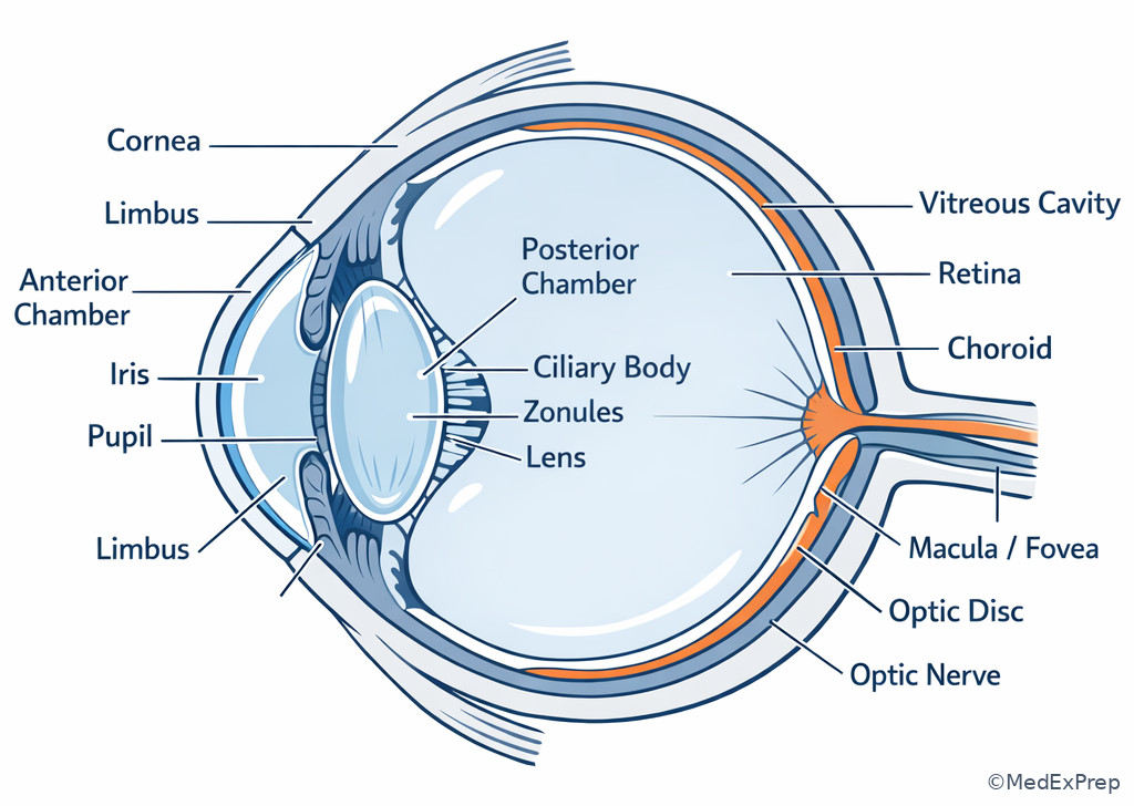
- Future topics build directly on this lesson: aqueous outflow requires ciliary body and angle anatomy; retinal physiology requires precise retinal layer and macular organization. See Fig 1.
Key Terms & Must-Know Facts
1.1.1.1 Key terms
- Cornea: transparent avascular anterior fibrous coat providing major refractive power.
- Sclera: opaque fibrous outer coat continuous with cornea.
- Limbus: corneoscleral junction; key surgical and stem landmark.
- Uvea: iris, ciliary body, and choroid.
- Anterior chamber: space between cornea and iris.
- Posterior chamber: narrow space between iris and lens/ciliary body.
- Vitreous cavity: posterior segment space behind lens.
- Pars plicata: anterior ciliary body with processes; aqueous production site.
- Pars plana: relatively avascular posterior ciliary body; important surgical entry zone.
- Ora serrata: anterior retinal termination.
- Macula: central retina specialized for high-acuity vision.
- Fovea: central macular depression with highest cone density.
- Tenon capsule: fascial sheath around globe.
- Orbital septum: fibrous barrier from orbital rim to tarsus; key in preseptal versus orbital disease.
- Tarsal plate: dense connective framework of eyelid.
1.1.1.2 Must-know facts
- The cornea has 5 classic layers on board exams: epithelium, Bowman layer, stroma, Descemet membrane, endothelium.
- Retina is neurosensory tissue; retinal pigment epithelium lies external to it.
- The optic nerve exits through the scleral canal at the posterior pole.
- Extraocular muscles insert anterior to the equator except superior oblique via trochlea and lateral/posterior functional paths.
- The orbital septum is the key exam boundary for eyelid infection triage.
- The lacrimal gland sits in the superotemporal orbit; the lacrimal sac sits inferomedially.
- The ciliary body both produces aqueous and anchors zonules.
- The choroid is highly vascular and lies between sclera and retina.
- The fovea lacks inner retinal layers, improving acuity.
- The optic disc has no photoreceptors and creates the physiologic blind spot.
| Structure | Location | Main board function | Classic tested association |
|---|---|---|---|
| Cornea | Anterior fibrous coat | Refraction, barrier | Keratitis, edema, abrasion |
| Iris | Anterior uvea | Pupil regulation | Anterior uveitis, synechiae |
| Ciliary body | Between iris and choroid | Aqueous production, accommodation | Glaucoma pharmacology, zonules |
| Lens | Behind iris | Fine focusing | Cataract, ectopia lentis |
| Vitreous | Posterior cavity | Structural support | PVD, hemorrhage |
| Retina | Inner posterior coat | Phototransduction | Detachment, vascular occlusion |
| Choroid | Between retina and sclera | Outer retinal blood supply | Uveal melanoma, choroiditis |
| Orbital septum | Lid-to-rim barrier | Compartment boundary | Preseptal vs orbital cellulitis |
Exam takeaway: Localize the lesion to the correct tissue layer or compartment before choosing diagnosis or next step.
flowchart LR
A[Outer coat] --> B[Cornea]
A --> C[Sclera]
D[Middle vascular coat] --> E[Iris]
D --> F[Ciliary body]
D --> G[Choroid]
H[Inner neural coat] --> I[Retina]
J[Spaces] --> K[Anterior chamber]
J --> L[Posterior chamber]
J --> M[Vitreous cavity]
Exam takeaway: Recognize the cornea, iris, ciliary body, lens, vitreous, retina, choroid, sclera, macula, and optic nerve in one view.
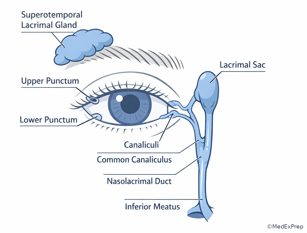
Exam takeaway: Use superotemporal gland versus inferomedial drainage anatomy to localize tearing and swelling complaints.
Exam takeaway: Distinguish the anterior chamber from the posterior chamber because board stems often misuse “behind the iris” to test this boundary.
flowchart TD
A[Cornea] --> B[Anterior chamber]
B --> C[Iris]
C --> D[Posterior chamber]
D --> E[Lens]
C --> F[Ciliary body]
F --> G[Zonules]
Core content
1.1.1.3 Globe wall layers, chambers, and landmark boundaries
A Explanation
The decision conflict in early anatomy questions is usually: Is the finding on the surface, within the wall, or in a chamber/cavity? The single rule that resolves most stems is to identify the nearest named boundary. Corneal pathology is anterior and transparent; scleral pathology is deeper, opaque, and often more painful. The anterior chamber is between cornea and iris; the posterior chamber is not the vitreous cavity but the narrow space between iris and lens/ciliary body. Many distractors exploit that terminology error. The uvea includes iris, ciliary body, and choroid; if a stem mentions vascular, pigmented, or inflammatory tissue in those regions, think uveal localization rather than neural retina or fibrous coat.
WHY THIS IS TESTED: board stems often ask you to choose between keratitis, scleritis, uveitis, angle pathology, lens pathology, and vitreoretinal disease using only one location clue from the exam.
Choose corneal localization when the lesion is visible on slit-lamp at the transparent anterior surface and fluorescein-related clues are present. Reject corneal localization when the pain is deep boring with violaceous hue and globe tenderness, which points deeper to the sclera. Choose anterior chamber localization when cells, flare, hypopyon, or layered blood are described. Reject “posterior chamber” when the finding is behind the lens; that is vitreous cavity or posterior segment, not posterior chamber. See Fig 2 for the cross-sectional relationships and Fig 1 for the layer map.
B Worked example
A 48-year-old man with rheumatoid arthritis, hypertension, and seasonal allergies presents with severe ocular pain and photophobia in the right eye. He says it “feels like a scratch,” and he mentions sleeping in contact lenses years ago, which is the tempting clue. Visual acuity is mildly reduced. The eye has a violaceous deep injection and marked tenderness to palpation. Fluorescein staining is negative. The board-style task is to identify the most likely anatomic site.
Reasoning chain: identify task: localization. Extract key facts: autoimmune history, deep boring pain, violaceous hue, no fluorescein uptake. Apply rule: surface abrasion/keratitis should give epithelial defect or infiltrate clues; deeper scleral inflammation gives severe tenderness and violaceous injection. Eliminate distractors: cornea is tempting because of pain and “scratch” description; conjunctiva is tempting because of redness; anterior chamber is tempting because of photophobia. Verify: the single best localization is sclera, consistent with scleritis.
C Exam trap
Exam trap: The common wrong answer is posterior chamber for anything “behind the iris.” Why it is tempting: learners remember that it is posterior to the iris and stop there. The single clue that eliminates it: if the stem places the finding behind the lens or in the posterior segment, the structure is vitreous cavity or retina/choroid, not posterior chamber.
D Checkpoint
A stem describes inflammatory cells layering inferiorly in the space between the corneal endothelium and the iris in a patient with pain and photophobia. Which anatomic location is most accurate?
- Anterior chamber
- Posterior chamber
- Vitreous cavity
- Subretinal space
Answer: A. Anterior chamber.
- A is tempting and correct because layered inflammatory material between cornea and iris is a hypopyon in the anterior chamber.
- B is tempting but wrong because “posterior” sounds like the better fit; however, the posterior chamber lies between iris and lens, not cornea and iris.
- C is tempting but wrong because vitritis can cause floaters and haze, not an inferior layer visible in the anterior segment.
- D is tempting but wrong because subretinal pathology is posterior and would not be described by slit-lamp layering in front of the iris.
1.1.1.4 Cornea, limbus, sclera, and conjunctival relationships
A Explanation
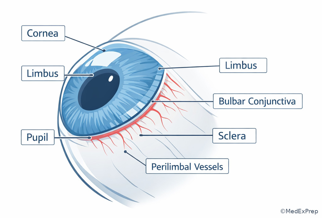
The decision conflict here is: When is a red painful eye superficial and when is it deep structural disease? The single resolving rule is the vascular pattern plus tissue transparency. Conjunctiva is mobile, superficial, and associated with discharge, papillae, follicles, or diffuse injection. Cornea is transparent and refractive, so pathology alters clarity, staining, contour, or sensation. Sclera is structural and deeper, so disease tends to cause severe pain, globe tenderness, and a violaceous hue. The limbus matters because it marks the transition from clear cornea to opaque sclera and is a landmark for neovascularization, stem cells, surgery, and angle-based conceptual questions.
WHY THIS IS TESTED: red-eye stems are packed with distractors that become easy once you know whether the process is conjunctival, corneal, limbal, or scleral.
Choose conjunctival disease when the eye is irritated rather than deeply painful and the cornea remains clear. Choose corneal disease when the symptom is pain plus photophobia and slit-lamp clues suggest epithelial or stromal involvement. Reject episcleritis or conjunctivitis when there is marked pain out of proportion, deep tenderness, or bluish-violet hue. Choose limbal importance when stems mention contact lens overuse, chemical injury, pannus, or surgery involving corneoscleral entry. Forward reference: limbal and angle relationships become essential in Aqueous Humor Dynamics.
B Worked example
A 29-year-old woman with asthma and atopy presents with unilateral redness, tearing, and foreign-body sensation after gardening. She also has migraine and takes oral antihistamines. A friend told her it is “pink eye,” the tempting clue. Vision is 20/20. There is diffuse injection and mild watery discharge. The cornea is clear without fluorescein uptake, and pain is mild rather than deep. The task is to choose the best tissue localization.
Reasoning chain: identify task: separate conjunctiva from cornea or sclera. Extract key facts: clear cornea, no epithelial defect, mild irritation, discharge, good vision. Apply rule: conjunctival pathology is superficial and often preserves corneal clarity and vision. Eliminate distractors: corneal abrasion would stain; scleritis would cause severe boring pain; limbal stem-cell issues would have chronic surface failure rather than isolated mild acute redness. Verify: conjunctival localization is best.
C Exam trap
Exam trap: The common wrong answer is episcleritis/scleritis whenever redness looks sectoral. Why it is tempting: examinees overvalue the pattern of redness and undervalue pain. The single clue that eliminates it: a comfortable patient with preserved vision and a clear cornea usually has superficial disease, not deep scleral inflammation.
D Checkpoint
A patient has severe ocular pain, tenderness to touch, and violaceous discoloration without epithelial staining. Which structure is the most likely primary site?
- Conjunctiva
- Sclera
- Corneal epithelium
- Canaliculus
Answer: B. Sclera.
- A is tempting but wrong because conjunctival disease causes redness, yet it typically lacks severe boring pain and marked tenderness.
- B is tempting and correct because deep pain plus violaceous hue localizes to scleral inflammation.
- C is tempting but wrong because epithelial injury causes foreign-body sensation and staining, not classically deep tenderness.
- D is tempting but wrong because canalicular disease causes localized medial lid symptoms and tearing rather than diffuse globe pain.
- The space between the cornea and iris is the anterior chamber.
- True or False: The posterior chamber is the same as the vitreous cavity. False.
- Name the 3 uveal structures: iris, ciliary body, choroid.
Exam takeaway: Pain depth, corneal clarity, and vessel pattern separate conjunctival, corneal, and scleral disease faster than symptom labels do.
flowchart TD
A[Red eye] --> B{Severe deep pain?}
B -->|Yes| C[Sclera]
B -->|No| D{Corneal staining or opacity?}
D -->|Yes| E[Cornea]
D -->|No| F{Discharge and mobile superficial injection?}
F -->|Yes| G[Conjunctiva]
F -->|No| H[Reassess episclera or uvea] Exam takeaway: Use the limbus as the transition landmark when stems ask where corneal pathology ends and scleral or conjunctival pathology begins.
1.1.1.5 Uveal tract, lens support, and ciliary body relationships
A Explanation
The decision conflict in this section is: When does a stem point to iris, ciliary body, choroid, lens, or zonules? The rule is to follow function. Pupil shape and anterior segment inflammation suggest iris involvement. Accommodation, aqueous production, and zonular attachment point to the ciliary body. Highly vascular posterior pigmented lesions suggest choroid. Lens position problems implicate zonules and ciliary body relationships. This matters because board questions often mix neighboring structures with similar names but different functions.
WHY THIS IS TESTED: understanding these relationships turns glaucoma, uveitis, ectopia lentis, and trauma questions from memorization into localization.
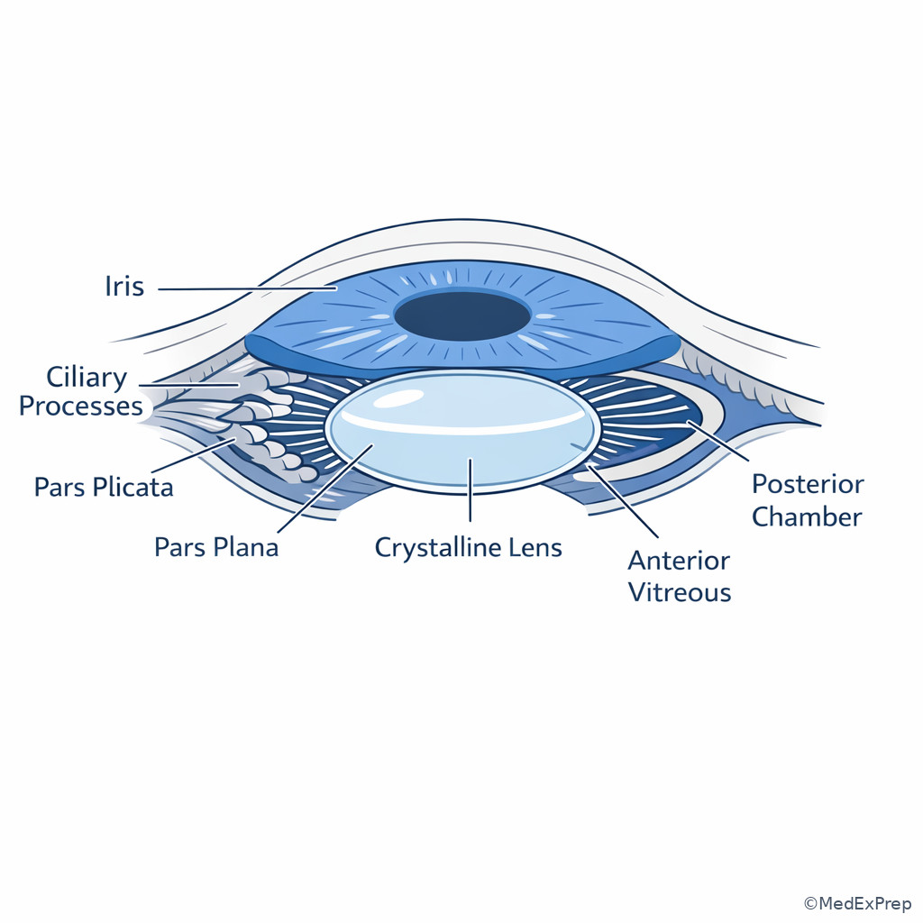
The ciliary body has pars plicata anteriorly and pars plana posteriorly. Pars plicata carries ciliary processes and aqueous production; pars plana is a relatively safer posterior entry landmark in vitreoretinal surgery because it is more avascular and avoids the lens and retina. The iris is the most anterior uveal component, while the choroid is the posterior vascular layer between sclera and retina. Zonules suspend the lens from the ciliary body; therefore, pseudoexfoliation, trauma, Marfan syndrome, homocystinuria, or hypermature cataract-related instability all turn on this support system. See Fig 4.
B Worked example
A 67-year-old woman with pseudoexfoliation, hypertension, and mild COPD presents for cataract evaluation. She reports monocular blur but no pain. On slit lamp, the lens appears mildly decentered with phacodonesis. A distracting clue is her use of inhalers, which may make candidates think about glaucoma medications rather than anatomy. The task is to identify the anatomic support structure most likely compromised.
Reasoning chain: identify task: lens support localization. Extract key facts: pseudoexfoliation, lens decentration, phacodonesis. Apply rule: zonules attach the lens to the ciliary body. Eliminate distractors: iris changes can alter pupil shape but do not suspend the lens; corneal edema can blur vision but does not cause lens wobble; choroid is posterior vascular tissue unrelated to lens stability. Verify: zonular weakness is the most likely anatomic problem.
C Exam trap
Exam trap: The common wrong answer is iris when the pupil looks abnormal in uveitis or trauma. Why it is tempting: the pupil is the visible feature. The single clue that eliminates it: if the question is about accommodation, aqueous production, or lens support, the ciliary body-zonule complex is the key structure, not the iris alone.
D Checkpoint
Which structure both contributes to aqueous production and provides attachment for the suspensory fibers of the lens?
- Iris stroma
- Ciliary body
- Choroid
- Retinal pigment epithelium
Answer: B. Ciliary body.
- A is tempting but wrong because the iris sits adjacent to the pupil and is part of the uvea, yet it is not the source of aqueous production or zonular origin.
- B is tempting and correct because ciliary processes produce aqueous and the ciliary body anchors zonules.
- C is tempting but wrong because the choroid is vascular uvea posteriorly, not the structure governing aqueous production or lens suspension.
- D is tempting but wrong because the RPE supports photoreceptors and is posterior, not part of lens support mechanics.
Exam takeaway: If the stem tests accommodation, aqueous production, or lens instability, the ciliary body and zonules are the key anatomic link.
Exam takeaway: Separate anterior pupil clues from posterior vascular clues by assigning each to iris, ciliary body, or choroid.
flowchart LR
A[Uvea] --> B[Iris]
A --> C[Ciliary body]
A --> D[Choroid]
B --> E[Pupil shape and synechiae]
C --> F[Aqueous production]
C --> G[Zonules and accommodation]
D --> H[Outer retinal blood supply]
1.1.1.6 Retina, choroid, optic disc, and macular landmarks
A Explanation
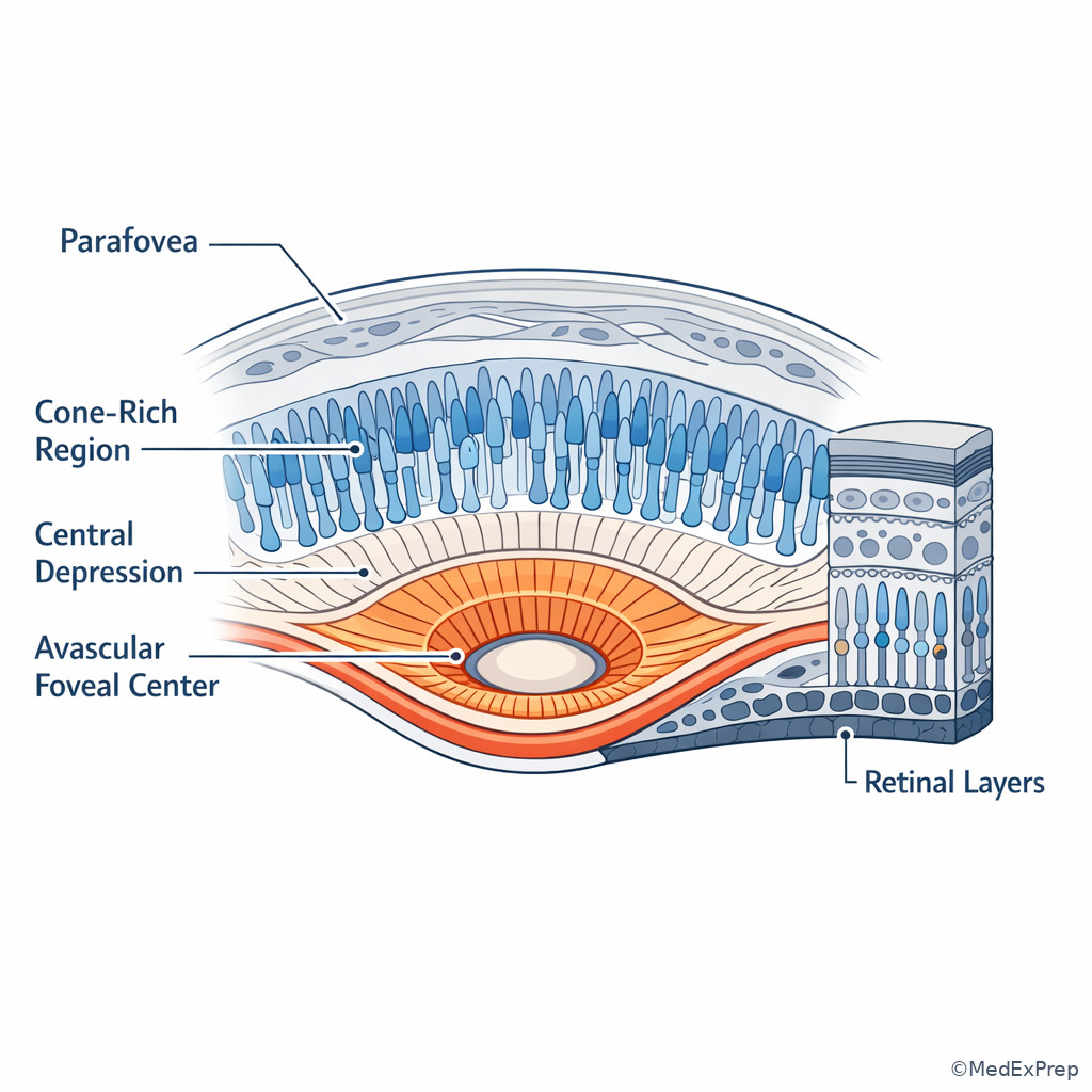
The decision conflict is: Is the posterior finding retinal, subretinal/RPE-choroidal, or optic nerve-related? The rule is to anchor yourself to function and location. Macular/foveal pathology threatens central acuity and metamorphopsia. Peripheral retinal pathology threatens flashes, floaters, and field loss. The optic disc is the nerve head and physiologic blind spot. Choroid lies external to the retina and nourishes the outer retina; lesions there are vascular and pigmented rather than neural. The fovea is specialized: densely packed cones, absent inner retinal layers, and avascular center, which explains its exquisite acuity and vulnerability to central distortion.
WHY THIS IS TESTED: board stems often present identical symptoms but ask you to localize to macula, retina, disc, or choroid.
Choose macular localization when the complaint is reduced central acuity, metamorphopsia, or central scotoma with otherwise preserved peripheral function. Choose optic disc/nerve head when the question centers on disc edema, pallor, cupping, or blind-spot-related field change. Choose peripheral retina when flashes, floaters, curtain, or horseshoe tear cues appear. Choose choroid when a pigmented posterior lesion is described as lying beneath the retina. See Fig 5. Forward reference: the photoreceptor and afferent significance of these regions will be built further in Retinal and Visual Physiology.
B Worked example
A 72-year-old man with hyperlipidemia, smoking history, and prior cataract surgery reports that straight lines look bent in his left eye. He also mentions chronic floaters, a tempting but wrong clue toward peripheral vitreoretinal disease. Visual acuity is reduced centrally, and peripheral fields are grossly intact. Fundus exam shows changes centered temporal to the disc. The task is to identify the most likely key anatomic region involved.
Reasoning chain: identify task: central visual complaint localization. Extract key facts: metamorphopsia, central acuity loss, preserved periphery. Apply rule: central distortion localizes to macula/fovea rather than peripheral retina or optic disc. Eliminate distractors: floaters may reflect vitreous change but do not explain metamorphopsia; optic disc lesions do not typically present with isolated line distortion; choroidal terms are tempting because of age-related pathology, but the functional clue points first to macula. Verify: macular localization is best.
C Exam trap
Exam trap: The common wrong answer is optic disc whenever central vision is reduced. Why it is tempting: the optic nerve is intuitively linked to all vision. The single clue that eliminates it: metamorphopsia is a macular clue, not a disc clue.
D Checkpoint
Which retinal region is most specifically specialized for highest acuity and cone-dominant central vision?
- Ora serrata
- Fovea
- Optic disc
- Peripheral retina
Answer: B. Fovea.
- A is tempting but wrong because it is a named retinal landmark, but it is the anterior termination of the retina rather than the acuity center.
- B is tempting and correct because the fovea is the center of maximal cone-mediated acuity.
- C is tempting but wrong because the disc is an important landmark, yet it has no photoreceptors.
- D is tempting but wrong because peripheral retina supports field detection and motion more than fine central detail.
- The most common symptom clue for macular involvement is metamorphopsia.
- True or False: The optic disc contains photoreceptors. False.
- Name the 3 key posterior landmarks in a routine fundus view: disc, macula, vascular arcades.
Exam takeaway: Distinguish disc, macula, fovea, vascular arcades, and peripheral retina because symptom localization depends on these landmarks.
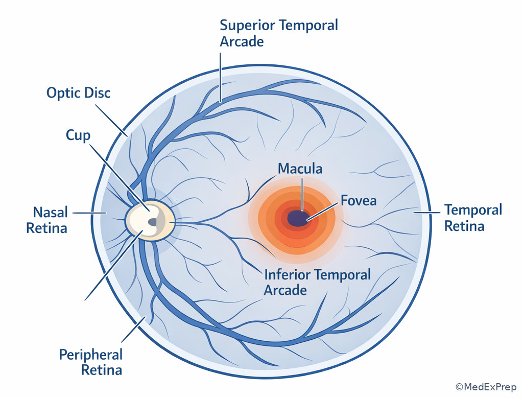
Exam takeaway: Remember that foveal specialization explains why small central lesions can cause disproportionate acuity loss.
1.1.1.7 Eyelids, septum, orbit, lacrimal system, and extraocular muscle context
A Explanation
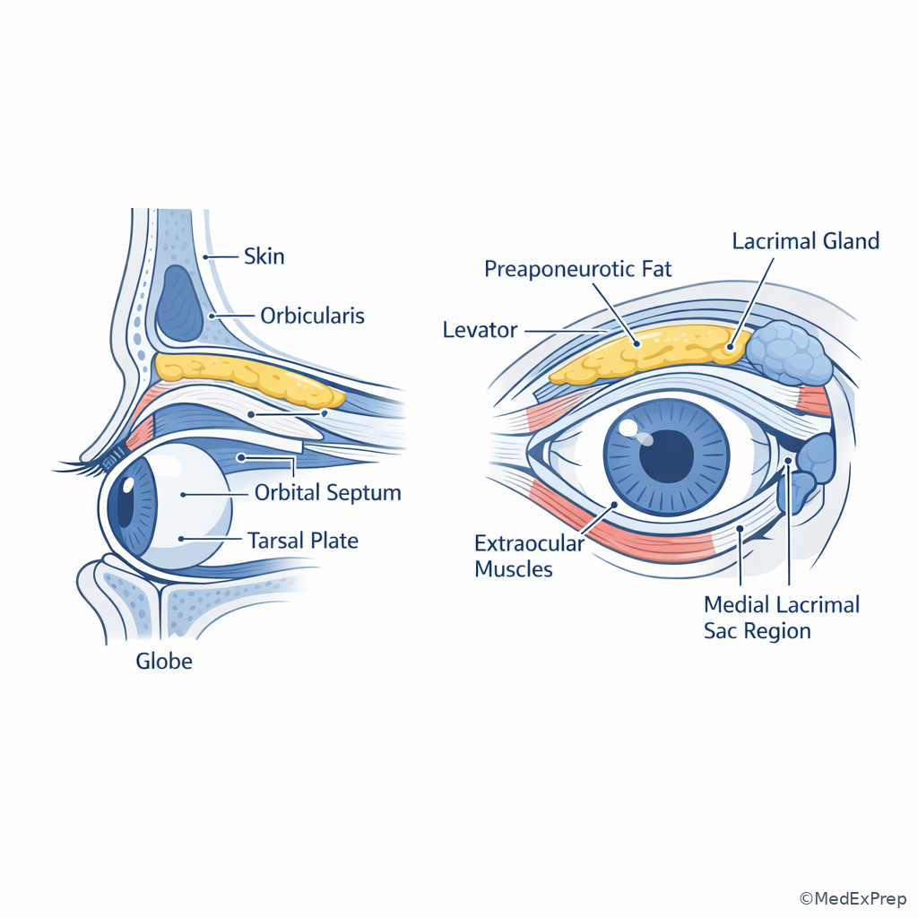
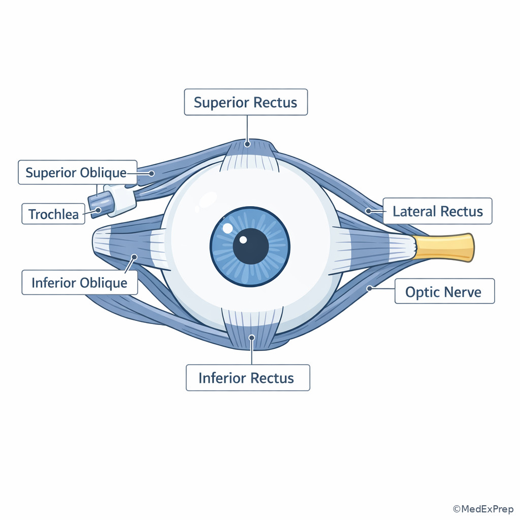
The decision conflict in adnexal stems is usually: Is the problem preseptal, orbital, lacrimal gland, lacrimal drainage, lid margin, or extraocular muscle related? The single best discriminator is the orbital septum plus motility findings. Preseptal processes stay anterior to the septum and do not impair ocular motility, create proptosis, or reduce vision through orbital inflammation. Orbital disease does. The lacrimal gland sits superotemporally; the lacrimal sac and drainage system sit inferomedially. So superotemporal swelling suggests dacryoadenitis or gland mass; inferomedial swelling near the medial canthus suggests sac disease or dacryocystitis. Tarsal plates support the lids, and the meibomian glands lie within them. Extraocular muscles arise from the annulus of Zinn except inferior oblique and have predictable insertions relevant to motility and surgery.
WHY THIS IS TESTED: eyelid swelling questions often test urgent triage, and the correct answer hinges on anatomy rather than organism or drug choice.
Choose preseptal disease when the patient has lid erythema and swelling but preserved vision, full motility, and no proptosis. Reject preseptal disease when pain with eye movements, ophthalmoplegia, afferent findings, or proptosis is present—those are orbital clues. Choose lacrimal sac disease with inferomedial tenderness/tearing. Choose lacrimal gland disease with superotemporal fullness causing inferonasal globe displacement. See Fig 6. This localization framework will reappear later in orbital cellulitis and thyroid eye disease topics.
B Worked example
A 9-year-old boy with recent sinusitis, asthma, and eczema develops a swollen red left eyelid. His mother reports fever and tearing. A distracting clue is crusting on the lashes, which could make blepharitis seem appealing. Exam shows pain with eye movement, mild proptosis, and limited abduction. Vision is slightly reduced. The task is to choose the most likely anatomic compartment that determines urgency.
Reasoning chain: identify task: preseptal versus orbital localization. Extract key facts: sinusitis, fever, pain with eye movement, proptosis, motility restriction. Apply rule: these are postseptal/orbital findings. Eliminate distractors: lid crusting can coexist but does not explain proptosis; dacryocystitis is inferomedial; chalazion is focal and nonurgent. Verify: orbital compartment involvement is the urgent localization.
C Exam trap
Exam trap: The common wrong answer is preseptal cellulitis whenever a child has a red swollen lid. Why it is tempting: preseptal disease is common and visually obvious. The single clue that eliminates it: pain with eye movement or ophthalmoplegia means the process is orbital until proven otherwise.
D Checkpoint
A patient has inferomedial tenderness with epiphora and swelling just below the medial canthal tendon. Which structure is most likely involved?
- Lacrimal gland
- Lacrimal sac
- Frontal sinus
- Lateral rectus muscle
Answer: B. Lacrimal sac.
- A is tempting but wrong because lacrimal anatomy causes tearing symptoms, yet the gland lies superotemporally, not inferomedially.
- B is tempting and correct because the lacrimal sac is an inferomedial drainage structure near the medial canthus.
- C is tempting but wrong because sinus disease can contribute to orbital processes, but the stem gives a classic lacrimal sac location.
- D is tempting but wrong because extraocular muscle pathology causes motility symptoms, not focal medial canthal epiphora.
Exam takeaway: Motility pain, ophthalmoplegia, proptosis, or vision change move a swollen-lid stem posterior to the septum and raise urgency.
flowchart TD
A[Lid swelling] --> B{Pain with eye movement, proptosis, or ophthalmoplegia?}
B -->|No| C[Preseptal compartment likely]
B -->|Yes| D[Orbital compartment likely]
C --> E[Anterior to orbital septum]
D --> F[Posterior to orbital septum]
F --> G[Urgent imaging and treatment pathway] Exam takeaway: Visualizing the septum helps separate routine anterior lid disease from orbital emergencies.
Exam takeaway: Know the muscle-globe relationship because motility deficits localize orbital disease and common cranial nerve patterns.
Exam Traps & Differentiators
Most common wrong answer and why: “posterior chamber” is the classic anatomy distractor. It sounds like the catch-all posterior eye space, but it is only the narrow space between iris and lens/ciliary body.
| Looks similar | Actually differs by | Think this instead | Board clue |
|---|---|---|---|
| Posterior chamber | Behind lens is not in this space | Vitreous cavity | “Behind the lens,” floaters, vitritis |
| Conjunctivitis | Deep pain and tenderness absent | Scleritis | Violaceous hue, autoimmune history |
| Preseptal cellulitis | No orbital signs | Orbital cellulitis | Pain with EOM, proptosis, ophthalmoplegia |
| Lacrimal gland swelling | Superotemporal location | Lacrimal sac disease | Inferomedial canthal tenderness, epiphora |
| Optic nerve problem | Central distortion is not a nerve clue | Macular lesion | Metamorphopsia |
If the stem says ___, think ___
- If the stem says “deep boring pain with violaceous injection”, think sclera.
- If the stem says “cells and flare between cornea and iris”, think anterior chamber.
- If the stem says “lens wobble or decentration”, think zonules/ciliary body relationship.
- If the stem says “metamorphopsia”, think macula/fovea.
- If the stem says “medial canthal swelling with tearing”, think lacrimal sac.
- If the stem says “pain with eye movement and proptosis”, think orbital disease posterior to the septum.
Related future traps: in Aqueous Humor Dynamics, candidates confuse ciliary body aqueous production with trabecular outflow anatomy; in Retinal and Visual Physiology, candidates confuse anatomy of fovea with function of rods versus cones. Use this lesson’s localization rules first, then add physiology.
| Differential focus | Location | Most useful discriminator | Urgency implication |
|---|---|---|---|
| Conjunctivitis vs keratitis | Conjunctiva vs cornea | Corneal staining/opacity and photophobia | Keratitis escalates |
| Episcleritis/scleritis vs conjunctivitis | Deep fibrous coat vs superficial covering | Severe pain and tenderness | Scleritis may indicate systemic disease |
| Preseptal vs orbital cellulitis | Anterior vs posterior to septum | Painful EOM/proptosis/ophthalmoplegia | Orbital cellulitis is urgent |
| Macular vs optic nerve dysfunction | Central retina vs nerve head/pathway | Metamorphopsia versus disc/field clues | Depends on cause, but localization changes workup |
| Lacrimal gland vs sac disorder | Superotemporal vs inferomedial | Exact quadrant of swelling | Guides cause and referral pathway |
Algorithm / Approach
Exam takeaway: Use one high-yield clue to sort most ophthalmic anatomy stems before you think about detailed pathology.
flowchart TD
A[Chief finding] --> B{Red eye}
A --> C{Swelling}
A --> D{Vision distortion}
B --> E[Surface vs sclera vs chamber]
C --> F[Preseptal vs orbital vs lacrimal]
D --> G[Macula vs optic disc vs peripheral retina]
E --> H[Choose exam set]
F --> H
G --> H Rapid Review
- Anterior chamber → between cornea and iris, not behind the lens
- Posterior chamber → between iris and lens/ciliary body, not vitreous cavity
- Cornea → transparent refractive surface; staining/clarity changes localize here
- Sclera → deep fibrous coat; boring pain and tenderness point here
- Limbus → cornea-sclera transition; key landmark for surgery and surface disease
- Uvea → iris, ciliary body, choroid
- Ciliary body → aqueous production plus zonular attachment
- Pars plana → posterior ciliary body region, surgically important entry zone
- Macula → central vision function; distortion localizes here
- Fovea → highest acuity, cone-dominant center
- Optic disc → blind spot, no photoreceptors
- Choroid → vascular layer external to retina, nourishes outer retina
- Orbital septum → key boundary for lid infection triage
- Lacrimal gland → superotemporal swelling
- Lacrimal sac → inferomedial tearing/swelling
Self-check quiz
1.1.1.8 Question 1
A 34-year-old woman undergoing routine examination asks which structure lies directly between the cornea and the iris. She has myopia and migraine, but no active eye disease. Which is the most appropriate answer?
- Anterior chamber
- Posterior chamber
- Vitreous cavity
- Suprachoroidal space
1.1.1.9 Question 2
A 61-year-old man with diabetes and hypertension reports that straight lines appear distorted in one eye. Which structure is most likely responsible for this central visual complaint?
- Ora serrata
- Fovea
- Optic disc
- Canaliculus
1.1.1.10 Question 3
A 43-year-old woman with rheumatoid arthritis, hypothyroidism, and dry mouth presents with severe unilateral eye pain. She says the eye feels “scratchy,” and she recently used artificial tears, a distracting detail. Exam shows violaceous injection, marked tenderness, and no corneal staining. Which anatomic site best localizes the primary process?
- Conjunctiva
- Corneal epithelium
- Sclera
- Lacrimal gland
1.1.1.11 Question 4
A 70-year-old patient with pseudoexfoliation, hypertension, and prior laser peripheral iridotomy is noted to have phacodonesis during cataract evaluation. A mildly irregular pupil makes iris pathology tempting. Which structure requires the most attention because it is the key support for the lens?
- Zonules attached to the ciliary body
- Trabecular meshwork
- Descemet membrane
- Choroid
1.1.1.12 Question 5
A 7-year-old boy with recent ethmoid sinusitis, allergic rhinitis, and eczema presents with fever and eyelid swelling. He has pain with eye movement and mild proptosis. Crusting on the lashes is also present. Which anatomic boundary best distinguishes the urgent compartment involved?
- Orbital septum
- Limbal arcade
- Ora serrata
- Bowman layer
1.1.1.13 Question 6
A 65-year-old woman with sarcoidosis, pseudophakia, and chronic floaters is found to have inflammatory cells visible between the corneal endothelium and the iris. A resident suggests this is posterior chamber inflammation because it is “behind the pupil.” Which is the most appropriate localization?
- Anterior chamber
- Posterior chamber
- Anterior vitreous
- Subretinal space
1.1.1.14 Question 7
A 58-year-old woman with chronic tearing, rosacea, and prior punctal plug placement presents with painful swelling near the medial canthus. The lesion is inferomedial, and pressure increases reflux from the punctum. She has no pain with eye movement or proptosis. Which structure is most likely involved?
- Lacrimal gland
- Lacrimal sac
- Medial rectus muscle
- Frontal lobe extension into the orbit
1.1.1.15 Question 8
A 74-year-old man with hyperlipidemia, smoking history, and posterior vitreous detachment describes a new central gray spot and distortion of straight lines. He also reports longstanding peripheral floaters, making a peripheral retinal process tempting. Which location best explains the current most vision-threatening symptom?
- Peripheral retina near the ora serrata
- Macula/fovea
- Optic disc cup
- Canal of Schlemm
Answer key
1.1.1.16 Question 1 answer: A. Anterior chamber
Why the correct answer is correct: The anterior chamber is the space between the cornea and iris. This is a classic board-tested anatomic boundary and is often contrasted with the posterior chamber. Source support: American Academy of Ophthalmology, Basic and Clinical Science Course (BCSC), Section 2 and Section 4.
- A. Anterior chamber — Tempting because it is the straightforward spatial answer, and it is correct for this exact border. Discriminating clue: “between the cornea and the iris.” Source: AAO BCSC.
- B. Posterior chamber — Tempting because it also involves the iris, but it is incorrect here because it lies between the iris and lens/ciliary body. Discriminating clue: cornea is named, not lens. Source: AAO BCSC.
- C. Vitreous cavity — Tempting because it is a larger posterior eye space, but it lies behind the lens. Discriminating clue: the question asks for the space directly anterior to the iris border. Source: Yanoff and Duker.
- D. Suprachoroidal space — Tempting because it is a real anatomic potential space, but it is external to the choroid and irrelevant to the cornea-iris interval. Discriminating clue: wrong compartment entirely. Source: AAO BCSC.
1.1.1.17 Question 2 answer: B. Fovea
Why the correct answer is correct: Central distortion points to the macula, specifically the foveal region for highest acuity function. Source support: AAO BCSC Section 2; Ryan’s Retina.
- A. Ora serrata — Tempting because it is a named retinal landmark, but it is peripheral and not responsible for central distortion. Discriminating clue: “central visual complaint.” Source: AAO BCSC.
- B. Fovea — Tempting and correct because the fovea mediates finest central vision and line distortion localizes there. Discriminating clue: metamorphopsia. Source: Ryan’s Retina.
- C. Optic disc — Tempting because it is visually central in fundus drawings, but it lacks photoreceptors and does not cause metamorphopsia as the typical primary clue. Discriminating clue: distortion, not blind-spot or nerve-head finding. Source: AAO BCSC.
- D. Canaliculus — Tempting only because it is an eye structure, but it belongs to the lacrimal drainage system and does not affect central retinal image quality. Discriminating clue: complaint is visual distortion, not tearing. Source: Kanski’s Clinical Ophthalmology.
1.1.1.18 Question 3 answer: C. Sclera
Why the correct answer is correct: Severe deep pain, tenderness, violaceous injection, and lack of corneal staining are classic for scleral inflammation. Source support: AAO BCSC Section 8; Kanski’s Clinical Ophthalmology.
- A. Conjunctiva — Tempting because redness and irritation are common conjunctival symptoms, but conjunctivitis usually does not cause severe boring pain and marked tenderness. Discriminating clue: violaceous deep injection. Source: Kanski.
- B. Corneal epithelium — Tempting because the patient says “scratchy,” but epithelial disease should usually give staining or a visible surface lesion. Discriminating clue: no staining. Source: AAO BCSC.
- C. Sclera — Tempting and correct because the systemic autoimmune background plus deep pain fit scleritis. Discriminating clue: tenderness to palpation. Source: AAO BCSC.
- D. Lacrimal gland — Tempting if one overreads the autoimmune dry-eye history, but lacrimal gland pathology causes superotemporal swelling rather than violaceous globe injection. Discriminating clue: no focal superotemporal mass/fullness. Source: Rootman and AAO BCSC Section 7.
1.1.1.19 Question 4 answer: A. Zonules attached to the ciliary body
Why the correct answer is correct: Phacodonesis and lens instability indicate zonular weakness, and zonules insert into the ciliary body. Pseudoexfoliation is a classic board clue. Source support: AAO BCSC Section 11; Kanski’s Clinical Ophthalmology.
- A. Zonules attached to the ciliary body — Tempting and correct because lens support depends on this system. Discriminating clue: phacodonesis. Source: AAO BCSC.
- B. Trabecular meshwork — Tempting because pseudoexfoliation also affects glaucoma risk, but this structure governs outflow, not lens suspension. Discriminating clue: the problem is lens wobble, not pressure regulation. Source: AAO BCSC Section 10.
- C. Descemet membrane — Tempting because cataract surgery often discusses it, but it is a corneal layer unrelated to lens support. Discriminating clue: wrong tissue plane. Source: Krachmer, Mannis, and Holland.
- D. Choroid — Tempting because it is a nearby internal eye layer, but it has no role in maintaining lens centration. Discriminating clue: support function absent. Source: AAO BCSC.
1.1.1.20 Question 5 answer: A. Orbital septum
Why the correct answer is correct: The orbital septum is the key boundary separating preseptal from orbital infections. Pain with eye movement and proptosis indicate disease posterior to the septum. Source support: AAO BCSC Section 7; Rootman’s orbital texts.
- A. Orbital septum — Tempting and correct because the clinical decision is preseptal versus orbital. Discriminating clue: pain with EOM and proptosis. Source: AAO BCSC.
- B. Limbal arcade — Tempting because inflammation and vascular landmarks can sound ocularly relevant, but lid swelling triage is not determined by the corneal limbus. Discriminating clue: adnexal/orbital stem, not corneal stem. Source: AAO BCSC.
- C. Ora serrata — Tempting because it is a memorable boundary, but it is a retinal boundary unrelated to eyelid swelling. Discriminating clue: posterior segment term in an orbital problem. Source: Ryan’s Retina.
- D. Bowman layer — Tempting because it is a known corneal layer, but again it does not separate lid from orbit. Discriminating clue: wrong anatomic region. Source: Krachmer, Mannis, and Holland.
1.1.1.21 Question 6 answer: A. Anterior chamber
Why the correct answer is correct: Cells between corneal endothelium and iris are in the anterior chamber. The resident’s “behind the pupil” heuristic is the exact trap tested on boards. Source support: AAO BCSC Section 9 and Section 2.
- A. Anterior chamber — Tempting and correct because the space named in the stem is anatomically the anterior chamber. Discriminating clue: “between the corneal endothelium and the iris.” Source: AAO BCSC.
- B. Posterior chamber — Tempting because it sounds more posterior and the pupil is involved conceptually, but it lies between the iris and lens/ciliary body. Discriminating clue: corneal endothelium is named. Source: AAO BCSC.
- C. Anterior vitreous — Tempting because the patient has floaters, but the visible cells are not behind the lens. Discriminating clue: anterior segment view and exact anatomic borders. Source: Yanoff and Duker.
- D. Subretinal space — Tempting because sarcoid can involve posterior structures, but nothing in the stem places this finding posteriorly. Discriminating clue: cells are seen in front of the iris. Source: AAO BCSC.
1.1.1.22 Question 7 answer: B. Lacrimal sac
Why the correct answer is correct: Inferomedial swelling with reflux from the punctum strongly localizes to the lacrimal sac/drainage system. Absence of orbital signs helps exclude a deeper orbital process. Source support: AAO BCSC Section 7; Kanski’s Clinical Ophthalmology.
- A. Lacrimal gland — Tempting because tearing suggests lacrimal anatomy, but the gland is superotemporal. Discriminating clue: inferomedial location. Source: AAO BCSC.
- B. Lacrimal sac — Tempting and correct because sac disease causes medial canthal swelling, epiphora, and reflux. Discriminating clue: pressure-related punctal reflux. Source: AAO BCSC.
- C. Medial rectus muscle — Tempting because it is nearby anatomically, but extraocular muscle disease would cause diplopia or motility pain rather than reflux through the punctum. Discriminating clue: drainage-system sign. Source: Rootman.
- D. Frontal lobe extension into the orbit — Tempting only as a deep-orbit possibility, but the stem specifically lacks orbital signs and gives classic lacrimal drainage localization. Discriminating clue: no proptosis or motility deficit. Source: AAO BCSC.
1.1.1.23 Question 8 answer: B. Macula/fovea
Why the correct answer is correct: The current symptom that matters is central gray spot with metamorphopsia, which localizes to the macula/fovea even though peripheral floaters create noise. Source support: Ryan’s Retina; AAO BCSC Section 12.
- A. Peripheral retina near the ora serrata — Tempting because the patient has a history of PVD and floaters, but peripheral tears cause flashes, floaters, and curtain-type symptoms rather than central distortion as the lead feature. Discriminating clue: metamorphopsia. Source: Ryan’s Retina.
- B. Macula/fovea — Tempting and correct because this region subserves fine central vision. Discriminating clue: new central gray spot and bent lines. Source: AAO BCSC.
- C. Optic disc cup — Tempting because older patients may have glaucoma-related concerns, but disc cupping does not classically produce metamorphopsia. Discriminating clue: distortion rather than arcuate field loss. Source: AAO BCSC Section 10.
- D. Canal of Schlemm — Tempting because it is an eye structure and glaucoma is common in older adults, but it is an outflow pathway unrelated to central distortion symptoms. Discriminating clue: visual complaint localizes posteriorly, not to the angle. Source: AAO BCSC.
Keep going with the full ABO Ophthalmology Prep Course (Ophth)
You just studied the way members do, in the same lesson interface. Full access unlocks every lesson, the question bank and flashcards for a full year.
One payment · 1 year of access · No auto-renewal