1.1.1 - Cortical Functional Anatomy
Cortical Functional Anatomy
Topic 1.1.1 — Core Neuroscience → Neuroanatomy and Localization → Cortical Functional Anatomy
What you'll learn
- DOMAIN → Objective → Subskill: Basic Science → Neuroanatomy → Map major cortical lobes, gyri, and sulci to testable functions.
- DOMAIN → Objective → Subskill: Cranial → Localization reasoning → Use deficits in language, praxis, neglect, and visual fields to localize cortical lesions.
- DOMAIN → Objective → Subskill: Tumor → Operative selection → Recognize eloquent cortex and when mapping changes management.
- DOMAIN → Objective → Subskill: Functional → Seizure localization → Relate cortical region to common seizure semiology.
- DOMAIN → Objective → Subskill: Neurocritical Care → Urgent next steps → Identify cortical patterns requiring rapid imaging, decompression, or vascular consideration.
- Core Neuroscience → Neuroanatomy and Localization → Cortical lobe function and lesion localization
- Cranial → Eloquent cortex → Language, motor, sensory, vision, and executive networks
- Tumor / Epilepsy / Functional overlap → Preoperative functional boundaries → Mapping-sensitive cortex
Why it matters
- Cortical localization is the first filter for neurosurgical diagnosis: before MRI pattern recognition, you should already suspect the lobe and hemisphere.
- Board stems often hide the answer in a single cortical clue such as aphasia, neglect, apraxia, gaze preference, or a congruous visual field cut.
- Operative risk-benefit discussions depend on whether pathology involves primary motor, primary sensory, language cortex, supplementary motor area, insula, or visual pathways.
- Cortical anatomy links directly to upcoming topics: brainstem localization uses long-tract plus cranial nerve patterns, while spinal syndromes use tract patterns without cortical signs.
Key Terms & Must-Know Facts
1.1.1.1 Key terms
- Precentral gyrus
- Postcentral gyrus
- Central sulcus
- Sylvian fissure
- Dominant hemisphere
- Broca area
- Wernicke area
- Arcuate fasciculus
- Primary motor cortex
- Primary somatosensory cortex
- Supplementary motor area
- Frontal eye fields
- Gerstmann syndrome
- Apraxia
- Hemispatial neglect
1.1.1.2 Must-know facts
- Dominant inferior frontal gyrus lesions cause nonfluent aphasia; dominant posterior superior temporal lesions cause fluent aphasia with poor comprehension.
- Nondominant parietal lesions classically cause neglect rather than aphasia.
- Primary motor cortex lesions produce contralateral weakness with somatotopic organization.
- Medial frontal/paracentral lobule lesions disproportionately affect leg function.
- Occipital cortical lesions cause contralateral homonymous visual field defects, often with macular sparing in PCA infarction.
- Frontal eye field lesions drive gaze toward the lesion; irritative seizures may drive gaze away.
- Dominant angular gyrus injury may produce Gerstmann syndrome: agraphia, acalculia, finger agnosia, left-right confusion.
- Supplementary motor area injury can produce transient akinesia or mutism despite intact primary motor pathways.
| Region | Core function | Classic deficit if injured | High-yield localization clue |
|---|---|---|---|
| Precentral gyrus | Primary motor output | Contralateral focal weakness | Somatotopic pattern; face/arm lateral, leg medial |
| Postcentral gyrus | Primary somatic sensation | Contralateral cortical sensory loss | Astereognosis if higher-order integration involved |
| Dominant inferior frontal | Motor language | Broca aphasia | Nonfluent speech, intact comprehension relatively preserved |
| Dominant posterior temporal | Language comprehension | Wernicke aphasia | Fluent but nonsensical output, poor repetition/comprehension |
| Nondominant parietal | Spatial attention | Neglect | Ignores contralateral body/space, extinction |
| Occipital cortex | Vision | Homonymous hemianopia | Congruous field defect |
| Mesial frontal/SMA | Initiation/planning of movement | Akinesia, speech initiation deficit | Transient postoperative SMA syndrome |
Core content
Exam takeaway: Start with the deficit pattern, then choose lobe and hemisphere before choosing pathology.
flowchart TD
A[Patient deficit] --> B{Motor or sensory?}
B -->|Motor| C[Frontal lobe]
B -->|Sensory| D[Parietal lobe]
A --> E{Language abnormality?}
E -->|Yes| F[Usually dominant hemisphere]
F --> G[Inferior frontal = Broca]
F --> H[Posterior temporal = Wernicke]
A --> I{Spatial neglect?}
I -->|Yes| J[Nondominant parietal]
A --> K{Visual field cut?}
K -->|Homonymous and congruous| L[Occipital cortex]
A --> M{Behavior/executive change?}
M -->|Yes| N[Prefrontal cortex] Exam takeaway: Use a lobe-based map to separate frontal, parietal, temporal, and occipital presentations quickly.
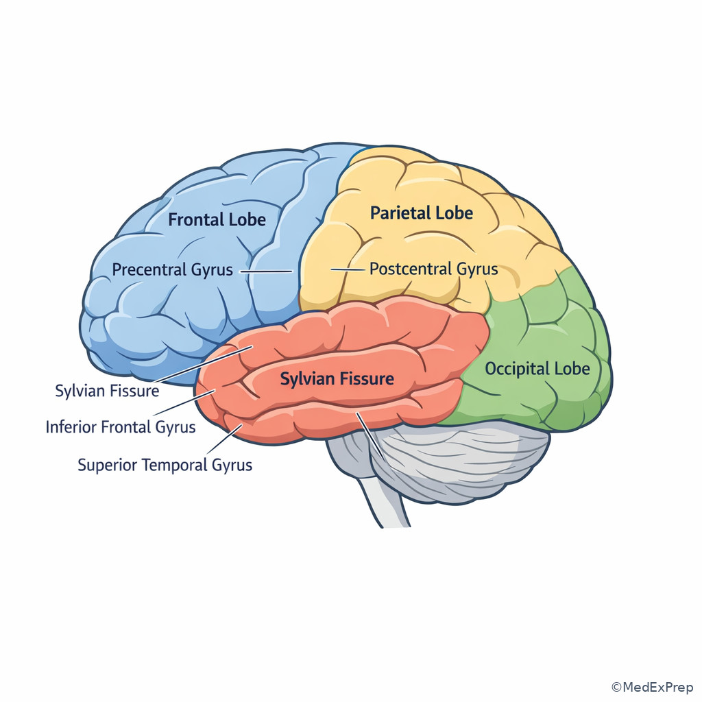
1.1.1.3 Foundational cortical map: lobes, sulci, and somatotopy
A Explanation
The first decision conflict in cortical localization is whether a stem is asking you to choose a lobe or a specific functional strip. The single rule that resolves this is to ask: Is the deficit primary and modality-specific, or higher-order and integrative? Pure contralateral weakness points to precentral gyrus; pure contralateral cortical sensory loss points to postcentral gyrus; combined executive, motor-planning, and initiation deficits push you more anterior into frontal association cortex.
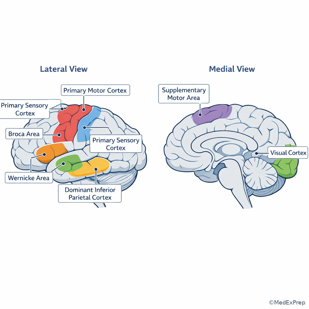
WHY THIS IS TESTED: board questions frequently distract with broad terms like “frontal mass” when the actual localizer is a narrow precentral, supplementary motor, or opercular lesion. The examinee must choose the most precise localization compatible with the deficit pattern. See Fig 1.
Remember the cortical homunculus: face and arm are lateral; leg is medial along the paracentral lobule. This matters because parasagittal meningioma, ACA territory infarct, or falcine mass often present with leg-predominant deficits rather than arm-face patterns. In neurosurgical practice, that distribution affects both lesion localization and surgical corridor planning.
The central sulcus separates motor from sensory cortex; the Sylvian fissure helps orient lateral frontal versus temporal structures. If you know only two landmarks under time pressure, know those two. A lesion just anterior to the central sulcus produces weakness; just posterior produces sensory deficits and cortical sensory phenomena. If the lesion extends medially, expect leg emphasis. If it extends inferiorly in the dominant frontal operculum, language may be at risk.
Exam takeaway: Decide first whether the stem reflects primary motor, primary sensory, or association cortex before naming a lobe.
B Worked example
A 63-year-old right-handed man with hypertension and atrial fibrillation develops abrupt left leg weakness greater than arm weakness while mowing the lawn. He has mild urinary urgency, no aphasia, and no facial droop. CT head is initially negative; glucose is 198 mg/dL, which is distracting but not explanatory. The stem asks for the most likely localization.
Reasoning chain: identify the task: localization, not etiology. Extract key facts: sudden onset, contralateral weakness, leg predominance, no language deficit, no crossed findings, no sensory level. Apply the rule: medial motor cortex/paracentral lobule controls the leg. Eliminate distractors: internal capsule usually gives dense face-arm-leg weakness; lateral frontal lesions often involve face/arm more than leg; spinal cord causes tract-pattern findings or a sensory level. Verify: a right ACA cortical region best matches.
C Exam trap
D Checkpoint
Question: A 54-year-old woman with hyperlipidemia and migraine presents with new right hand numbness, astereognosis, and impaired graphesthesia in the right hand. Strength is preserved. Which localization is most likely?
- Left postcentral/parietal cortical region
- Left precentral gyrus
- Right inferior parietal lobule
- Left internal capsule posterior limb
- A. Tempting because contralateral sensory cortical deficits fit; correct because cortical sensory processing deficits with preserved strength localize posterior to the central sulcus.
- B. Tempting because precentral and postcentral lesions are adjacent; wrong because primary motor cortex injury should produce weakness, not isolated cortical sensory loss.
- C. Tempting because parietal lobe is involved; wrong because right parietal lesions usually affect left-sided spatial attention, not right-hand contralateral sensory integration.
- D. Tempting because internal capsule lesions can cause contralateral symptoms; wrong because they produce dense motor/sensory pathway deficits, not higher-order cortical sensory signs like astereognosis.
Exam takeaway: Leg-predominant deficits point medial, while face-arm deficits point lateral cortex.
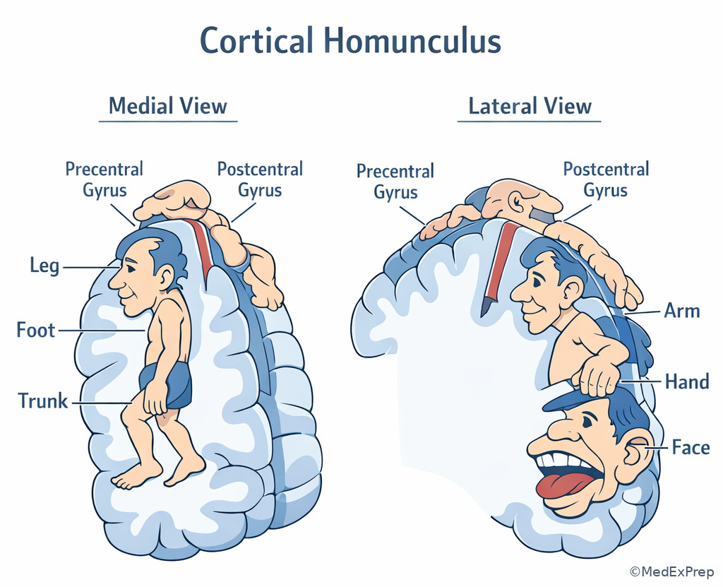
1.1.1.4 Dominant versus nondominant hemisphere: language, praxis, neglect
A Explanation
The key decision conflict here is whether a patient with impaired communication has aphasia, dysarthria, or neglect/inattention masquerading as confusion. The single finding that resolves this is whether language formulation/comprehension is impaired rather than articulation or attention alone. Dominant hemisphere lesions produce aphasic syndromes; nondominant parietal lesions produce neglect, extinction, dressing apraxia, or poor awareness of deficit.
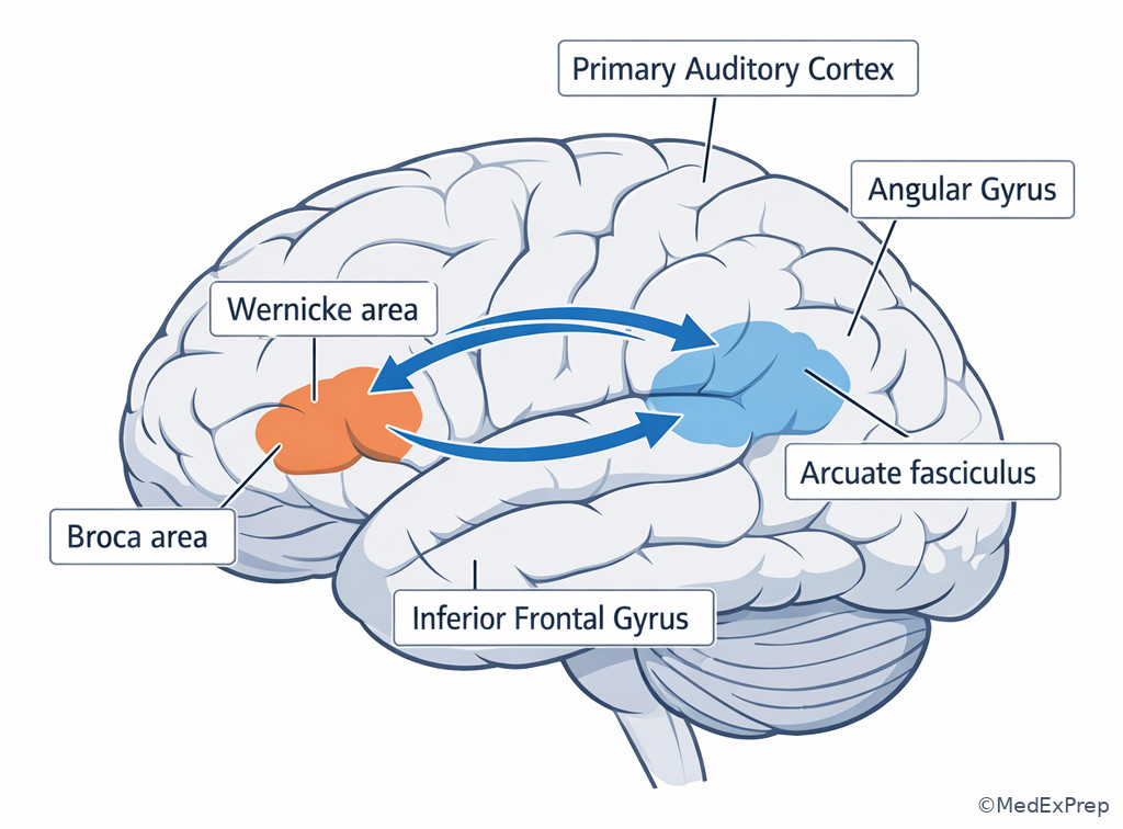
WHY THIS IS TESTED: exam stems often mix reduced output, gaze deviation, and inattention. You must reject “aphasia” if comprehension and naming are intact but speech is slurred, and reject “confusion” if the patient ignores one side of space. Dominant hemisphere is usually left in right-handed patients, but do not overstate certainty in every individual case. See Fig 3.
Broca aphasia arises from dominant inferior frontal injury: nonfluent, effortful output with relatively preserved comprehension. Wernicke aphasia arises from dominant posterior superior temporal injury: fluent but paraphasic output with impaired comprehension. Conduction aphasia implicates the arcuate fasciculus: fluent speech and preserved comprehension but poor repetition. Dominant inferior parietal injury may produce Gerstmann syndrome or ideomotor apraxia. In contrast, the classic nondominant parietal syndrome is hemispatial neglect, especially after right inferior parietal damage.
For neurosurgical planning, any lesion near dominant perisylvian cortex raises the question of awake mapping, tractography, or task-based functional correlation. If the lesion is in the nondominant parietal lobe, counseling and risk assessment should emphasize spatial deficits rather than aphasia. See Fig 1 for the first-pass branching logic.
Exam takeaway: Aphasia localizes to the dominant hemisphere, whereas neglect points to the nondominant parietal cortex.
flowchart LR
A[Communication or behavior deficit] --> B{Language impaired?}
B -->|Nonfluent, effortful| C[Dominant inferior frontal]
B -->|Fluent, poor comprehension| D[Dominant posterior temporal]
B -->|Poor repetition only| E[Arcuate fasciculus/perisylvian]
A --> F{Ignores one side of space?}
F -->|Yes| G[Nondominant parietal]
A --> H{Motor planning problem with intact strength?}
H -->|Yes| I[Dominant parietal/premotor praxis network] B Worked example
A 71-year-old right-handed woman with atrial fibrillation, type 2 diabetes, and a remote left mastectomy is brought in for “confusion.” She speaks fluently but gives nonsensical answers, cannot follow two-step commands, and repeatedly says she feels fine. She has a right superior quadrantanopia. Sodium is 130 mEq/L, a tempting but insufficient explanation for altered mentation. The question asks for the most likely cortical localization.
Reasoning chain: identify task: localization. Extract key facts: fluent output, poor comprehension, right visual field deficit, right-handed patient. Apply the rule: dominant posterior temporal/perisylvian lesion causes Wernicke aphasia and may involve optic radiations. Eliminate distractors: nondominant parietal lesions cause neglect rather than classic aphasia; frontal opercular lesions cause nonfluent speech; dysarthria would preserve comprehension and content. Verify: left posterior superior temporal region is best.
C Exam trap
D Checkpoint
Question: A 60-year-old man after resection of a right parietal convexity mass consistently eats food only from the right side of the tray and denies weakness despite bumping his left arm against the bedrail. Which deficit best explains this pattern?
- Left hemispatial neglect
- Broca aphasia
- Ideomotor apraxia from dominant parietal injury
- Pseudobulbar dysarthria
- A. Tempting because the syndrome is spatial and lateralized; correct because right nondominant parietal injury causes neglect of the contralateral side and anosognosia.
- B. Tempting because frontal language syndromes can reduce purposeful behavior; wrong because speech production is not the core issue and the lesion side is inconsistent.
- C. Tempting because parietal lesions can impair learned motor acts; wrong because ideomotor apraxia is usually dominant hemisphere-related and does not explain environmental inattention.
- D. Tempting because dysarthria can make patients seem cognitively impaired; wrong because dysarthria affects articulation, not spatial awareness.
- The classic syndrome from dominant inferior frontal injury is Broca aphasia.
- True or False: A patient with fluent but meaningless speech and poor comprehension most likely has nondominant parietal injury. False.
- Name the 4 components of Gerstmann syndrome: agraphia, acalculia, finger agnosia, left-right confusion.
Exam takeaway: Inferior frontal and posterior temporal language nodes create distinct aphasia patterns that are highly testable.
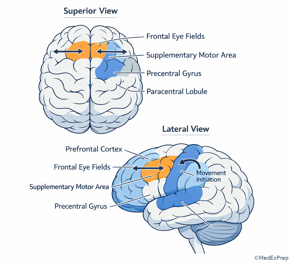
1.1.1.5 Frontal and parietal association cortex: executive control, gaze, praxis, and sensory integration
A Explanation
The central decision conflict in frontal-parietal association lesions is whether the deficit reflects initiation/planning or perception/integration. The single finding that resolves this is the pattern of failure: abulia, disinhibition, motor impersistence, grasp reflexes, or gaze preference suggest frontal systems; astereognosis, agraphesthesia, apraxia, neglect, or constructional failure suggest parietal systems.
WHY THIS IS TESTED: many neurosurgical lesions involve association cortex rather than primary cortex, especially meningiomas, low-grade gliomas, metastases, and edema patterns. These patients may not present with dense weakness or numbness, and the board-style question asks you to choose the more subtle cortical localization. Frontal eye fields are high-yield: destructive frontal eye field lesions cause eyes to deviate toward the lesion; irritative seizures may drive gaze away.
The supplementary motor area deserves special emphasis for neurosurgeons. Lesions or resections here can cause transient contralateral akinesia, difficulty initiating movement, or mutism if the dominant mesial frontal region is involved. This can look dramatic postoperatively but often improves over days to weeks. The exam tests whether you can distinguish this from a new capsular stroke, operative hematoma, or persistent primary motor injury.
Parietal association cortex integrates somatosensory input into meaningful perception. That is why astereognosis and graphesthesia require intact primary sensation plus intact integration. Dominant parietal lesions cause praxis and calculation/writing syndromes; nondominant lesions impair body schema and space. For lesion planning, convexity lesions near superior parietal or inferior parietal regions may demand different counseling and postoperative expectations.
B Worked example
A 48-year-old woman with a known left parasagittal frontal low-grade glioma, hypothyroidism, and chronic migraine undergoes subtotal resection. On postoperative day 1 she is awake, follows commands, and has marked difficulty initiating speech and movement of the right leg; facial strength is preserved. CT shows expected postoperative change without hematoma. A mildly elevated temperature leads one team member to worry about sepsis, but the stem asks for the most likely explanation.
Reasoning chain: identify the task: postoperative localization syndrome. Extract key facts: mesial frontal surgery, akinesia/initiation deficit, no expanding lesion on CT, preserved alertness. Apply rule: supplementary motor area syndrome causes transient initiation deficits and sometimes mutism. Eliminate distractors: capsular stroke should produce a denser fixed deficit; postoperative hematoma would show mass effect; infection does not selectively cause contralateral leg akinesia with clean imaging. Verify with anatomy: mesial frontal/paracentral-SMA region fits.
C Exam trap
D Checkpoint
Question: A 66-year-old man with a right frontal convexity meningioma has eyes deviated to the right after acute hemorrhage into the lesion. Which localization rule best explains this finding?
- Destructive frontal eye field lesion causes gaze toward the lesion
- Occipital lesion causes gaze toward the lesion
- Temporal lobe lesion causes gaze away from the lesion
- Parietal lesion causes vertical gaze palsy
- A. Tempting because frontal eye fields control contralateral saccades; correct because a destructive right frontal eye field lesion leaves unopposed left frontal eye field drive, pulling gaze rightward.
- B. Tempting because visual systems can alter eye behavior; wrong because occipital lesions cause visual field defects, not the classic horizontal gaze preference localizer.
- C. Tempting because temporal seizures can cause eye version; wrong because the stem describes destructive frontal pathology, not an irritative temporal focus.
- D. Tempting because parietal lesions affect attention; wrong because vertical gaze palsy is a midbrain, not cortical parietal, localizer.
Exam takeaway: Mesial frontal lesions alter initiation, while frontal eye field lesions alter conjugate gaze direction.
1.1.1.6 Temporal and occipital cortex: memory, auditory-language integration, and visual field localization
A Explanation
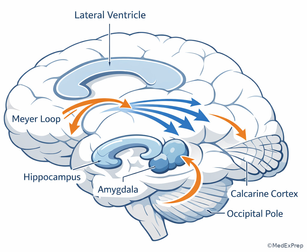
The main decision conflict in temporal-occipital stems is whether to localize a deficit to language/auditory association, mesial temporal memory, or primary visual cortex/optic radiations. The single discriminator is usually the type of deficit: episodic memory and seizure aura suggest mesial temporal structures; receptive language deficits suggest dominant posterior superior temporal cortex; congruous homonymous field cuts suggest occipital cortex; superior quadrantanopia suggests temporal optic radiations.
WHY THIS IS TESTED: exam writers often pair a temporal lobe lesion with a visual field defect or aphasia clue and ask for the most likely cortical region or operative risk. You must separate temporal neocortex from mesial temporal structures and from occipital cortex. Mesial temporal lesions are especially important in epilepsy localization, while occipital lesions more cleanly produce visual syndromes.
Occipital cortical lesions cause contralateral homonymous hemianopia that is often highly congruous. PCA infarction may spare the macula because of collateral supply. Temporal lobe lesions involving Meyer loop cause contralateral superior quadrantanopia. Dominant posterior temporal lesions may combine receptive aphasia with field deficits. Mesial temporal structures support declarative memory, and dominant mesial temporal surgery raises verbal memory concerns, whereas nondominant surgery raises nonverbal/visuospatial memory concerns.
In neurosurgical practice, this anatomy matters for tumor resection, epilepsy surgery, and PCA-territory stroke assessment. A patient with “confusion” and a field cut may actually have a posterior cortical syndrome, not diffuse encephalopathy. See Fig 4.
Exam takeaway: A congruous homonymous field cut points posteriorly, while language or memory clues move the localizer forward into temporal cortex.
flowchart TD
A[Temporal or visual symptom] --> B{Main deficit}
B -->|Poor comprehension, fluent speech| C[Dominant posterior temporal]
B -->|Episodic memory, aura, automatisms| D[Mesial temporal]
B -->|Superior quadrantanopia| E[Temporal optic radiations]
B -->|Congruous homonymous hemianopia| F[Occipital cortex]
F --> G[Consider PCA territory or occipital mass]
D --> H[Consider seizure focus or mesial lesion] B Worked example
A 39-year-old right-handed teacher with medically refractory focal seizures, depression, and hypothyroidism reports a rising epigastric sensation followed by behavioral arrest and lip-smacking. MRI shows a left mesial temporal lesion; formal field testing is normal. A tiny nonspecific occipital white matter focus is noted on imaging and is tempting but likely incidental. The question asks for the most likely seizure onset localization.
Reasoning chain: identify task: seizure localization. Extract key facts: epigastric aura, automatisms, behavioral arrest, mesial temporal MRI lesion, no isolated visual syndrome. Apply the rule: classic focal impaired-awareness seizure semiology points to mesial temporal structures. Eliminate distractors: occipital onset causes visual phenomena first; frontal onset is brief, often hypermotor; dominant posterior temporal neocortex would more likely produce language disturbance. Verify with concordant imaging: left mesial temporal onset is best.
C Exam trap
D Checkpoint
Question: A 58-year-old man with atrial fibrillation presents with sudden inability to see the right visual field in either eye. Speech, strength, and sensation are intact. The field deficit is highly congruous. Which localization is most likely?
- Left occipital cortex
- Left optic nerve
- Right temporal lobe
- Left frontal eye field
- A. Tempting because occipital cortical lesions cause contralateral homonymous field loss; correct because a congruous right homonymous hemianopia with otherwise preserved exam localizes to the left occipital cortex.
- B. Tempting because monocular vision loss can be vascular; wrong because optic nerve lesions do not produce homonymous bilateral field defects.
- C. Tempting because temporal lobe can cause visual deficits via Meyer loop; wrong because this would more classically cause a contralateral superior quadrantanopia, not a full congruous hemianopia.
- D. Tempting because frontal eye fields influence visual behavior; wrong because they cause gaze preference, not isolated visual field loss.
- The most localizing visual field clue for occipital cortex is a congruous homonymous hemianopia.
- True or False: Meyer loop lesions classically cause inferior quadrantanopia. False.
- Name the seizure clue that strongly suggests mesial temporal onset: epigastric rising aura with automatisms.
Exam takeaway: Temporal lesions can create superior quadrantanopia, whereas occipital lesions more often create congruous hemianopia.
1.1.1.7 Integration for neurosurgery: eloquent cortex, lesion localization, and operative decision-making
A Explanation
The final integration conflict is not merely “where is the lesion?” but does this localization change what I do next as a neurosurgical candidate? The single rule that resolves most stems is: if the lesion abuts or invades eloquent cortex or critical subcortical pathways, functional risk must explicitly shape the next step. That may mean urgent decompression for mass effect, but it may also mean awake mapping, staged resection, tract-informed planning, seizure workup, or biopsy rather than maximal resection in the first move.
WHY THIS IS TESTED: ABNS-style questions move beyond textbook localization into action. A frontal convexity meningioma over noneloquent cortex is different from an insular glioma near dominant language pathways. A dominant temporal lesion in a teacher with subtle aphasia is different from a nondominant occipital metastasis causing field loss. You are expected to integrate anatomy, symptoms, imaging, and risk-benefit judgment.
Primary motor cortex, primary sensory cortex, dominant perisylvian language areas, mesial frontal SMA, visual cortex, and deep white matter tracts are high-risk zones. Motor strip lesions may still be resectable, but the preoperative plan changes: detailed exam, MRI/tractography correlation, sometimes cortical stimulation, and specific consent about expected deficits. Dominant language-area lesions often trigger awake language mapping if feasible. Posterior lesions with field deficits may be approached differently depending on whether the symptom is already established and whether the lesion is surgically accessible. When the syndrome does not match the image, pause and reassess; edema, seizure, hemorrhage, or noncortical extension may be the real explanation.
Local protocols vary; confirm with your institution. The exam, however, usually tests the principle rather than institutional technique details: know when eloquence changes operative aggressiveness or demands more functional definition. See Fig 5.
Exam takeaway: When lesion location and deficit imply eloquent cortex involvement, functional preservation must shape the next neurosurgical step.
flowchart TD
A[Cortical lesion identified] --> B{Eloquent cortex nearby?}
B -->|No| C[Standard resection planning]
B -->|Yes| D[Define exact function at risk]
D --> E{Motor/sensory?}
E -->|Yes| F[Map strip and subcortical pathways]
D --> G{Language dominant hemisphere?}
G -->|Yes| H[Consider language mapping strategy]
D --> I{Visual or memory network?}
I -->|Yes| J[Counsel on field or memory risk]
F --> K[Choose safest corridor and extent]
H --> K
J --> K
K --> L[Verify benefit outweighs deficit risk] B Worked example
A 45-year-old right-handed software engineer with focal seizures, mild word-finding pauses, and well-controlled hypertension is found to have a nonenhancing left posterior inferior frontal-insular mass. He has normal strength, mild naming difficulty on detailed testing, and no neglect. The family asks whether the best next step is immediate maximal asleep resection, stereotactic biopsy, observation, or functional mapping-based planning. A small incidental chronic cerebellar infarct on MRI is present but irrelevant.
Reasoning chain: identify the task: safest next neurosurgical planning step. Extract key facts: dominant hemisphere, language-adjacent lesion, subtle aphasia, seizure presentation, insular/perisylvian location. Apply rule: eloquent cortex involvement changes planning; define function before aggressive resection. Eliminate distractors: observation ignores symptomatic likely neoplasm; biopsy may be chosen in some cases but does not answer the immediate need to assess resectability near eloquent cortex; asleep maximal resection without language strategy risks avoidable deficit. Verify: mapping-informed operative planning is the best next step.
C Exam trap
D Checkpoint
Question: A 57-year-old right-handed woman with a left parietal enhancing lesion has mild acalculia, finger agnosia, and word-finding difficulty but intact strength. Which consideration most strongly argues that operative planning must account for dominant eloquent cortex?
- Presence of Gerstmann features suggesting dominant inferior parietal involvement
- Absence of hemiparesis suggesting noneloquent cortex
- Enhancement alone indicating motor strip invasion
- Age older than 50 years indicating poor mapping reliability
- A. Tempting because the syndrome is classic and localization-specific; correct because Gerstmann features imply dominant parietal association cortex where function-preserving planning matters.
- B. Tempting because lack of weakness can falsely reassure; wrong because eloquence includes language and calculation/praxis networks, not just motor function.
- C. Tempting because enhancement sounds aggressive; wrong because enhancement says little by itself about exact functional location.
- D. Tempting because age affects some operative considerations; wrong because age alone does not negate the need to recognize eloquent cortex.
Exam takeaway: Language, motor, sensory, and visual cortices are not interchangeable operative territory and should alter the next planning step.
Exam Traps & Differentiators
- Most common wrong answer and why: internal capsule or thalamus for any unilateral deficit. Tempting because these are compact pathway lesions; wrong when higher-order cortical processing is impaired.
- Looks similar but isn't: dysarthria versus aphasia. Dysarthria preserves language content and comprehension.
- If the stem says “ignores the left side of the plate,” think right nondominant parietal neglect.
- If the stem says “fluent speech but follows no commands,” think dominant posterior temporal aphasia.
- If the stem says “leg worse than arm and face,” think medial frontal/paracentral cortex or ACA territory.
- If the stem says “congruous homonymous hemianopia,” think occipital cortex more than diffuse temporal disease.
- If the stem says “postoperative akinesia after mesial frontal resection with clean CT,” think supplementary motor area syndrome before assuming hematoma or capsular stroke.
| Confusable pair | Why they look similar | Single discriminator | Likely localizer |
|---|---|---|---|
| Aphasia vs dysarthria | Both can present as “speech problem” | Comprehension/naming/repetition are impaired only in aphasia | Dominant perisylvian cortex for aphasia |
| Neglect vs visual field cut | Both may cause patients to miss one side | Neglect includes inattention/extinction and poor awareness | Nondominant parietal cortex |
| Medial frontal lesion vs internal capsule lesion | Both can cause contralateral weakness | Leg predominance favors medial cortex | Paracentral lobule/ACA territory |
| Occipital lesion vs temporal optic radiation lesion | Both can affect contralateral visual fields | Full congruous hemianopia favors occipital; superior quadrantanopia favors temporal | Occipital cortex or temporal lobe |
| SMA syndrome vs postoperative stroke | Both may cause immediate postoperative deficit | Initiation deficit with expected imaging favors SMA syndrome | Mesial frontal cortex |
| Syndrome | Dominant symptom | Closest distractor | Why distractor fails |
|---|---|---|---|
| Broca aphasia | Nonfluent language | Dysarthria | Dysarthria preserves grammar/content and comprehension |
| Wernicke aphasia | Fluent incomprehensible language | Delirium | Focal language breakdown plus localizing signs beats generalized confusion |
| Neglect | Spatial inattention | Homonymous hemianopia | Neglect includes extinction and anosognosia |
| Gerstmann syndrome | Acalculia/agraphia/finger agnosia/left-right confusion | Diffuse cognitive decline | Cluster is focal and dominant parietal |
| Occipital hemianopia | Congruous field cut | Optic nerve disease | Optic nerve lesions are monocular |
Related trap for upcoming topics: in Brainstem And Cranial Nerves, gaze abnormalities and language deficits are not interchangeable; brainstem gaze disorders produce ocular motor patterns plus crossed long-tract findings, whereas cortical gaze syndromes lack cranial nerve nucleus localization. Likewise, in Spinal Cord Tract Syndromes, a sensory level or bilateral tract pattern argues against isolated cortical localization.
Algorithm / Approach
Rapid Review
- Precentral gyrus → contralateral weakness with somatotopy
- Paracentral lobule → leg-predominant deficit
- Postcentral cortex → cortical sensory loss without primary weakness
- Dominant inferior frontal → nonfluent aphasia
- Dominant posterior temporal → fluent aphasia with poor comprehension
- Arcuate fasciculus → poor repetition with relatively preserved fluency/comprehension
- Nondominant inferior parietal → hemispatial neglect and extinction
- Dominant inferior parietal → Gerstmann features and praxis problems
- Frontal eye field destruction → gaze toward lesion
- Mesial frontal/SMA → initiation failure, transient postop akinesia or mutism
- Temporal Meyer loop → contralateral superior quadrantanopia
- Occipital cortex → congruous homonymous hemianopia
- Mesial temporal structures → memory and focal impaired-awareness seizure semiology
- Cortical sign present → reject pure brainstem or spinal localization first
Self-check quiz
1. A 62-year-old right-handed man with hypertension presents with abrupt nonfluent speech and preserved ability to follow simple commands. He has mild right face-arm weakness and no neglect. Which cortical region is the most likely localization?
- Left inferior frontal gyrus
- Left occipital cortex
- Right inferior parietal lobule
- Left postcentral gyrus
2. A 55-year-old woman with hyperlipidemia reports isolated inability to see the left visual field in both eyes after sudden symptom onset. Examination is otherwise normal, and the defect is highly congruous. Which cortical region is the most likely localization?
- Right occipital cortex
- Right frontal eye field
- Left temporal neocortex
- Right inferior parietal lobule
3. A 47-year-old right-handed woman with focal seizures and mild word-finding pauses has a left posterior inferior frontal lesion near the operculum. Strength is normal. The neurosurgical team must choose the most appropriate next step. Which is best?
- Proceed directly to asleep maximal resection without further functional planning
- Plan language-focused functional assessment and mapping-based operative strategy
- Observe with serial MRI only because strength is intact
- Assume nondominant hemisphere language representation because she is under age 50
4. A 68-year-old man with diabetes and atrial fibrillation develops right leg weakness greater than right arm weakness. He has no aphasia, no facial droop, and no sensory level. The question asks for the most likely localization. Which is best?
- Left medial frontal/paracentral cortex
- Left internal capsule posterior limb
- Right cervical spinal cord hemicord
- Left lateral temporal cortex
5. A 73-year-old woman after resection of a right parietal meningioma ignores her left arm, leaves food on the left side of the tray, and denies any problem. Strength is near baseline. Which deficit best explains the syndrome?
- Left hemispatial neglect
- Left homonymous hemianopia alone
- Broca aphasia
- Pure sensory stroke
6. A 42-year-old right-handed man with medically refractory seizures describes a rising epigastric sensation followed by behavioral arrest and oral automatisms. MRI shows a left hippocampal lesion and an incidental tiny right occipital T2 focus. Which localization is the most likely seizure onset zone?
- Left mesial temporal structures
- Right occipital cortex
- Left frontal supplementary motor area
- Right inferior parietal cortex
7. A 50-year-old right-handed woman undergoes resection of a left mesial frontal glioma. On postoperative day 1 she is awake, follows commands, and has marked difficulty initiating speech and right leg movement. CT shows expected postoperative changes without hemorrhage or infarct. Which diagnosis is the best explanation?
- Supplementary motor area syndrome
- Left internal capsule infarct
- Dominant posterior temporal aphasia
- Acute right parietal neglect syndrome
8. A 59-year-old right-handed accountant has a left parietal lesion and demonstrates acalculia, finger agnosia, left-right confusion, and agraphia, but no dense weakness. The team asks what this finding requires immediate clarification of before resection planning. Which is best?
- Dominant inferior parietal eloquent involvement
- Nondominant frontal eye field dysfunction
- Primary motor strip invasion because enhancement predicts paresis
- Brainstem localization because calculation is not cortical
Answer key
1. Correct answer: A. Left inferior frontal gyrus.
- A. Tempting because nonfluent speech and mild contralateral face-arm weakness fit a dominant frontal opercular lesion; correct because Broca-area injury causes effortful output with relatively preserved comprehension, and nearby motor cortex can explain right face-arm weakness. The single discriminating clue is nonfluent speech with preserved comprehension. Source: Greenberg; Blumenfeld.
- B. Tempting because cortical lesions can be subtle and occipital disease is a common cortical distractor; incorrect because occipital lesions produce visual field deficits, not a primary expressive language syndrome. The single clue is language production impairment. Source: Blumenfeld.
- C. Tempting because parietal lesions can affect communication indirectly through attention; incorrect because right inferior parietal injury more typically causes left neglect, not dominant expressive aphasia. The single clue is no neglect, clear aphasic pattern. Source: Greenberg.
- D. Tempting because precentral and postcentral regions are adjacent and central sulcus localization can be confused; incorrect because isolated postcentral injury causes sensory deficits, not nonfluent aphasia. The single clue is speech pattern outweighs sensory localization. Source: Blumenfeld.
2. Correct answer: A. Right occipital cortex.
- A. Tempting because occipital cortex is the classic cortical visual localizer; correct because a left homonymous field deficit in both eyes that is highly congruous points to the contralateral occipital cortex. The single discriminating clue is congruous homonymous hemianopia with otherwise normal exam. Source: Blumenfeld; Adams and Victor.
- B. Tempting because frontal eye fields affect visual behavior; incorrect because frontal eye field lesions produce gaze preference, not isolated field loss. The clue is field defect rather than eye deviation. Source: Greenberg.
- C. Tempting because temporal lesions can affect vision through Meyer loop; incorrect because temporal lesions more classically produce contralateral superior quadrantanopia, not a full congruous hemianopia. The clue is full congruous field cut. Source: Blumenfeld.
- D. Tempting because parietal lesions can make patients miss one side; incorrect because neglect reflects inattention, often with extinction and poor awareness, not a pure congruous field defect. The clue is isolated visual loss without neglect behavior. Source: Adams and Victor.
3. Correct answer: B. Plan language-focused functional assessment and mapping-based operative strategy.
- A. Tempting because early resection can be beneficial for symptomatic cortical tumors; incorrect because a dominant posterior inferior frontal lesion with word-finding symptoms is near eloquent language cortex, so functional definition should precede aggressive resection strategy. The clue is dominant language-adjacent location. Source: Greenberg; Dea et al. consensus principles in neurosurgical practice.
- B. Tempting because it may feel less decisive than immediate surgery; correct because language-risk lesions require mapping-oriented planning to balance extent of resection and functional preservation. The clue is mild aphasia near dominant operculum. Source: Greenberg; contemporary neuro-oncology operative principles.
- C. Tempting because strength is intact and some low-grade lesions are observed briefly; incorrect because symptomatic seizure-producing dominant frontal lesions generally require more active evaluation, not passive observation alone. The clue is symptomatic lesion with eloquent adjacency. Source: Greenberg.
- D. Tempting because language lateralization varies; incorrect because age does not justify assuming nondominance. The clue is right-handed patient with language symptoms. Source: Blumenfeld.
4. Correct answer: A. Left medial frontal/paracentral cortex.
- A. Tempting because medial frontal cortex controls leg function; correct because leg-predominant contralateral weakness with sparing of face and language points to the paracentral lobule/ACA cortical territory. The clue is leg greater than arm weakness. Source: Blumenfeld; Greenberg.
- B. Tempting because internal capsule lesions cause pure motor deficits; incorrect because capsular lesions usually affect face, arm, and leg more uniformly due to compact fiber arrangement. The clue is disproportionate leg involvement. Source: Adams and Victor.
- C. Tempting because hemicord lesions can weaken one leg; incorrect because hemicord syndromes also produce tract findings such as a sensory level or dissociated deficits, which are absent. The clue is no spinal tract pattern. Source: Greenberg.
- D. Tempting because temporal cortex is a cortical distractor from the same organ system; incorrect because lateral temporal lesions localize with language, auditory, memory, or quadrantanopic findings, not isolated leg weakness. The clue is motor somatotopy. Source: Blumenfeld.
5. Correct answer: A. Left hemispatial neglect.
- A. Tempting because the behavior is classic for spatial inattention; correct because right parietal injury commonly causes neglect of the left side of space and poor awareness of deficit. The clue is ignoring the left side with anosognosia-like behavior. Source: Blumenfeld; Adams and Victor.
- B. Tempting because visual field loss can cause patients to miss one side of space; incorrect because a pure hemianopia does not usually cause denial of deficit or generalized left-sided environmental neglect. The clue is denies any problem and ignores the left arm. Source: Adams and Victor.
- C. Tempting because postoperative cortical syndromes can affect communication; incorrect because the lesion is right parietal and the syndrome is spatial rather than expressive language. The clue is eating from one side of the tray. Source: Greenberg.
- D. Tempting because a sensory deficit may impair awareness of a limb; incorrect because pure sensory stroke does not explain environmental inattention and denial. The clue is multimodal spatial neglect. Source: Blumenfeld.
6. Correct answer: A. Left mesial temporal structures.
- A. Tempting because the semiology is classic for mesial temporal epilepsy; correct because epigastric rising aura, behavioral arrest, and oral automatisms strongly localize to mesial temporal limbic circuits, especially with concordant hippocampal imaging. The clue is epigastric aura plus automatisms. Source: Greenberg; standard epilepsy localization texts.
- B. Tempting because the MRI mentions an occipital lesion and occipital seizures are cortical; incorrect because occipital onset classically begins with visual symptoms, not visceral aura and automatisms. The clue is nonvisual seizure semiology. Source: Blumenfeld.
- C. Tempting because frontal seizures can be brief and dramatic; incorrect because SMA seizures are often hypermotor and abrupt rather than featuring classic mesial temporal aura/automatisms. The clue is limbic aura pattern. Source: Greenberg.
- D. Tempting because parietal seizures can produce unusual sensory symptoms; incorrect because inferior parietal onset would not be the best fit for this stereotyped mesial temporal sequence. The clue is hippocampal concordance. Source: epilepsy surgery literature and Greenberg.
7. Correct answer: A. Supplementary motor area syndrome.
- A. Tempting because the syndrome fits the operative site exactly; correct because postoperative initiation failure affecting speech and contralateral movement after mesial frontal surgery with reassuring imaging is classic for SMA syndrome. The clue is mesial frontal resection plus clean CT. Source: Greenberg; neurosurgical operative outcome literature.
- B. Tempting because postoperative strokes are dangerous and must be considered; incorrect because a capsular infarct would more likely produce fixed dense weakness rather than disproportionate initiation failure with intact awareness and no imaging correlate. The clue is akinesia/initiation deficit over dense pyramidal deficit. Source: Greenberg.
- C. Tempting because speech initiation difficulty can resemble aphasia; incorrect because posterior temporal aphasia impairs comprehension and language content, not mainly initiation of speech after mesial frontal surgery. The clue is follows commands and lesion location. Source: Blumenfeld.
- D. Tempting because neglect causes reduced spontaneous use of one side; incorrect because the operative site is left mesial frontal and the syndrome includes speech initiation problems rather than spatial inattention. The clue is no neglect behavior. Source: Adams and Victor.
8. Correct answer: A. Dominant inferior parietal eloquent involvement.
- A. Tempting because the symptom cluster is textbook Gerstmann syndrome; correct because acalculia, agraphia, finger agnosia, and left-right confusion localize to dominant inferior parietal cortex, which is eloquent and materially changes operative planning. The clue is the full Gerstmann cluster. Source: Blumenfeld; Greenberg.
- B. Tempting because cortical localization questions often invoke frontal eye fields; incorrect because none of the findings concern gaze or frontal executive eye movement control. The clue is calculation/writing/body schema pattern. Source: Greenberg.
- C. Tempting because enhancement can worry surgeons about aggressive disease; incorrect because enhancement alone does not define motor strip invasion, and the symptoms point to dominant parietal association cortex instead. The clue is association-cortex syndrome without paresis. Source: Greenberg.
- D. Tempting because difficult cognitive findings can trigger overbroad localization; incorrect because calculation and writing are cortical association functions, not brainstem signs. The clue is higher cortical dysfunction without crossed findings. Source: Adams and Victor.
Keep going with the full ABNS Neurological Surgery Prep Course (Neurosurg)
You just studied the way members do, in the same lesson interface. Full access unlocks every lesson, the question bank and flashcards for a full year.
One payment · 1 year of access · No auto-renewal