1.1.1 - Lung Volumes And Compliance
Lung Volumes And Compliance
What you'll learn
- Pulmonary Physiology and Diagnostic Testing: Lung Volumes And Compliance -> Distinguish static lung volumes and identify what increases or decreases TLC, RV, FRC, and VC in obstructive versus restrictive disease.
- Pulmonary Physiology and Diagnostic Testing: Lung Volumes And Compliance -> Interpret compliance patterns by connecting pressure-volume behavior to emphysema, fibrosis, chest wall disease, and obesity.
- Pulmonary Physiology and Diagnostic Testing: Lung Volumes And Compliance -> Apply mechanics to board vignettes using symptoms, PFT data, and imaging clues to choose the best diagnosis or next test.
- Pulmonary Physiology and Diagnostic Testing: Lung Volumes And Compliance -> Recognize mixed or misleading patterns and avoid overcalling obstruction or restriction from incomplete data.
- Pulmonary Physiology and Diagnostic Testing: Lung Volumes And Compliance -> Connect respiratory mechanics to later topics including V/Q mismatch and DLCO interpretation.
- Domain: Pulmonary Physiology and Diagnostic Testing → Respiratory mechanics → static lung volumes
- Domain: Pulmonary Physiology and Diagnostic Testing → Respiratory mechanics → lung and chest wall compliance
- Domain: Pulmonary Physiology and Diagnostic Testing → Pulmonary function interpretation → restrictive versus obstructive physiology
- Domain: Pulmonary Physiology and Diagnostic Testing → Clinical reasoning → choosing confirmatory PFT components beyond spirometry
Illustrations
Exam takeaway: Use this visual to recall the key testable points for Lung Volumes And Compliance.
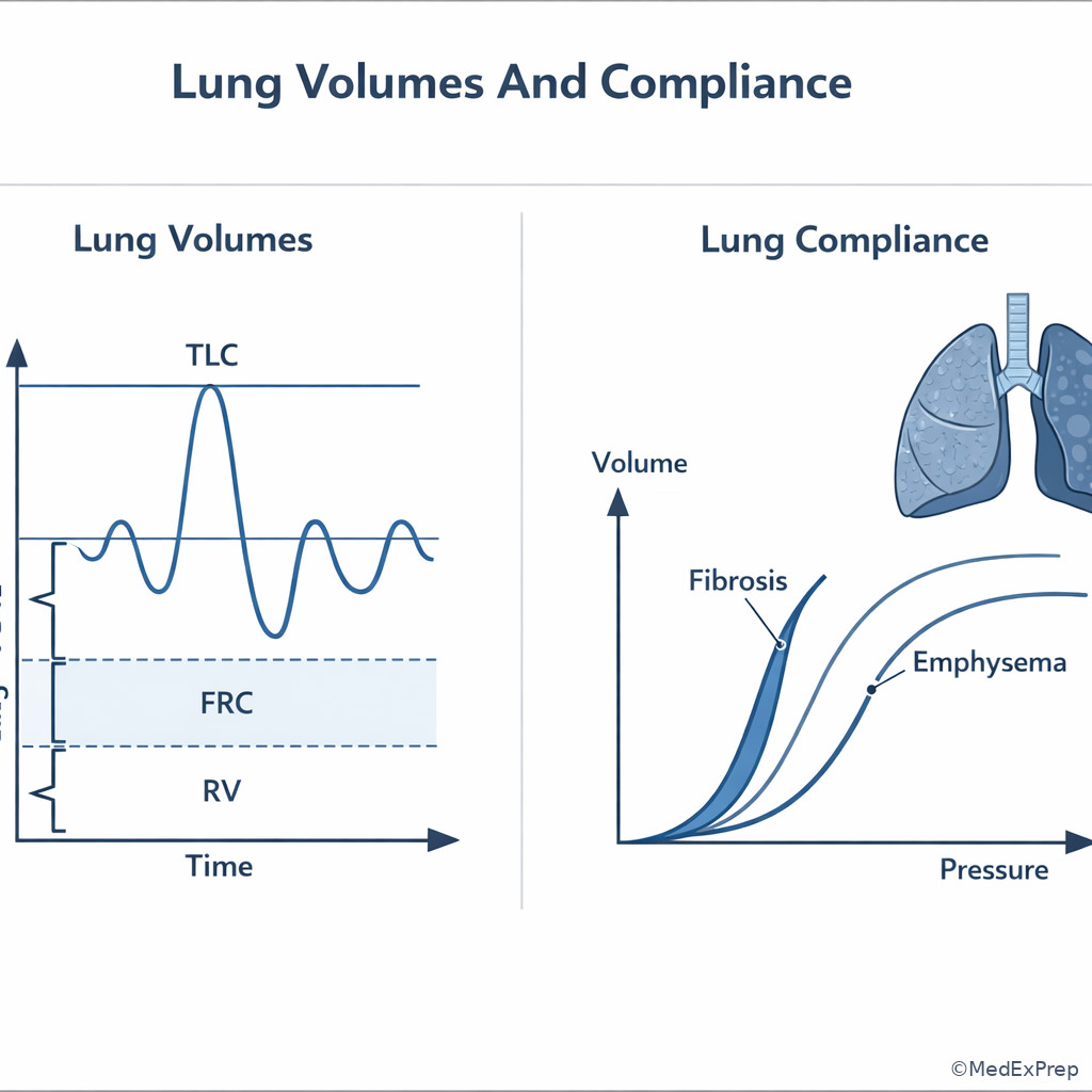
Why it matters
- Board questions often hide the real task: not naming a disease, but deciding whether the pattern is obstruction with air trapping, true restriction, pseudorestriction, or altered compliance.
- Lung volumes explain why two dyspneic patients can have similar spirometry but very different mechanisms and management pathways.
- Compliance is the bridge from physiology to imaging and bedside behavior: emphysema lungs inflate easily but empty poorly; fibrotic lungs resist inflation and produce rapid shallow breathing.
- This topic sets up upcoming interpretation tasks in Fig 4 and Fig 5, especially V/Q mismatch and DLCO pattern recognition.
Key Terms & Must-Know Facts
1.1.1.1 Key terms
- TLC: Total lung capacity
- RV: Residual volume
- FRC: Functional residual capacity
- VC: Vital capacity
- IC: Inspiratory capacity
- ERV: Expiratory reserve volume
- Air trapping: Increased RV due to incomplete exhalation
- Hyperinflation: Increased TLC, often with increased FRC and RV
- Compliance: Change in volume per change in pressure, \( C = \Delta V / \Delta P \)
- Elastance: Tendency to recoil; inverse of compliance
- Static compliance: Compliance measured without airflow
- Dynamic compliance: Compliance during breathing; affected by airway resistance
- Pseudorestriction: Low FVC from severe air trapping with normal or high TLC
- Closing volume: Volume at which dependent airways begin to close during exhalation
1.1.1.2 Must-know facts
- Spirometry cannot measure RV or TLC; body plethysmography or gas dilution techniques are needed.
- Restriction requires reduced TLC; low FVC alone is not enough.
- Obstruction often raises RV first; TLC may be normal early and high later.
- Emphysema increases compliance because elastic recoil is lost.
- Pulmonary fibrosis decreases compliance because lungs become stiff.
- Obesity lowers FRC, often before TLC falls.
- Neuromuscular weakness lowers VC; RV may increase due to incomplete exhalation and weak expiratory muscles.
- Chest wall restriction reduces respiratory system compliance even when intrinsic lung tissue is normal.
- Rapid shallow breathing is mechanically favored in low-compliance states.
- Barrel chest and flattened diaphragms suggest hyperinflation, not just obstruction on spirometry.
| Volume or capacity | What it includes | Measured by spirometry? | High-yield test implication |
|---|---|---|---|
| TV | Tidal breath | Yes | Rarely the discriminating board variable |
| IRV | Extra inhaled after tidal inspiration | Yes | Falls when inspiratory reserve is limited |
| ERV | Extra exhaled after tidal expiration | Yes | Often reduced early in obesity |
| RV | Air left after maximal exhalation | No | Key marker of air trapping |
| VC | IRV + TV + ERV | Yes | Can fall in both restriction and severe obstruction |
| IC | TV + IRV | Yes | Falls when hyperinflation encroaches on inspiratory reserve |
| FRC | ERV + RV | No | Raised in hyperinflation; reduced in obesity and supine position |
| TLC | All lung volume compartments | No | Required to confirm restriction |
Exam takeaway: Use TLC first to confirm restriction and RV first to identify air trapping when FVC is low.
flowchart TD
A[Low FVC or dyspnea on PFT stem] --> B{Is TLC low?}
B -->|Yes| C[True restriction]
B -->|No| D{Is RV high?}
D -->|Yes| E[Pseudorestriction from air trapping]
D -->|No| F[Poor effort or mixed/other pattern]
C --> G[Think ILD chest wall obesity neuromuscular]
E --> H[Think asthma COPD severe obstruction]
F --> I[Need full PFT quality review] Exam takeaway: Distinguish which compartments include RV because those cannot be measured by simple spirometry.
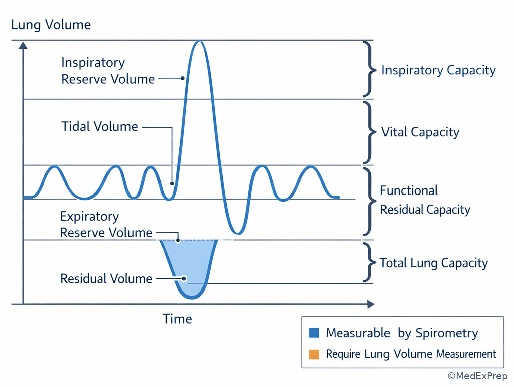
Core content
1.1.1.3 Foundational lung volumes: what changes first, and what actually proves restriction?
A Explanation
The central decision conflict is low FVC: restriction versus air trapping. The single rule that resolves it is simple: restriction requires a reduced TLC. If the stem gives only spirometry with low FVC, you do not yet know whether the patient has restrictive disease. Severe obstruction can lower FVC because RV expands and steals from the exhalable volume, creating pseudorestriction. On ABIM-style questions, the examiner often wants you to choose full lung volumes rather than prematurely labeling a restrictive defect.
Normal volume relationships matter because disease shifts them in recognizable directions. Obstruction tends to increase RV first, then FRC, and in more advanced hyperinflation can raise TLC. Restriction reduces TLC and usually reduces all subdivided volumes proportionally, although the exact pattern depends on the mechanism. Obesity often lowers ERV and FRC early; neuromuscular disease may lower VC while RV is preserved or elevated. WHY THIS IS TESTED: the testable pattern is a low FVC with reduced FEV1/FVC-normalized uncertainty, where only TLC clarifies the true physiology.
B Worked example
A 62-year-old man with 45 pack-years of smoking, coronary artery disease, and gastroesophageal reflux is referred for exertional dyspnea. He has occasional wheeze and bibasilar crackles are absent. Spirometry shows FEV1 52% predicted, FVC 61% predicted, and FEV1/FVC 0.68. A clinic note labels this “mixed obstructive-restrictive disease.” Chest radiograph mentions mild hyperlucency; BMI is 31 kg/m2, which is a tempting distraction toward obesity-related restriction. The most appropriate next step is full lung volume testing.
Reasoning chain: Identify task: determine whether restriction is truly present. Extract key facts: obstructive ratio, smoker, hyperlucency, low FVC. Apply rule: low FVC does not prove restriction; need TLC. Eliminate distractors: obesity can reduce FRC and ERV, but an obstructive ratio plus smoking history makes pseudorestriction plausible; empiric fibrosis workup is premature without low TLC. Verify: if TLC returns normal/high with elevated RV, this is obstruction with air trapping, not mixed disease.
C Exam trap
Why it is tempting: Learners correctly recognize obstruction and then assume the reduced FVC must represent an additional restrictive process.
Single clue that eliminates it: No TLC was provided. Without reduced TLC, you cannot diagnose restriction.
D Checkpoint
Question: A 58-year-old woman with chronic cough, obesity, and prior smoking has FEV1/FVC 0.64, FEV1 58% predicted, and FVC 63% predicted. Which finding would most strongly support pseudorestriction rather than true mixed disease?
- Reduced TLC to 68% predicted
- Elevated RV to 165% predicted
- Reduced DLCO to 48% predicted
- Bibasilar reticulation on CT
- A: Tempting because low TLC confirms restrictive physiology, but that argues against pseudorestriction and supports true mixed disease.
- B: Tempting because RV is not always emphasized, and it is correct here because marked RV elevation identifies air trapping as the cause of low FVC.
- C: Tempting because low DLCO can coexist with emphysema, but it does not by itself explain low FVC or distinguish true restriction from air trapping.
- D: Tempting because parenchymal abnormality suggests restriction, but CT reticulation points toward an additional interstitial process rather than pseudorestriction.
1.1.1.4 Compliance and recoil: choose emphysema versus fibrosis by the mechanics
A Explanation
The decision conflict here is hyperexpanded lungs that are easy to inflate versus stiff lungs that resist inflation. The single finding that resolves it is the direction of compliance change. Compliance is \( \Delta V / \Delta P \). High compliance means a small pressure change produces a large volume change; low compliance means the lungs are stiff. Emphysema destroys alveolar walls and elastic tissue, so the lung becomes highly compliant but loses recoil. Fibrosis increases elastic recoil and makes the lung difficult to inflate, so compliance falls.
This matters because the same symptom—dyspnea—arises from opposite mechanics. The emphysema patient has trouble exhaling and traps gas. The fibrosis patient has trouble inflating and compensates with rapid shallow breathing. Pressure-volume curves shift accordingly: emphysema shows a steeper curve and larger resting volumes; fibrosis shows a flatter curve and lower volumes. WHY THIS IS TESTED: board questions present chest imaging, body habitus, and symptom tempo, then ask you to identify the mechanical abnormality or predict lung volume/compliance changes. See Fig 2.
Exam takeaway: High compliance with low recoil points to emphysema, whereas low compliance with high recoil points to fibrosis.
flowchart LR
A[Dyspnea with suspected mechanics problem] --> B{Compliance}
B -->|High| C[Easy inflation]
C --> D[Low elastic recoil]
D --> E[Air trapping and hyperinflation]
E --> F[Emphysema pattern]
B -->|Low| G[Stiff lung]
G --> H[Higher recoil pressure]
H --> I[Rapid shallow breathing]
I --> J[Fibrotic restriction pattern]
Do not confuse compliance of the lung with compliance of the total respiratory system. Chest wall problems, obesity, ascites, and kyphoscoliosis can lower overall respiratory system compliance even if intrinsic lung tissue is normal. That distinction matters when the stem points to obesity hypoventilation or neuromuscular/chest wall restriction rather than ILD.
B Worked example
A 69-year-old man with 50 pack-years of smoking, hypertension, and stable ischemic heart disease reports progressive exertional dyspnea. He is thin, uses accessory muscles, and has decreased breath sounds without crackles. CT from an outside hospital notes hyperinflation and bullous change; a distracting note mentions a positive ANA at low titer. The stem asks for the most likely compliance change. The answer is increased lung compliance with reduced elastic recoil.
Reasoning chain: Identify task: determine mechanics, not etiology of autoimmune disease. Extract key facts: smoker, hyperinflation, bullae, absent crackles. Apply rule: emphysema destroys elastic tissue, increasing compliance. Eliminate distractors: low-titer ANA is common and not enough to reclassify the patient into connective tissue disease-associated ILD; fibrosis would produce crackles, reduced volumes, and low compliance. Verify: emphysema explains easy inflation, gas trapping, and hyperexpanded chest.
C Exam trap
Why it is tempting: Test-takers overgeneralize that any abnormal lung is stiffer than normal.
Single clue that eliminates it: Hyperinflation with bullae means loss of elastic recoil, the hallmark of increased compliance.
D Checkpoint
Question: Which patient most likely has decreased lung compliance as the dominant physiologic abnormality?
- A 64-year-old man with centrilobular emphysema and increased RV/TLC
- A 57-year-old woman with bibasilar reticulation, reduced TLC, and fine inspiratory crackles
- A 48-year-old man with acute bronchospasm and marked wheezing but normal TLC between exacerbations
- A 61-year-old woman with chronic bronchitis and preserved lung volumes
- A: Tempting because emphysema causes major physiologic abnormality, but the abnormality is increased compliance, not decreased compliance.
- B: Tempting because crackles can be underweighted, and it is correct because fibrotic interstitial disease produces stiff lungs with low compliance and low TLC.
- C: Tempting because wheezing suggests difficult breathing, but acute airway resistance is not the same as chronically decreased lung compliance.
- D: Tempting because chronic bronchitis is chronic lung disease, but compliance is often not the defining abnormality when lung volumes are preserved.
- The most reliable volume to confirm restriction is TLC.
- True or False: Emphysema decreases elastic recoil. True.
- Name the 3 volume changes most associated with hyperinflation: RV, FRC, and often TLC.
1.1.1.5 Restrictive patterns: intrinsic lung disease versus extraparenchymal restriction
A Explanation
The decision conflict is small lungs from parenchymal stiffness versus small lungs from an external mechanical limit. The single resolving step is to integrate TLC pattern plus clinical context. Both intrinsic and extrinsic restriction reduce TLC, but they differ in associated clues. Intrinsic restrictive disease such as ILD lowers compliance within lung tissue, often reduces DLCO, and produces parenchymal imaging abnormalities. Extraparenchymal restriction—obesity, kyphoscoliosis, pleural disease, or neuromuscular weakness—limits chest expansion or respiratory muscle performance; imaging may show low volumes without interstitial abnormalities, and DLCO may be preserved when corrected for alveolar volume depending on the condition.
Board stems often disguise this distinction by emphasizing dyspnea and low FVC but slipping in a clue such as severe scoliosis, morbid obesity, elevated hemidiaphragm, or myasthenia symptoms. In obesity, FRC and ERV commonly fall first; TLC may remain normal until obesity is more severe. In neuromuscular disease, VC falls and RV may rise because weak muscles cannot fully exhale. WHY THIS IS TESTED: the exam wants you to choose the correct mechanism and the best next diagnostic focus—parenchymal imaging versus respiratory muscle assessment versus body habitus/chest wall explanation. See Fig 3.
Exam takeaway: Low TLC with abnormal parenchymal clues suggests intrinsic restriction, whereas low TLC with obesity, chest wall, or weakness clues suggests extraparenchymal restriction.
flowchart TD
A[Low TLC confirmed] --> B{Parenchymal clues?}
B -->|Reticulation crackles low DLCO| C[Intrinsic lung restriction]
C --> D[Think ILD or alveolar process]
B -->|Obesity scoliosis weakness pleural disease| E[Extraparenchymal restriction]
E --> F[Check chest wall diaphragm or neuromuscular cause]
F --> G[DLCO may be less reduced than in ILD] B Worked example
A 54-year-old woman with BMI 44 kg/m2, hypertension, and diabetes presents with exertional dyspnea. She has no crackles, chest radiograph shows low lung volumes without infiltrates, and spirometry reveals reduced FVC. A note from urgent care suggests “possible pulmonary fibrosis.” Her TLC is 82% predicted, ERV is markedly reduced, and RV is normal. A mildly elevated ESR is the distracting clue. The best interpretation is obesity-related reduction in ERV/FRC without true restriction.
Reasoning chain: Identify task: determine whether low FVC means restrictive lung disease. Extract key facts: severe obesity, low-volume radiograph, absent crackles, normal TLC, low ERV. Apply rule: obesity reduces ERV and FRC early and may lower FVC without reducing TLC enough to diagnose restriction. Eliminate distractors: fibrosis would require low TLC and supporting imaging/parenchymal exam; elevated ESR is nonspecific noise. Verify: the pattern is extraparenchymal mechanical limitation from obesity, not ILD.
C Exam trap
Why it is tempting: Low-volume films and dyspnea superficially resemble restriction from lung fibrosis.
Single clue that eliminates it: TLC is normal; true restriction is not established.
D Checkpoint
Question: A 66-year-old man with progressive dyspnea, severe kyphoscoliosis, and no smoking history has low TLC and normal chest CT parenchyma. Which interpretation is most appropriate?
- Intrinsic restrictive disease due to occult ILD
- Extraparenchymal restriction due to reduced chest wall compliance
- Obstruction with pseudorestriction from air trapping
- Normal physiology because CT is unrevealing
- A: Tempting because low TLC suggests restriction, but the normal parenchyma and major chest wall deformity argue against intrinsic ILD.
- B: Tempting because chest wall mechanics are easy to overlook, and it is correct because kyphoscoliosis reduces respiratory system compliance and causes extraparenchymal restriction.
- C: Tempting because pseudorestriction is common, but low TLC—not just low FVC—proves true restriction rather than obstruction with air trapping.
- D: Tempting because CT seems reassuring, but normal lung parenchyma does not exclude nonparenchymal restriction.
1.1.1.6 Air trapping, hyperinflation, and pseudorestriction in obstructive disease
A Explanation
The decision conflict is does a low FVC in obstructive disease mean mixed pathology, or is it just severe air trapping? The single best discriminator is the relationship between TLC and RV. Air trapping means RV is elevated because the patient cannot empty fully. Hyperinflation means the resting operating volume is shifted upward, usually with increased FRC and often increased TLC. In advanced obstruction, especially emphysema, the inspiratory reserve shrinks because the lungs already sit near higher volumes; the patient becomes mechanically disadvantaged. See Fig 4.
This explains classic bedside features: flattened diaphragms, barrel chest, reduced inspiratory capacity, and dyspnea with exertion due to dynamic hyperinflation. On a board stem, the examiner may not use the words “dynamic hyperinflation” but will describe severe COPD, tachypnea, and worsening breathlessness during exertion because expiratory time is too short to empty each breath. At rest on formal PFTs, static hyperinflation shows high RV and FRC; during exercise or exacerbation, dynamic hyperinflation worsens symptoms further. WHY THIS IS TESTED: ABIM stems often ask you to reject false mixed disease and to link hyperinflation to symptom burden or therapy response.
Exam takeaway: Flattened diaphragms and enlarged residual volume identify obstructive air trapping rather than true restriction.
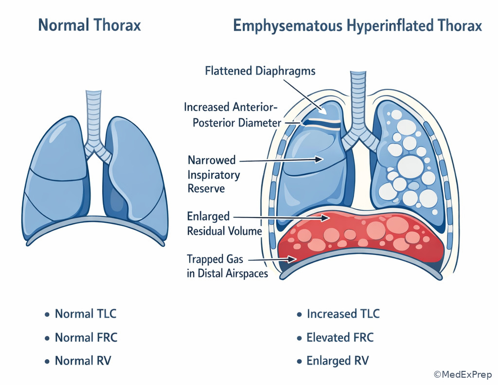
B Worked example
A 63-year-old woman with severe COPD, osteoporosis, and anxiety reports marked dyspnea climbing one flight of stairs. Spirometry shows severe obstruction; lung volumes reveal TLC 122% predicted, FRC 148% predicted, and RV 185% predicted. She also has mild ankle edema, a distracting clue that can pull attention toward heart failure. The best explanation for her exertional breathlessness is hyperinflation with reduced inspiratory capacity due to air trapping.
Reasoning chain: Identify task: explain symptom mechanism. Extract key facts: severe obstruction, strikingly high RV/FRC/TLC. Apply rule: hyperinflated lungs operate near high resting volume, leaving little room for tidal expansion during exertion. Eliminate distractors: mild edema alone does not explain this highly characteristic lung-volume pattern; restrictive disease is excluded by high TLC. Verify: dynamic hyperinflation during exercise worsens dyspnea even when oxygenation is not yet profoundly abnormal.
C Exam trap
Why it is tempting: FVC is commonly misused as a stand-alone marker of restriction.
Single clue that eliminates it: TLC is elevated, which rules out pure restriction and supports hyperinflation.
D Checkpoint
Question: In a patient with established COPD, which PFT change most directly reflects air trapping?
- Decreased TLC
- Increased RV
- Increased VC
- Decreased FRC
- A: Tempting because severe lung disease often lowers some values, but decreased TLC suggests restriction rather than air trapping.
- B: Tempting because several volumes change in COPD, and it is correct because retained gas after maximal exhalation defines elevated RV.
- C: Tempting because patients may improve VC after bronchodilator therapy, but increased VC is not the defining static marker of air trapping.
- D: Tempting if one confuses obesity with obstruction, but FRC usually rises rather than falls in hyperinflation.
- The most common static volume abnormality in early obstruction is increased RV.
- True or False: High TLC excludes pure restrictive disease. True.
- Name the 2 bedside imaging clues of hyperinflation: flattened diaphragms and increased retrosternal airspace/barrel chest.
1.1.1.7 Integrating mechanics with test selection and next-step interpretation
A Explanation
The final decision conflict is which additional pulmonary test best resolves the stem’s ambiguity? The single rule is to order the test that measures the missing physiologic compartment. If the question hinges on whether restriction is present, order lung volumes. If the question hinges on gas transfer, upcoming lessons will favor DLCO. If hypoxemia mechanism is unclear, later lessons on V/Q and A-a gradient become the next step. In this lesson, do not overreach: lung volumes and compliance solve mechanical questions, but they do not answer every gas-exchange question.
A common ABIM design is progressive data release. First, spirometry suggests a pattern. Next, lung volumes confirm or refute restriction. Then DLCO or imaging refines the disease class. You score well when you answer the current question, not the eventual full diagnosis. WHY THIS IS TESTED: pulmonary boards reward disciplined sequencing—identify the missing variable, choose the right test, and avoid premature closure. See Fig 5 and Fig 6.
Exam takeaway: Choose the next pulmonary test based on the unresolved physiologic question rather than ordering the entire workup reflexively.
flowchart TD
A[Spirometry completed] --> B{Question left unanswered}
B -->|Need to confirm restriction| C[Order lung volumes]
B -->|Need gas transfer clue| D[Order DLCO]
B -->|Need hypoxemia mechanism| E[Use ABG or oxygenation framework]
C --> F[TLC low = restriction confirmed]
D --> G[Low or high DLCO refines disease class]
E --> H[VQ mismatch shunt dead space logic] Exam takeaway: Steeper curves indicate high compliance and flatter curves indicate low compliance when board stems ask about recoil.
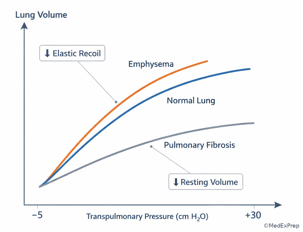
B Worked example
A 71-year-old woman with rheumatoid arthritis, prior tobacco use, and chronic anemia presents with progressive dyspnea. Spirometry shows preserved FEV1/FVC and low FVC. CT from 2 years earlier was normal; current exam reveals mild crackles. A tempting wrong move is to order DLCO immediately because rheumatoid arthritis and anemia both affect it. The best next step is to obtain lung volumes first, because the unresolved question is whether restriction is actually present.
Reasoning chain: Identify task: confirm physiologic category. Extract key facts: low FVC, preserved ratio, systemic disease clues, anemia noise. Apply rule: low FVC alone does not prove restriction, so TLC comes before DLCO interpretation. Eliminate distractors: DLCO may indeed be useful later, but anemia would confound it and it does not replace TLC for confirming restriction. Verify: once low TLC is proven, you can then pursue ILD evaluation more confidently.
C Exam trap
Why it is tempting: Low DLCO is strongly associated with many pulmonary diseases and feels more disease-specific.
Single clue that eliminates it: The stem’s unresolved question is whether the patient is truly restrictive, and only TLC answers that directly.
D Checkpoint
Question: A patient has low FVC with preserved FEV1/FVC. The stem asks for the best test to confirm a restrictive ventilatory defect. What is the best answer?
- Repeat spirometry after bronchodilator
- Lung volume measurement with TLC
- DLCO measurement alone
- Six-minute walk test
- A: Tempting because bronchodilator response can clarify obstruction, but it does not confirm or exclude restriction.
- B: Tempting because it seems less disease-specific, and it is correct because reduced TLC is required to diagnose restriction.
- C: Tempting because DLCO often falls in ILD, but DLCO alone cannot confirm restrictive physiology.
- D: Tempting because functional limitation matters clinically, but exercise testing does not establish the mechanical category.
Exam Traps & Differentiators
Most common wrong answer and why: Calling “restriction” from low FVC alone. This is attractive because many board-takers overvalue spirometry while forgetting that RV and TLC are invisible to basic spirometry.
| Looks similar | Why it tempts | Single discriminator | Best board move |
|---|---|---|---|
| True restriction vs pseudorestriction | Both can have low FVC | TLC low only in true restriction | Order or interpret lung volumes |
| Emphysema vs fibrosis | Both cause dyspnea and low DLCO later | Compliance high in emphysema, low in fibrosis | Use imaging plus volume pattern |
| Obesity vs ILD | Both can produce low lung volumes on chest radiograph | Obesity lowers ERV/FRC early; ILD usually has parenchymal findings | Do not overcall ILD without TLC/imaging support |
| Neuromuscular weakness vs parenchymal restriction | Both can lower VC and TLC | Weakness may raise RV and has muscle/positional clues | Assess respiratory muscle function |
| Hyperinflation vs air trapping | Often used interchangeably in casual teaching | Air trapping = high RV; \(hyperinflation = high FRC and often high TLC\) | Name the exact abnormality asked |
- If the stem says low FVC with no TLC provided, think “restriction is not yet proven.”
- If the stem says bullae, hyperlucency, flattened diaphragms, think increased compliance and reduced recoil.
- If the stem says severe obesity with low ERV/FRC and normal imaging, think extraparenchymal mechanics before ILD.
- If the stem says low TLC plus normal CT parenchyma with scoliosis or neuromuscular clues, think chest wall or muscle restriction.
- If the stem says smoker with obstructive ratio and low FVC, think air trapping until TLC proves otherwise.
- If the stem says exertional dyspnea with marked RV/TLC elevation, think hyperinflation-driven mechanical limitation.
Tables
| Condition | TLC | RV | FRC | Compliance | Classic clue |
|---|---|---|---|---|---|
| Emphysema | Normal to high | High | High | High | Hyperinflation, bullae, reduced recoil |
| Asthma with air trapping | Normal to high | High | High | Usually not the primary defect | Reversible obstruction |
| ILD/fibrosis | Low | Low to normal | Low | Low | Crackles, reticulation |
| Obesity | Often normal early; low if severe | Normal | Low | Respiratory system compliance low | Markedly low ERV |
| Neuromuscular weakness | Low | Normal to high | Variable | Lung tissue may be normal | Weak cough, positional VC change |
| Kyphoscoliosis | Low | Variable | Low | Respiratory system compliance low | Chest wall deformity |
| Feature | Intrinsic restriction | Extraparenchymal restriction |
|---|---|---|
| Main problem | Stiff lung tissue | Limited chest expansion or weak respiratory muscles |
| TLC | Low | Low |
| Compliance | Lung compliance low | Respiratory system compliance low; lung tissue may be normal |
| Imaging | Parenchymal abnormality common | Often normal parenchyma |
| DLCO tendency | Often reduced | May be preserved or less reduced depending on cause |
| Exam clues | Crackles, connective tissue disease, fibrosis pattern | Obesity, scoliosis, weakness, pleural disease |
| Best next focus | HRCT/ILD workup | Body habitus, chest wall, diaphragm, muscle assessment |
| Misleading stem feature | Wrong conclusion | Correct interpretation |
|---|---|---|
| Low FVC on spirometry | Definite restriction | Need TLC to confirm restriction |
| Low lung volumes on radiograph in obesity | Fibrotic lung disease | Obesity commonly lowers ERV/FRC without ILD |
| Low DLCO in smoker | Must be fibrosis | Emphysema also lowers DLCO |
| Dyspnea with wheeze and low FVC | Mixed disease | Could be obstruction with air trapping |
| Normal CT in low TLC patient | No pulmonary abnormality exists | Chest wall or neuromuscular restriction may explain it |
| Severe COPD with small inspiratory reserve | Weak respiratory muscles as primary problem | Hyperinflation can mechanically limit inspiration |
Algorithm / Approach
Rapid Review
- Restriction → requires low TLC, not just low FVC
- Pseudorestriction → low FVC with normal/high TLC and high RV
- Air trapping → best static marker is elevated RV
- Hyperinflation → increased FRC and often increased TLC
- Emphysema → high compliance, low recoil
- Fibrosis → low compliance, high recoil
- Obesity → ERV and FRC fall early
- Neuromuscular weakness → low VC with possible high RV
- Kyphoscoliosis → extraparenchymal restriction from chest wall mechanics
- Bullae plus flattened diaphragms → choose hyperinflation, not fibrosis
- Normal parenchymal CT with low TLC → think chest wall or muscle cause
- Steeper pressure-volume curve → increased compliance
- Flatter pressure-volume curve → decreased compliance
- Next test after uncertain low FVC → lung volumes
Self-check quiz
1. A 60-year-old man with chronic cough and prior smoking has low FVC on screening spirometry with preserved FEV1/FVC. Which measurement is most appropriate to confirm a restrictive ventilatory defect?
- TLC
- PEF
- FEV1
- ERV
2. A 67-year-old woman with progressive dyspnea, clubbing, and bibasilar crackles asks which mechanical change best explains her breathing pattern. Which is most likely?
- Increased lung compliance
- Decreased lung compliance
- Increased RV from air trapping as the primary defect
- Increased chest wall compliance
3. A 58-year-old man with obesity, hypertension, and exertional dyspnea has FEV1/FVC 0.82 and FVC 69% predicted. Chest radiograph shows low lung volumes without infiltrates. Which is the most appropriate next step to determine whether he has true restriction?
- Order lung volumes with TLC measurement
- Start inhaled bronchodilator therapy and repeat symptoms assessment
- Diagnose interstitial lung disease and order antifibrotic evaluation
- Order six-minute walk testing only
4. A 65-year-old woman with severe COPD, anxiety, and coronary disease has worsening exertional dyspnea. Spirometry shows severe obstruction; lung volumes reveal markedly elevated RV and increased TLC. Which mechanism best explains her symptom burden?
- Dynamic and static hyperinflation reducing inspiratory capacity
- Primary intrinsic restrictive disease lowering TLC
- Pulmonary edema causing acute low compliance as the main chronic process
- Upper airway obstruction causing fixed flattening as the dominant abnormality
5. A 52-year-old woman with rheumatoid arthritis, anemia, and chronic dyspnea has preserved FEV1/FVC with low FVC on spirometry. Fine crackles are present, but no lung volumes have been measured. Which test most appropriately clarifies the immediate physiologic question?
- TLC measurement
- DLCO alone
- Bronchoprovocation testing
- Repeat pulse oximetry after ambulation only
6. A 61-year-old man with 40 pack-years of smoking, weight loss, and chronic dyspnea has FEV1/FVC 0.63, FVC 64% predicted, TLC 118% predicted, RV 176% predicted, and DLCO 45% predicted. CT shows upper-lobe lucencies and no reticulation. Which is the most likely physiologic interpretation?
- Obstruction with air trapping and emphysematous hyperinflation
- True mixed obstructive-restrictive disease due to emphysema plus occult fibrosis
- Pure restrictive disease from smoking-related ILD
- Normal lung volumes with isolated diffusion abnormality
7. A 55-year-old woman with morbid obesity, obstructive sleep apnea, and diabetes reports dyspnea. Spirometry shows low FVC; TLC is 84% predicted, ERV is markedly reduced, RV is normal, and CT chest shows no interstitial abnormality. Which interpretation is most appropriate?
- Intrinsic fibrotic restriction
- Obesity-related mechanical reduction in ERV/FRC without confirmed restriction
- Severe air trapping from emphysema
- Neuromuscular weakness causing elevated RV
8. A 68-year-old man with progressive exertional dyspnea, rheumatoid arthritis, former smoking, and chronic kidney disease has TLC 63% predicted, RV 72% predicted, bibasilar crackles, and CT with subpleural reticulation. Which single physiologic statement is most accurate?
- Lung compliance is likely increased because total lung volume is reduced
- Lung compliance is likely decreased because the parenchyma is stiff
- The low TLC is most likely due to isolated obesity physiology
- The dominant abnormality is pseudorestriction from air trapping
Answer key
1.1.1.8 1. Answer: A. TLC
Why A is correct: The mechanism is that true restrictive ventilatory defects are defined by reduced total lung capacity. Low FVC alone may reflect poor effort, obesity, or pseudorestriction from air trapping. The single stem clue is preserved FEV1/FVC with low FVC, which leaves restriction unconfirmed. Source: Pellegrino et al.; ATS/ERS interpretive standards and standard pulmonary physiology texts.
Why B is tempting but wrong: Peak expiratory flow sounds like another volume-related measure, but it is effort-dependent airflow and does not define restriction. The discriminating clue is the need to measure a compartment containing RV, which PEF cannot do. Source: ATS/ERS standards.
Why C is tempting but wrong: FEV1 is familiar and often reported first, but it is still spirometric airflow, not a total lung volume. The clue is that the question asks to confirm restriction, which requires TLC. Source: Murray and Nadel’s Textbook of Respiratory Medicine.
Why D is tempting but wrong: ERV changes in obesity and some restrictive states, but isolated ERV cannot establish the presence of restriction. The discriminating clue is that ERV does not substitute for the full TLC definition. Source: ATS/ERS standards.
1.1.1.9 2. Answer: B. Decreased lung compliance
Why B is correct: Clubbing, bibasilar crackles, and progressive dyspnea point toward fibrotic interstitial lung disease, in which lung parenchyma is stiff and compliance falls. The discriminating clue is the crackles plus fibrotic clinical picture, not just dyspnea. Source: Harrison’s Principles of Internal Medicine; ATS/ERS ILD references.
Why A is tempting but wrong: Increased compliance is a famous pulmonary mechanics abnormality, but it belongs to emphysema with hyperinflation and reduced recoil rather than crackles and clubbing. The clue that rejects it is the fibrotic bedside phenotype. Source: Costanzo Physiology; Harrison’s.
Why C is tempting but wrong: Elevated RV is a real pulmonary abnormality and can cause dyspnea, but this stem lacks obstructive clues and instead suggests restrictive parenchymal disease. The single discriminator is inspiratory crackles rather than wheeze/hyperinflation. Source: Murray and Nadel’s.
Why D is tempting but wrong: Chest wall compliance matters in extraparenchymal conditions, but nothing here points to obesity, scoliosis, or another chest wall process. The stem clue favoring parenchymal stiffness is bibasilar crackles with clubbing. Source: Harrison’s.
1.1.1.10 3. Answer: A. Order lung volumes with TLC measurement
Why A is correct: The diagnosis-mechanism-treatment logic is to first confirm whether restrictive physiology truly exists. Low FVC with preserved ratio in an obese patient may reflect obesity mechanics rather than intrinsic restriction. The single stem clue is the unresolved physiologic category. Source: ATS/ERS interpretive standards.
Why B is tempting but wrong: Bronchodilator therapy seems reasonable for dyspnea, but it does not answer whether a restrictive defect is present. The discriminator is the preserved ratio without proof of obstruction. Source: ATS/ERS standards.
Why C is tempting but wrong: Low lung volumes on chest radiograph can suggest ILD, but obesity is a close-call mimic. The single clue that rejects immediate ILD labeling is absence of infiltrates and lack of confirmed low TLC. Source: Harrison’s.
Why D is tempting but wrong: Walk testing helps functional assessment and desaturation evaluation, but it does not define the mechanical pattern. The clue is that the question asks whether he has true restriction. Source: ATS/ERS standards.
1.1.1.11 4. Answer: A. Dynamic and static hyperinflation reducing inspiratory capacity
Why A is correct: Severe obstruction with markedly elevated RV and increased TLC indicates air trapping and hyperinflation. Mechanistically, the lungs operate at higher resting volumes, compressing inspiratory reserve and worsening exertional dyspnea. The discriminating clue is the combination of high RV and high TLC in COPD. Source: GOLD strategy documents; Murray and Nadel’s.
Why B is tempting but wrong: Restriction can cause dyspnea, but low TLC would be expected, not increased TLC. The single clue excluding it is hyperinflation rather than volume loss. Source: ATS/ERS standards.
Why C is tempting but wrong: Low compliance from edema causes dyspnea, but this is a chronic COPD physiology stem with established hyperinflation, not acute alveolar flooding. The discriminator is the chronic obstructive volume pattern. Source: Harrison’s.
Why D is tempting but wrong: Fixed upper airway obstruction can cause exertional symptoms, but the provided lung volumes strongly support lower-airway obstructive hyperinflation instead. The single clue is markedly elevated RV/TLC rather than flow-volume loop flattening data. Source: ATS/ERS PFT interpretation guidance.
1.1.1.12 5. Answer: A. TLC measurement
Why A is correct: Rheumatoid arthritis and crackles make ILD plausible, but the immediate unresolved question remains whether there is true restriction. TLC must be measured first. The discriminating clue is low FVC with preserved ratio but no lung volumes yet. Source: ATS/ERS standards; Harrison’s.
Why B is tempting but wrong: DLCO is attractive because RA-associated ILD and anemia both affect gas transfer. However, anemia can confound DLCO, and DLCO cannot replace TLC for proving restrictive physiology. The single clue is that the question asks for the immediate physiologic question. Source: ATS/ERS; pulmonary physiology texts.
Why C is tempting but wrong: Bronchoprovocation assesses airway hyperresponsiveness and does not address low FVC with preserved ratio. The clue excluding it is the absence of an asthma-confirmation task. Source: ATS/ERS standards.
Why D is tempting but wrong: Ambulatory oximetry may reveal exertional desaturation, but it does not resolve whether the spirometric pattern is restrictive. The discriminator is confirmation of mechanical category, not oxygen qualification. Source: ATS/ERS standards.
1.1.1.13 6. Answer: A. Obstruction with air trapping and emphysematous hyperinflation
Why A is correct: The mechanism is classic emphysema: obstructive ratio, elevated TLC, markedly elevated RV, low DLCO, and CT lucencies without reticulation. The single clue that discriminates from mixed disease is the high TLC, which argues against restrictive physiology. Source: GOLD; ATS/ERS interpretation standards; Harrison’s.
Why B is tempting but wrong: Weight loss and low DLCO can make occult fibrosis seem possible, but true mixed disease would require reduced TLC or convincing restrictive imaging. The discriminator is TLC 118% predicted plus absent reticulation. Source: ATS/ERS standards.
Why C is tempting but wrong: Smoking-related ILD exists, but pure restrictive disease would not produce an obstructive ratio with high RV and high TLC. The clue excluding it is the emphysematous volume pattern. Source: Harrison’s.
Why D is tempting but wrong: Isolated diffusion abnormality would not explain the abnormal FEV1/FVC or high RV. The discriminating clue is clear airflow obstruction with hyperinflation. Source: ATS/ERS standards.
1.1.1.14 7. Answer: B. Obesity-related mechanical reduction in ERV/FRC without confirmed restriction
Why B is correct: Severe obesity commonly lowers ERV and FRC, sometimes with reduced FVC, while TLC may remain near normal. The single clue is markedly reduced ERV with TLC still 84% predicted and no parenchymal CT abnormality. Source: Murray and Nadel’s; pulmonary physiology references.
Why A is tempting but wrong: Low FVC can suggest intrinsic fibrotic restriction, but normal CT and nonreduced TLC argue against it. The discriminating clue is the obesity-specific volume pattern. Source: Harrison’s.
Why C is tempting but wrong: Emphysema would more likely raise RV and often TLC, not leave RV normal while ERV is selectively depressed. The single clue excluding it is normal RV without imaging evidence of emphysema. Source: GOLD.
Why D is tempting but wrong: Neuromuscular weakness can lower VC, but RV is often normal to high and the stem highlights obesity/OSA rather than weakness signs. The key discriminator is profound ERV reduction with preserved RV. Source: ATS/ERS physiology guidance.
1.1.1.15 8. Answer: B. Lung compliance is likely decreased because the parenchyma is stiff
Why B is correct: Reduced TLC, bibasilar crackles, rheumatoid arthritis, and subpleural reticulation point to intrinsic fibrotic restriction. Mechanistically, fibrotic lung parenchyma is stiff, so compliance decreases. The single clue is CT-confirmed reticulation with low TLC. Source: ATS/ERS ILD references; Harrison’s.
Why A is tempting but wrong: Some learners mistakenly infer that lower total volume means the lung empties well and thus is more compliant, but the opposite is true in fibrosis. The discriminating clue is parenchymal stiffness on imaging. Source: Costanzo Physiology.
Why C is tempting but wrong: Obesity can reduce some volumes, but this patient has rheumatoid arthritis, crackles, and reticulation—far stronger evidence of intrinsic disease. The key clue excluding obesity-only physiology is abnormal parenchymal imaging. Source: Harrison’s.
Why D is tempting but wrong: Pseudorestriction requires normal or high TLC with high RV from air trapping. Here TLC and RV are both reduced or low-normal, excluding that mechanism. The discriminator is confirmed low TLC rather than isolated low FVC. Source: ATS/ERS standards.
Keep going with the full ABIM Pulmonary Disease Prep Course (Pulm/CC)
You just studied the way members do, in the same lesson interface. Full access unlocks every lesson, the question bank and flashcards for a full year.
One payment · 1 year of access · No auto-renewal