1.1.1 - GFR And Renal Blood Flow
GFR And Renal Blood Flow
What you'll learn
- Physiology, Pathology & Diagnostics: Explain the determinants of glomerular filtration rate and renal blood flow -> renal hemodynamic reasoning
- Physiology, Pathology & Diagnostics: Distinguish measured GFR, estimated GFR, renal plasma flow, and filtration fraction -> diagnostic interpretation
- AKI & Critical Care: Predict how afferent and efferent arteriolar changes alter GFR in common clinical settings -> hemodynamic AKI pattern recognition
- CKD: Choose the most appropriate filtration marker and understand the major limitations of creatinine-based estimates -> CKD assessment
- Physiology, Pathology & Diagnostics: Apply autoregulation principles to board-style vignettes involving NSAIDs, RAAS blockade, cirrhosis, and heart failure -> mechanism-to-management logic
- Domain: Physiology, Pathology & Diagnostics → renal hemodynamics, filtration, and clearance interpretation
- Domain: AKI & Critical Care → prerenal and hemodynamic kidney dysfunction mechanisms
- Domain: CKD → kidney function assessment using serum markers and estimating equations
Why it matters
- Board stems frequently test hemodynamic kidney injury by asking what happens to GFR after NSAIDs, ACE inhibitors, sepsis, cirrhosis, or renal artery stenosis.
- Many management choices in nephrology depend on whether a creatinine rise reflects expected physiology, reduced perfusion, or intrinsic damage.
- Renal blood flow and filtration fraction are the foundation for later topics in fractional excretion, tubular transport, urine concentration, and AKI classification.
- The exam often rewards the candidate who identifies the single arteriolar or Starling-force change that resolves an otherwise noisy vignette.
Key Terms & Must-Know Facts
1.1.1.1 Key terms
- GFR: volume of plasma filtered across glomeruli per unit time
- Renal blood flow (RBF): blood delivered to both kidneys per minute
- Renal plasma flow (RPF): plasma component of renal blood flow
- Filtration fraction (FF): fraction of renal plasma flow that becomes filtrate
- Autoregulation: maintenance of relatively stable renal blood flow and GFR across a range of perfusion pressures
- Myogenic response: afferent arteriolar smooth muscle constricts with stretch
- Tubuloglomerular feedback: macula densa-mediated adjustment of afferent tone and renin release
- Net filtration pressure: balance of hydrostatic and oncotic forces across the glomerular capillary
- Creatinine clearance: practical surrogate for GFR, slightly overestimates true GFR
- Inulin clearance: ideal measured GFR marker
- PAH clearance: approximates renal plasma flow
- Effective arterial blood volume: perfusion sensed by the kidney, not identical to total body water
- Afferent arteriole: vessel entering the glomerulus
- Efferent arteriole: vessel leaving the glomerulus
1.1.1.2 Must-know facts
- Kidneys receive about 20% of cardiac output.
- Normal adult GFR is roughly 90-120 mL/min/1.73 m2, but interpretation depends on age and context.
- Creatinine-based eGFR is less reliable in extremes of muscle mass, unstable kidney function, amputation, rhabdomyolysis, and cirrhosis.
- ACE inhibitors and ARBs preferentially dilate the efferent arteriole.
- NSAIDs reduce vasodilatory prostaglandins and therefore can cause afferent constriction.
- Severe hypovolemia lowers both renal plasma flow and GFR.
- Efferent constriction may initially preserve GFR despite reduced renal plasma flow.
- Filtration fraction equals \( \text{GFR} / \text{RPF} \).
- Renal plasma flow equals \( \text{RBF} \times (1 - \text{hematocrit}) \).
- Creatinine clearance tends to overestimate GFR because creatinine is also secreted.
| Parameter | What it reflects | Typical exam value | High-yield caveat |
|---|---|---|---|
| RBF | Total renal perfusion | ~1.0-1.2 L/min | Falls with low cardiac output or renal vasoconstriction |
| RPF | Perfusing plasma flow | ~600-700 mL/min | Approximated by PAH clearance |
| GFR | Filtered plasma per minute | ~90-120 mL/min/1.73 m2 | Best overall index of kidney function |
| FF | Fraction of plasma filtered | ~20% | Often rises with efferent constriction |
| Serum creatinine | Marker used to estimate GFR | Context dependent | Delayed and muscle-mass dependent |
| Cystatin C | Alternative filtration marker | Lab dependent | Useful when creatinine is misleading |
Core content
1.1.1.3 Foundational determinants: what sets GFR at the glomerulus?
A. Explanation
The first board decision conflict is this: Is the kidney problem one of filtration pressure, blood flow, or marker interpretation? The single rule that resolves the first layer is that GFR depends on glomerular capillary hydrostatic pressure, Bowman space hydrostatic pressure, plasma oncotic pressure, and the filtration coefficient. In practical test language, most stems reduce to a change in afferent tone, efferent tone, plasma flow, or obstruction.
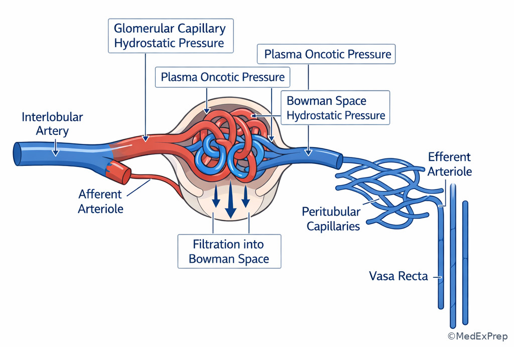
Think in Starling terms. Glomerular capillary hydrostatic pressure favors filtration. Plasma oncotic pressure opposes it and rises along the capillary as water is filtered. Bowman space hydrostatic pressure opposes filtration and increases in obstruction. The filtration coefficient reflects permeability and surface area; it falls in disease states that reduce functioning glomerular surface area.
The hemodynamic shortcut for exams: afferent dilation tends to increase both RBF and GFR; afferent constriction lowers both; efferent constriction lowers RPF and can initially maintain or raise GFR; efferent dilation tends to lower GFR. See Fig 1.
WHY THIS IS TESTED: the exam wants you to identify which single vascular or pressure change explains the creatinine change before moving to management.
Exam takeaway: Match the vignette to afferent tone, efferent tone, obstruction, or filtration surface loss before choosing the next step.
flowchart TD
A[Change in kidney function] --> B{What changed?}
B --> C[Afferent constriction]
B --> D[Afferent dilation]
B --> E[Efferent constriction]
B --> F[Efferent dilation]
B --> G[Obstruction / high Bowman pressure]
B --> H[Lower Kf / less filtering surface]
C --> C1[RBF down, GFR down]
D --> D1[RBF up, GFR up]
E --> E1[RPF down, FF up, GFR preserved or up early]
F --> F1[RPF up or stable, GFR down]
G --> G1[GFR down]
H --> H1[GFR down despite variable flow] B. Worked example
A 69-year-old man with ischemic cardiomyopathy, diabetes, and osteoarthritis is admitted for poor oral intake and dizziness after a viral illness. He takes lisinopril, furosemide, and daily ibuprofen. Blood pressure is 88/54 mm Hg. Creatinine rises from 1.2 to 2.1 mg/dL over 48 hours. Urinalysis is bland. A junior trainee focuses on trace proteinuria and suggests glomerulonephritis.
Identify task: explain the physiologic cause of the GFR drop. Extract key facts: hypotension, RAAS blockade, NSAID exposure, diuretic use, bland sediment. Apply rule: hypovolemia lowers renal perfusion; NSAIDs block afferent vasodilation; ACE inhibitor removes efferent support of glomerular pressure. Eliminate distractors: trace proteinuria is nonspecific; bland sediment argues against inflammatory intrinsic disease. Verify: the most likely mechanism is combined afferent constriction plus efferent dilation on a low-flow background causing marked fall in glomerular capillary pressure.
C. Exam trap
D. Checkpoint
Question: A patient with acute bilateral ureteral obstruction develops a sudden fall in GFR. Which physiologic change most directly explains this?
- Increased glomerular capillary hydrostatic pressure
- Decreased Bowman space hydrostatic pressure
- Increased Bowman space hydrostatic pressure
- Decreased plasma oncotic pressure
Answer: C
- A: Tempting because hydrostatic pressure is the main pro-filtration force, but obstruction lowers GFR by the opposite mechanism.
- B: Tempting if one remembers pressure gradients loosely, but Bowman space pressure rises, not falls, in obstruction.
- C: Correct because back pressure in Bowman space opposes filtration and abruptly lowers net filtration pressure.
- D: Tempting because oncotic changes affect GFR, but lower plasma oncotic pressure would favor filtration rather than reduce it.
1.1.1.4 Renal blood flow, renal plasma flow, and filtration fraction: the numbers behind the physiology
A. Explanation
The next decision conflict is which flow variable the question is actually asking about. Many candidates mix up renal blood flow, renal plasma flow, and GFR. The single resolving rule is that RBF includes cells and plasma; RPF includes only plasma; GFR is the filtered portion of RPF. Filtration fraction ties them together: \( \text{FF} = \text{GFR} / \text{RPF} \). Renal plasma flow can be estimated from blood flow using \( \text{RPF} = \text{RBF} \times (1 - \text{hematocrit}) \).
On exams, filtration fraction is most useful qualitatively. If efferent constriction lowers renal plasma flow but preserves glomerular capillary pressure, FF often rises. If both RPF and GFR fall proportionally, FF may remain unchanged. In classic prerenal states with neurohormonal compensation, FF may increase. In ATN, autoregulation and tubular mechanisms become less reliable, so the expected prerenal pattern may disappear.
Clearance concepts are tested as mechanism questions more often than heavy calculations. Inulin is the ideal marker because it is freely filtered and neither secreted nor reabsorbed. Creatinine is the clinical surrogate because it is easy to measure, but it slightly overestimates true GFR. PAH approximates renal plasma flow because it is filtered and avidly secreted, so almost all PAH delivered to the kidney is excreted in one pass at low concentrations. See Fig 2.
WHY THIS IS TESTED: the exam wants you to recognize which parameter changes in ACE inhibitor use, volume depletion, or efferent constriction rather than getting lost in terminology.
Exam takeaway: Translate every renal hemodynamic vignette into RBF, RPF, GFR, and FF rather than relying on memorized drug associations alone.
flowchart LR
A[Cardiac output] --> B[Renal blood flow]
B --> C[Subtract hematocrit]
C --> D[Renal plasma flow]
D --> E[Filtered portion]
E --> F[GFR]
F --> G[Filtration fraction = GFR / RPF]
H[Inulin clearance] --> F
I[PAH clearance] --> D
J[Creatinine clearance] --> K[Approximate GFR, slight overestimate]
Exam takeaway: Visualizing afferent inflow, the glomerular tuft, and efferent outflow helps predict whether a change primarily alters flow, pressure, or both.
B. Worked example
A 58-year-old woman with cirrhosis, tense ascites, and type 2 diabetes is admitted with rising creatinine after large-volume paracentesis without adequate albumin replacement. Hematocrit is 30%. A question asks which renal variable is most likely increased as compensatory angiotensin II levels rise. Bilirubin is markedly elevated, which distracts the team toward intrinsic tubular injury.
Identify task: determine the hemodynamic variable changed by angiotensin II. Extract key facts: low effective arterial blood volume, RAAS activation, cirrhosis. Apply rule: angiotensin II preferentially constricts the efferent arteriole. Eliminate distractors: RPF tends to fall, not rise; total renal blood flow is not the preserved variable; GFR may be maintained transiently. Verify: the best answer is an increase in filtration fraction.
C. Exam trap
D. Checkpoint
Question: Which clearance most closely approximates renal plasma flow in standard physiology testing conditions?
- Inulin clearance
- PAH clearance
- Creatinine clearance
- Urea clearance
Answer: B
- A: Tempting because inulin is the “ideal” marker, but it is ideal for GFR, not RPF.
- B: Correct because PAH is filtered and secreted, so nearly all delivered PAH is excreted, approximating effective renal plasma flow.
- C: Tempting because it is commonly used clinically, but creatinine estimates GFR and slightly overestimates it.
- D: Tempting because urea is filtered, but reabsorption makes it unsuitable as a reliable plasma-flow marker.
- Filtration fraction = ______ / ______.
- True or False: Creatinine clearance slightly underestimates true GFR.
- Name the classic marker that approximates renal plasma flow.
1.1.1.5 Autoregulation and vasoactive drugs: when perfusion changes but filtration is defended
A. Explanation
The board-level conflict here is when to expect the kidney to compensate successfully and when that compensation fails. The single rule is that the kidney normally autoregulates renal blood flow and GFR over a range of arterial pressures using the myogenic response and tubuloglomerular feedback. But this system depends on intact arteriolar responses and enough perfusion reserve.
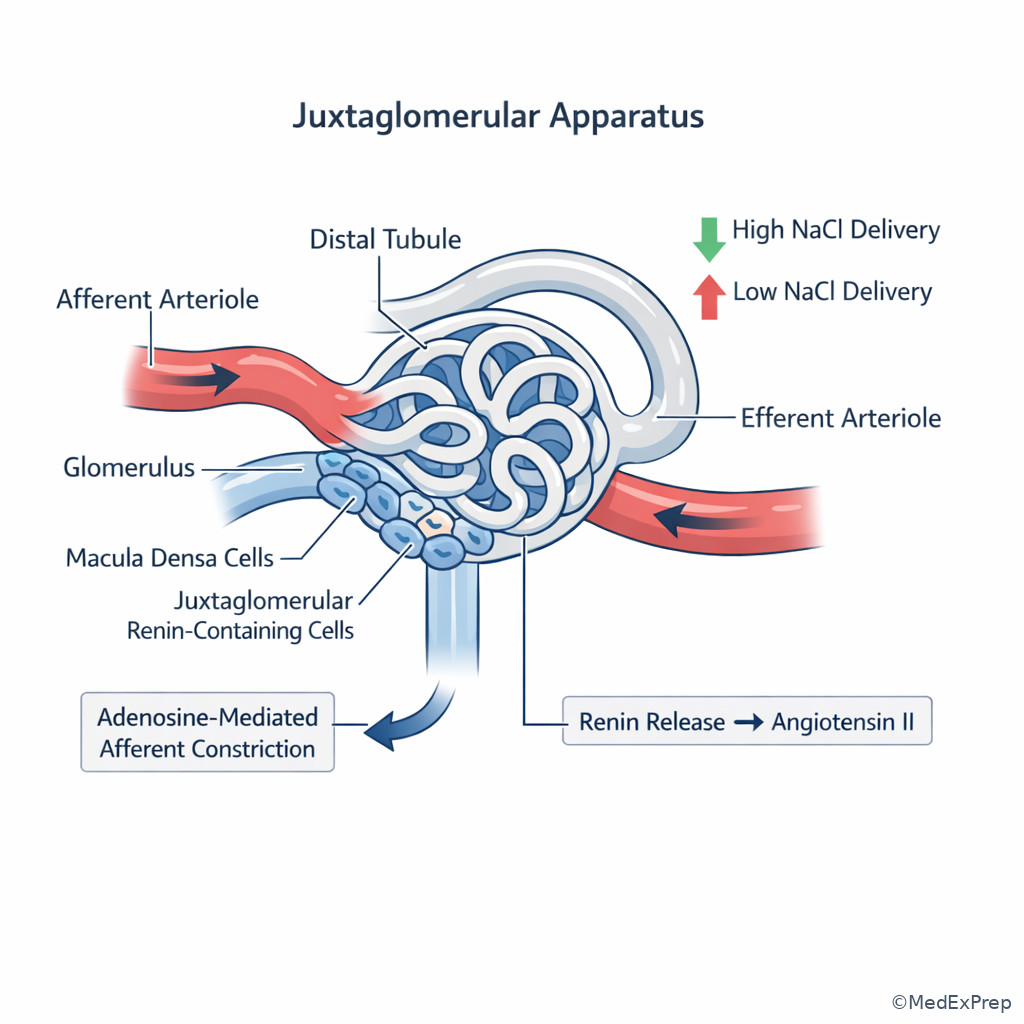
The myogenic response occurs when increased pressure stretches the afferent arteriole, causing reflex constriction to protect downstream capillaries. Tubuloglomerular feedback uses the macula densa: high distal sodium chloride delivery promotes adenosine-mediated afferent constriction; low sodium chloride delivery stimulates renin release and RAAS activation, increasing efferent tone. This is why modest hemodynamic stress may not immediately crash GFR.
Drug questions exploit these mechanisms. NSAIDs block prostaglandin-mediated afferent vasodilation; this matters most when the kidney is already relying on prostaglandins, such as in volume depletion, CHF, cirrhosis, or CKD. ACE inhibitors and ARBs reduce angiotensin II-mediated efferent constriction; this is beneficial long term in proteinuric disease but can acutely lower GFR when efferent support is needed, such as bilateral renal artery stenosis or severe volume depletion. See Fig 4.
WHY THIS IS TESTED: these are classic ABIM-style “best explanation for creatinine rise after medication” questions where the correct answer is a hemodynamic mechanism, not allergic interstitial nephritis or acute tubular injury.
Exam takeaway: NSAIDs threaten afferent support and RAAS blockers remove efferent support, especially when effective arterial blood volume is low.
flowchart TD
A[Low effective arterial blood volume] --> B[Kidney activates prostaglandins and RAAS]
B --> C[Prostaglandins dilate afferent arteriole]
B --> D[Angiotensin II constricts efferent arteriole]
C --> E[Preserve inflow]
D --> F[Preserve glomerular pressure]
G[NSAID] --> H[Block afferent support]
I[ACEi / ARB] --> J[Remove efferent support]
H --> K[GFR falls]
J --> K
Exam takeaway: Macula densa sodium chloride sensing links tubular delivery to afferent tone and renin release.
B. Worked example
A 76-year-old woman with long-standing hypertension, peripheral vascular disease, heart failure with reduced ejection fraction, and stage 3 CKD is started on lisinopril for albuminuric kidney disease. One week later, creatinine rises from 1.6 to 2.4 mg/dL and potassium is 5.3 mEq/L. She denies rash or fever. Urinalysis shows no blood and no pyuria. A renal bruit is noted, but the patient also reports poor oral intake after a dental procedure, making volume depletion a tempting alternative explanation.
Identify task: choose the most likely mechanism of the creatinine rise. Extract key facts: ACE inhibitor initiation, vascular disease, bruit, bland urine, mild hyperkalemia. Apply rule: efferent dilation after ACE inhibitor lowers glomerular capillary pressure; this effect is exaggerated when renal perfusion depends on angiotensin II, as in bilateral renal artery stenosis. Eliminate distractors: absence of pyuria/rash argues against AIN; no muddy brown casts are provided for ATI; pure dehydration is possible but does not explain the bruit clue as cleanly. Verify: the most likely mechanism is loss of angiotensin II-mediated efferent arteriolar constriction in renovascular disease.
C. Exam trap
D. Checkpoint
Question: In a patient with advanced cirrhosis and low effective arterial blood volume, which medication is most likely to cause an abrupt fall in GFR by preventing compensatory afferent vasodilation?
- Ibuprofen
- Lisinopril
- Spironolactone
- Amlodipine
Answer: A
- A: Correct because NSAIDs inhibit prostaglandin synthesis, removing afferent vasodilatory support in low-perfusion states.
- B: Tempting because ACE inhibitors can also lower GFR, but they do so primarily by efferent dilation rather than by blocking afferent prostaglandins.
- C: Tempting in cirrhosis because it affects hemodynamics and potassium, but it is not the classic afferent constrictor mechanism tested here.
- D: Tempting because it is a vasodilator, but it does not target the renal prostaglandin pathway responsible for this autoregulatory support.
1.1.1.6 Measuring kidney function: true GFR, estimated GFR, and when creatinine misleads you
A. Explanation
The decision conflict here is whether to trust the serum creatinine at face value. The single rule: creatinine reflects filtration and creatinine generation, distribution, and secretion. Therefore, creatinine is most reliable in steady state and least reliable when muscle mass is unusual or kidney function is changing rapidly.
Measured GFR is rarely needed on routine wards, but the physiology matters. Inulin clearance is ideal but impractical. Timed creatinine clearance is more available but imperfect because tubular secretion overestimates true GFR. Estimated GFR equations based on serum creatinine are extremely useful for outpatient CKD staging and medication adjustment, but not for abrupt AKI because creatinine lags behind the true fall in filtration. In a rapidly evolving injury, a “normal” creatinine may still coexist with a markedly reduced true GFR.
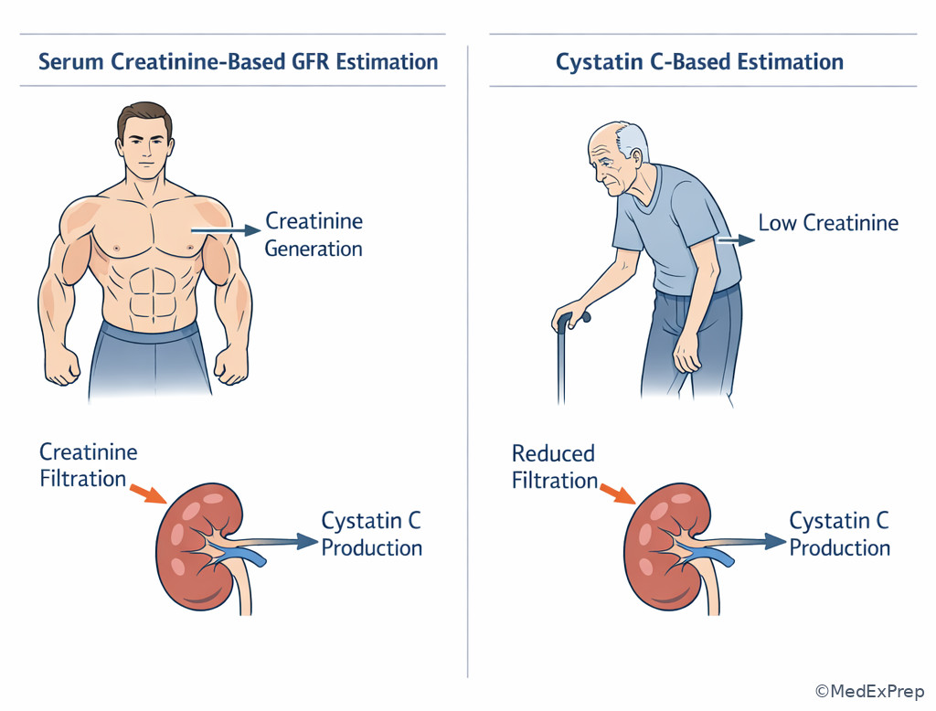
Cystatin C is useful when creatinine is unreliable, including low muscle mass, amputation, malnutrition, frailty, and sometimes cirrhosis. However, cystatin C is not perfect; inflammation, thyroid disease, and glucocorticoids can affect it. The exam usually rewards the broader principle rather than obscure exceptions. See Fig 5.
WHY THIS IS TESTED: many board stems ask for the best test to assess kidney function in cachexia, acute illness, obesity extremes, amputation, or cirrhosis.
Exam takeaway: Use creatinine-based eGFR in steady state, question it in unstable or low-muscle states, and escalate to alternative markers or measured approaches when the estimate is likely misleading.
flowchart TD
A[Need kidney function assessment] --> B{Steady state?}
B -->|Yes| C{Muscle mass typical?}
B -->|No| D[Do not rely on eGFR alone in AKI]
C -->|Yes| E[Creatinine-based eGFR usually acceptable]
C -->|No| F[Consider cystatin C or measured clearance]
D --> G[Use trend, urine data, and clinical context]
F --> H[Interpret with awareness of non-GFR influences] Exam takeaway: Low muscle mass can falsely reassure with creatinine, making cystatin C or measured approaches more informative.
B. Worked example
A 71-year-old woman with cirrhosis, severe sarcopenia, metastatic breast cancer, and chronic edema is referred for “normal kidney function” because her creatinine is 0.6 mg/dL. She is being considered for a renally cleared chemotherapy adjunct. Blood pressure is low-normal. Urinalysis is bland. A trainee is reassured by the low creatinine and wants standard dosing.
Identify task: determine whether creatinine-based eGFR is reliable. Extract key facts: severe sarcopenia, cirrhosis, malignancy, bland urinalysis, low creatinine. Apply rule: low creatinine generation can mask substantial reduction in true GFR. Eliminate distractors: bland urinalysis does not prove normal filtration; edema is not enough to classify renal function. Verify: the most appropriate next step is to use an alternative filtration assessment, often cystatin C-based estimation, before making dosing decisions.
C. Exam trap
D. Checkpoint
Question: Which setting most strongly limits the accuracy of creatinine-based eGFR for estimating true kidney function?
- Stable outpatient CKD with unchanged muscle mass over months
- Acute septic shock with rapidly rising creatinine over 12 hours
- Controlled hypertension with trace albuminuria
- Normal-weight adult with routine annual labs
Answer: B
- A: Tempting because CKD is where GFR matters, but steady-state outpatient CKD is precisely where eGFR performs best.
- B: Correct because creatinine lags behind abrupt changes in filtration, making formula-based eGFR unreliable in evolving AKI.
- C: Tempting because albuminuria complicates CKD care, but it does not inherently invalidate creatinine-based eGFR.
- D: Tempting because screening settings can hide mild disease, but the equation itself is generally more valid here than in unstable illness.
- The ideal measured GFR marker is ______.
- True or False: Creatinine clearance slightly overestimates true GFR.
- Name one setting where cystatin C may outperform creatinine.
1.1.1.7 Integration: hemodynamic patterns in real nephrology vignettes
A. Explanation
The final decision conflict is which hemodynamic pattern best explains the entire vignette and therefore what the most appropriate next step is. The single rule is to integrate the volume state, medication exposures, urine sediment, and comorbid conditions before labeling the problem as intrinsic renal disease.
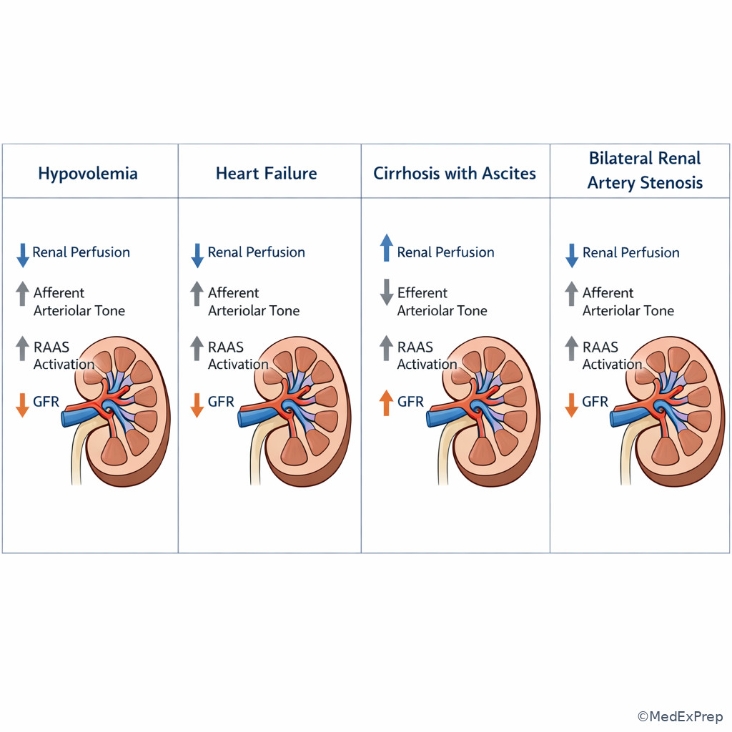
In effective arterial underfilling states such as heart failure and cirrhosis, total body fluid may be increased while the kidney senses low perfusion. This distinction is tested repeatedly. The kidney activates sympathetic tone, RAAS, and vasopressin; renal plasma flow falls and compensatory efferent constriction helps sustain filtration. Add NSAIDs or excessive diuresis and GFR falls. In bilateral renal artery stenosis, perfusion pressure distal to the stenosis is low, so angiotensin II support becomes essential; ACE inhibitor can therefore unmask a marked decline in GFR. In obstruction, the clue is rising Bowman space pressure rather than a vascular mechanism. In intrinsic glomerular or tubular disease, the sediment usually becomes more informative than the drug list alone.
The exam also likes “most important to monitor” questions. After starting RAAS blockade, monitor creatinine and potassium. After NSAID exposure in a vulnerable patient, monitor kidney function and volume status. In suspected hemodynamic AKI, the next step is often not biopsy but restoration of perfusion, drug review, and reassessment. See Fig 6.
WHY THIS IS TESTED: nephrology boards emphasize mechanism-to-management logic: know the physiologic reason first, then choose the safest next action.
Exam takeaway: Volume state, vasoactive medications, urine sediment, and obstruction clues separate hemodynamic AKI from intrinsic renal disease.
flowchart TD
A[Falling GFR / rising creatinine] --> B{Urine sediment active?}
B -->|Yes| C[Think intrinsic renal disease]
B -->|No| D{Hemodynamic clue present?}
D -->|NSAID + low EABV| E[Afferent constriction]
D -->|ACEi/ARB + renovascular state| F[Efferent dilation]
D -->|HF/cirrhosis/hypovolemia| G[Reduced renal perfusion with compensation]
D -->|Hydronephrosis / retention| H[Obstruction]
E --> I[Hold trigger, restore perfusion]
F --> I
G --> I
H --> J[Relieve obstruction urgently] Exam takeaway: Side-by-side visualization of hypovolemia, heart failure, cirrhosis, and bilateral renal artery stenosis helps identify which compensatory pathway the kidney is relying on.
Exam takeaway: Obstruction lowers GFR by increasing Bowman space pressure rather than by primary afferent or efferent arteriolar tone changes.
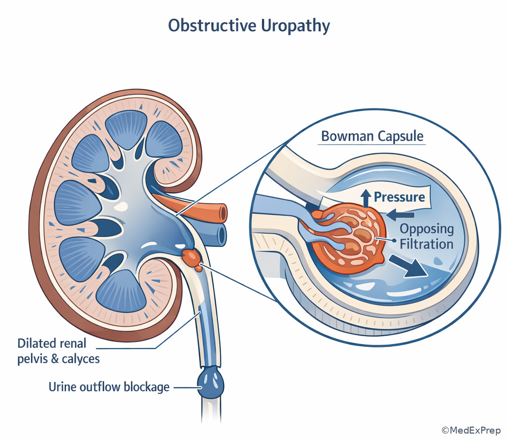
B. Worked example
A 64-year-old man with ischemic cardiomyopathy, BPH, and CKD stage 3 presents with dyspnea and edema. He receives aggressive diuresis and several doses of ketorolac for back pain. Creatinine rises from 1.8 to 2.7 mg/dL. He has mild suprapubic fullness but passes some urine. Urinalysis shows hyaline casts only. Ultrasound later shows mild bilateral hydronephrosis, while exam also suggests persistent venous congestion. The tempting wrong answer is “ATN from overdiuresis” because the creatinine rose after hospitalization.
Identify task: select the most appropriate first-pass mechanism and next step. Extract key facts: CHF with low effective arterial blood volume, NSAID exposure, BPH, bland sediment, possible obstruction. Apply rule: bland sediment plus multiple hemodynamic/obstructive hits argues against primary intrinsic tubular injury. Eliminate distractors: hyaline casts are nonspecific; absence of muddy brown casts makes ATI less compelling. Verify: the best next step is to evaluate and relieve obstruction while stopping NSAIDs and reassessing hemodynamics, because obstruction and hemodynamic compromise are both more likely than intrinsic necrosis.
C. Exam trap
D. Checkpoint
Question: A patient with decompensated heart failure has edema, low urine sodium, bland urine sediment, and rising creatinine after ibuprofen exposure. What is the most likely immediate mechanism of the GFR decline?
- Loss of prostaglandin-mediated afferent vasodilation
- Immune complex deposition in the mesangium
- Myoglobin-induced tubular obstruction
- Decreased creatinine production from sarcopenia
Answer: A
- A: Correct because low effective arterial blood volume makes the kidney prostaglandin dependent, and NSAID exposure removes afferent vasodilatory support.
- B: Tempting because kidney dysfunction can reflect glomerular disease, but the bland sediment and drug/volume clues point away from inflammatory pathology.
- C: Tempting because tubular obstruction is a renal mechanism, but there is no rhabdomyolysis context or pigmented casts.
- D: Tempting because creatinine interpretation matters, but decreased production would falsely lower creatinine rather than explain its acute rise.
Exam Traps & Differentiators
- Most common wrong answer: intrinsic renal disease when the real problem is hemodynamic. Why: creatinine rose and the stem feels “renal.” Fix: first ask whether the urine sediment is bland and whether a drug/volume explanation is sitting in front of you.
- Looks similar but isn't: cirrhosis and heart failure can present with edema yet still produce low effective arterial blood volume; the kidney responds as if underperfused.
- False reassurance trap: low creatinine in sarcopenia or cirrhosis. The exam wants you to distrust a deceptively normal creatinine when muscle mass is low.
- Drug-mechanism swap: candidates reverse NSAID and ACE inhibitor effects. NSAIDs hit the afferent rescue pathway; ACE inhibitors remove efferent rescue.
| Look-alike scenario | Why it tempts you | Decisive clue | Think |
|---|---|---|---|
| CHF with rising creatinine after NSAID | Hospitalized patient with AKI could be ATI | Bland sediment, low effective arterial volume, NSAID timing | Afferent constriction/hemodynamic AKI |
| Renal bruit and creatinine rise after ACE inhibitor | Medication-related AKI may sound nonspecific | Vascular disease plus marked creatinine increase after RAAS blockade | Loss of efferent support in renovascular disease |
| Edema with low urine sodium in cirrhosis | Edema suggests volume overload only | Kidney senses low effective arterial blood volume despite excess total body fluid | Perfusion-dependent RAAS activation |
| Low serum creatinine in cachexia | Low creatinine feels “good” | Severe muscle loss reduces creatinine generation | eGFR may overestimate true kidney function |
| AKI with hydronephrosis | Could still have medications on board | Back-pressure clues or bladder outlet symptoms | Obstructive fall in GFR |
| If the stem says... | Think... |
|---|---|
| “Started ibuprofen during GI illness” | Loss of prostaglandin-mediated afferent vasodilation |
| “Creatinine rose after ACE inhibitor and there is a renal bruit” | Bilateral renal artery stenosis or severe renovascular dependence on angiotensin II |
| “Massive edema but kidney behaves as if underfilled” | Low effective arterial blood volume, not true adequate perfusion |
| “Bland sediment and clear hemodynamic insult” | Choose hemodynamic AKI before intrinsic pathology |
| “Sarcopenic patient with very low creatinine” | Creatinine may overestimate GFR; consider alternative markers |
| “Hydronephrosis or urinary retention” | Increased Bowman space pressure lowering GFR |
Related trap from upcoming topics: when you later study Fractional Excretion Reasoning, do not overread FeNa in patients on diuretics or mixed hemodynamic-intrinsic states. The hemodynamic logic from this lesson comes first; urine indices refine it.
Algorithm / Approach
Exam takeaway: A structured medication-volume-sediment review often answers the stem before advanced testing is needed.
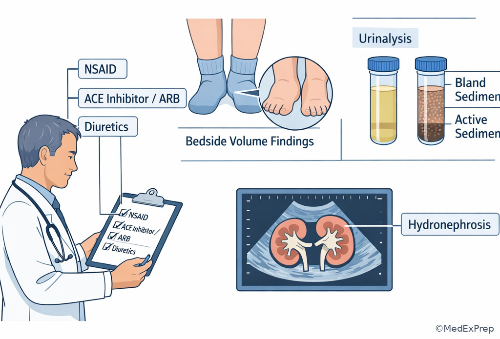
Rapid Review
- Afferent constriction → RBF down, GFR down
- Afferent dilation → RBF up, GFR up
- Efferent constriction → RPF down, FF up
- Efferent dilation → intraglomerular pressure down, GFR down
- Obstruction → Bowman space pressure up
- PAH clearance → approximates RPF, not GFR
- Inulin clearance → ideal measured GFR marker
- Creatinine clearance → slight GFR overestimate from secretion
- AKI in non–steady state → eGFR formula unreliable
- Sarcopenia/cirrhosis → creatinine may overestimate kidney function
- NSAID-associated AKI → prostaglandin-dependent afferent support lost
- ACEi/ARB creatinine rise → efferent support withdrawn; watch for renovascular disease
- CHF/cirrhosis edema → low effective arterial blood volume can still dominate renal behavior
- Bland sediment → prioritize hemodynamic/obstructive causes before inflammatory intrinsic disease
Self-check quiz
1. A 45-year-old man undergoing physiology testing has a substance infused that is freely filtered and neither reabsorbed nor secreted. Which clearance most directly measures GFR?
- Inulin clearance
- PAH clearance
- Creatinine clearance
- Urea clearance
2. A 52-year-old woman with no kidney disease asks which vascular change most directly lowers GFR by reducing blood delivery into the glomerulus. Which is the best answer?
- Afferent arteriolar constriction
- Afferent arteriolar dilation
- Efferent arteriolar constriction
- Efferent arteriolar dilation
3. A 67-year-old man with HFrEF, diabetes, and osteoarthritis develops AKI after gastroenteritis. Medications are lisinopril, furosemide, and ibuprofen. Blood pressure is 90/56 mm Hg, urinalysis is bland, and creatinine rises from 1.1 to 2.0 mg/dL. What is the most appropriate physiologic explanation?
- Immune-mediated interstitial inflammation from ibuprofen
- Combined loss of afferent and efferent support of glomerular pressure in a low-perfusion state
- Postinfectious glomerulonephritis causing abrupt reduction in filtration coefficient
- Rhabdomyolysis causing pigmented cast nephropathy
4. A 74-year-old woman with peripheral vascular disease, resistant hypertension, and CKD stage 3 is started on losartan. One week later, creatinine rises by 35% and potassium is 5.4 mEq/L. She has a renal bruit and bland urine sediment. What is the most appropriate interpretation?
- Expected acute interstitial nephritis from ARB exposure
- Loss of angiotensin II-mediated efferent arteriolar constriction in renovascular disease
- Mesangial immune-complex disease unmasked by blood pressure control
- PAH secretion failure causing pseudoelevation of creatinine
5. A 70-year-old woman with cirrhosis, sarcopenia, and chronic edema has a creatinine of 0.5 mg/dL before planned dosing of a renally cleared drug. Blood pressure is 98/60 mm Hg, urinalysis is bland, and there is marked muscle wasting. What is the most appropriate next step for assessing kidney function?
- Assume normal GFR because creatinine is low
- Use creatinine-based eGFR alone because cirrhosis does not affect creatinine generation
- Consider cystatin C-based estimation because creatinine may overestimate GFR in severe sarcopenia
- Delay all assessment until a kidney biopsy is performed
6. A 63-year-old man with decompensated cirrhosis, tense ascites, and CKD stage 2 undergoes large-volume paracentesis without adequate albumin replacement. Creatinine rises over 24 hours, urine sediment is bland, and urine sodium is low. Which renal hemodynamic pattern is most likely present early in this course?
- Increased renal plasma flow and decreased filtration fraction
- Decreased renal plasma flow with preserved or relatively maintained GFR from efferent constriction
- Increased Bowman space pressure from occult obstruction
- Normal renal plasma flow with decreased GFR from isolated fall in filtration coefficient
7. A 68-year-old woman with CHF, BPH, CKD stage 3, and chronic back pain presents with rising creatinine after aggressive diuresis and ketorolac exposure. She has mild suprapubic fullness, still makes urine, and urinalysis shows only hyaline casts. Kidney ultrasound reveals new bilateral hydronephrosis. What is the most appropriate next step?
- Start high-dose corticosteroids for presumed interstitial nephritis
- Order urgent measures to relieve obstruction while stopping hemodynamically harmful medications
- Continue NSAIDs because the bland urine excludes hemodynamic injury
- Proceed directly to kidney biopsy because hydronephrosis does not affect GFR
8. A 60-year-old man with bilateral renal artery stenosis, diabetes, and albuminuric CKD is started on an ACE inhibitor. His creatinine rises substantially, but urine sediment remains bland and there is no hypotension. Which single physiologic detail best distinguishes this from simple volume depletion?
- The ACE inhibitor causes afferent constriction instead of efferent dilation
- Renal perfusion beyond the stenoses depends on angiotensin II to maintain intraglomerular pressure
- Creatinine rises only in intrinsic tubular necrosis, not hemodynamic states
- Filtration fraction must decrease in all RAAS-activated states
Answer key
1. Correct answer: A — Inulin clearance.
- A: Tempting because it is the classic textbook answer, and it is correct here because inulin is freely filtered without reabsorption or secretion, making its clearance equal to GFR. Single clue: “freely filtered and neither reabsorbed nor secreted.” Source: Brenner and Rector’s The Kidney; Harrison’s Principles of Internal Medicine.
- B: Tempting because PAH is another famous renal physiology marker, but it approximates effective renal plasma flow due to filtration plus secretion. Single clue: the stem asks for direct measurement of GFR, not plasma flow. Source: Brenner and Rector; Costanzo Physiology.
- C: Tempting because creatinine is used clinically to estimate GFR, but secretion makes creatinine clearance a slight overestimate rather than the ideal direct measure. Single clue: “directly measures” excludes the imperfect surrogate. Source: KDIGO CKD guideline; Harrison’s.
- D: Tempting because urea is filtered and familiar in nephrology, but variable tubular reabsorption makes it unreliable for direct GFR measurement. Single clue: reabsorption violates the ideal marker rule. Source: Costanzo Physiology; Brenner and Rector.
2. Correct answer: A — Afferent arteriolar constriction.
- A: Tempting because it is the simplest inflow mechanism, and it is correct because narrowing the vessel entering the glomerulus reduces renal blood flow and glomerular capillary hydrostatic pressure. Single clue: “reducing blood delivery into the glomerulus.” Source: Costanzo Physiology; Harrison’s.
- B: Tempting because arteriolar changes often alter GFR, but afferent dilation increases inflow and usually raises GFR. Single clue: dilation has the opposite direction of effect. Source: Brenner and Rector.
- C: Tempting because efferent constriction certainly changes filtration dynamics, but mild-to-moderate efferent constriction tends to preserve or raise glomerular pressure rather than directly reducing inflow. Single clue: the stem emphasizes delivery into, not pressure distal to, the glomerulus. Source: Harrison’s; Costanzo.
- D: Tempting because efferent dilation lowers GFR, but it does so by lowering intraglomerular pressure, not by reducing inflow into the tuft. Single clue: vessel leaving the glomerulus is not the inflow resistor. Source: Brenner and Rector.
3. Correct answer: B — Combined loss of afferent and efferent support of glomerular pressure in a low-perfusion state.
- A: Tempting because ibuprofen can cause AIN, but the absence of pyuria, rash, fever, and active sediment makes immune interstitial injury less likely in this specific case. Single clue: bland urinalysis with hypotension and combined NSAID/ACEi exposure. Source: Harrison’s; UpToDate review of NSAID nephrotoxicity.
- B: Correct because hypovolemia lowers renal perfusion, ibuprofen blocks prostaglandin-dependent afferent vasodilation, and lisinopril removes angiotensin II-mediated efferent constriction; together these sharply reduce glomerular filtration pressure. Single clue: gastroenteritis plus ibuprofen plus lisinopril with bland sediment. Source: KDIGO AKI guideline; Brenner and Rector.
- C: Tempting because infection precedes some glomerulonephritides, but there is no nephritic sediment, edema pattern, or complement clue here. Single clue: bland sediment argues against inflammatory glomerular injury. Source: Harrison’s.
- D: Tempting because AKI can result from pigmented casts, but the stem offers no muscle injury, dark urine, or hyperCKemia clue. Single clue: the medication-volume story fully explains the presentation. Source: KDIGO AKI guideline; Harrison’s.
4. Correct answer: B — Loss of angiotensin II-mediated efferent arteriolar constriction in renovascular disease.
- A: Tempting because any new medication can cause drug-related AKI, but ARBs are not classic triggers of AIN, and bland urine with vascular clues points elsewhere. Single clue: renal bruit and resistant hypertension. Source: Harrison’s; AHA/ACC hypertension guidance.
- B: Correct because in bilateral renovascular disease, downstream perfusion pressure is low and angiotensin II-dependent efferent constriction helps sustain GFR; blocking this causes a marked creatinine rise. Single clue: rise after losartan in a patient with vascular disease and renal bruit. Source: Brenner and Rector; Harrison’s.
- C: Tempting because CKD can coexist with glomerular disease, but there is no hematuria, proteinuria pattern, or active sediment supporting immune-complex pathology. Single clue: the timing after ARB initiation fits a hemodynamic explanation far better. Source: Harrison’s.
- D: Tempting because secretion pathways matter for renal markers, but PAH is a physiology test substance and not the mechanism of this clinical creatinine change. Single clue: real-world ARB exposure with renovascular clues. Source: Costanzo Physiology.
5. Correct answer: C — Consider cystatin C-based estimation because creatinine may overestimate GFR in severe sarcopenia.
- A: Tempting because a low creatinine superficially suggests preserved filtration, but severe sarcopenia lowers creatinine production and can mask major kidney dysfunction. Single clue: marked muscle wasting. Source: KDIGO CKD guideline; Harrison’s.
- B: Tempting because creatinine-based eGFR is commonly used, but cirrhosis and sarcopenia both reduce reliability of creatinine-based estimation. Single clue: the stem intentionally pairs cirrhosis with muscle loss. Source: KDIGO; Brenner and Rector.
- C: Correct because alternative filtration markers such as cystatin C can be more informative when creatinine generation is abnormally low. Single clue: low creatinine in the setting of profound sarcopenia. Source: KDIGO CKD guideline; Harrison’s.
- D: Tempting because definitive evaluation sounds thorough, but biopsy assesses kidney pathology, not first-line estimation of filtration in this context. Single clue: the question asks kidney function assessment for drug dosing, not tissue diagnosis. Source: KDIGO; Harrison’s.
6. Correct answer: B — Decreased renal plasma flow with preserved or relatively maintained GFR from efferent constriction.
- A: Tempting because hyperdynamic cirrhosis can confuse total circulatory concepts, but the kidney in low effective arterial volume does not experience increased renal plasma flow. Single clue: low urine sodium and post-paracentesis underfilling imply renal vasoconstrictive compensation. Source: Harrison’s; UpToDate review of hepatorenal physiology.
- B: Correct because RAAS activation lowers renal plasma flow while angiotensin II-mediated efferent constriction initially helps maintain filtration pressure, often increasing filtration fraction. Single clue: early bland AKI after inadequate albumin replacement in cirrhosis. Source: Brenner and Rector; Harrison’s.
- C: Tempting because obstruction lowers GFR with bland sediment, but there is no retention or hydronephrosis clue; the timing after paracentesis points to circulatory underfilling. Single clue: procedure-related effective volume loss. Source: Harrison’s.
- D: Tempting because filtration coefficient can fall in intrinsic disease, but the vignette is hemodynamic rather than glomerular inflammatory. Single clue: low urine sodium and cirrhotic underfilling. Source: KDIGO AKI guideline; Harrison’s.
7. Correct answer: B — Order urgent measures to relieve obstruction while stopping hemodynamically harmful medications.
- A: Tempting because NSAIDs can cause AIN, but hydronephrosis and outlet symptoms supply a more direct and urgent explanation for falling GFR. Single clue: new bilateral hydronephrosis. Source: KDIGO AKI guideline; Harrison’s.
- B: Correct because obstructive AKI can coexist with hemodynamic injury, and urgent relief of obstruction plus removal of NSAIDs addresses the highest-yield reversible causes first. Single clue: bilateral hydronephrosis in a patient with BPH and rising creatinine. Source: KDIGO; Brenner and Rector.
- C: Tempting because bland urine does suggest hemodynamic rather than intrinsic injury, but it does not make NSAIDs safe; they remain harmful in this low-perfusion state. Single clue: ketorolac exposure in CHF and AKI. Source: Harrison’s.
- D: Tempting because biopsy can clarify AKI, but obstruction is a known, reversible cause and hydronephrosis absolutely can lower GFR by increasing Bowman space pressure. Single clue: structural back-pressure is already demonstrated. Source: Harrison’s; Brenner and Rector.
8. Correct answer: B — Renal perfusion beyond the stenoses depends on angiotensin II to maintain intraglomerular pressure.
- A: Tempting because students mix up arteriolar drug effects, but ACE inhibitors cause efferent dilation, not afferent constriction. Single clue: RAAS blockade acts on angiotensin II-mediated efferent tone. Source: Costanzo Physiology; Harrison’s.
- B: Correct because the stenotic kidneys rely on angiotensin II-mediated efferent constriction to preserve glomerular capillary hydrostatic pressure despite reduced inflow pressure. Single clue: bilateral renal artery stenosis with bland sediment after ACE inhibitor initiation. Source: Brenner and Rector; AHA/ACC guidance.
- C: Tempting because large creatinine rises feel like structural injury, but hemodynamic states can produce substantial increases without tubular necrosis. Single clue: bland sediment argues against intrinsic tubular injury. Source: KDIGO AKI guideline.
- D: Tempting because filtration fraction is a hemodynamic concept linked to RAAS, but it does not necessarily decrease in all RAAS-activated states and often rises with efferent constriction. Single clue: the discriminating issue is dependence on angiotensin II, not a universal FF direction. Source: Costanzo; Harrison’s.
Keep going with the full ABIM Nephrology Prep Course (Nephrology)
You just studied the way members do, in the same lesson interface. Full access unlocks every lesson, the question bank and flashcards for a full year.
One payment · 1 year of access · No auto-renewal