1.1.1 - Acute Coronary Syndrome Patterns
Acute Coronary Syndrome Patterns
What you'll learn
- DOMAIN: Cardiovascular Medicine → Chest pain evaluation → Pattern recognition — Distinguish unstable angina, NSTEMI, and STEMI using symptoms, ECG findings, and troponin behavior.
- DOMAIN: Cardiovascular Medicine → Diagnostic reasoning → Data interpretation — Interpret the first ECG and troponin in context rather than overcalling or undercalling ACS.
- DOMAIN: Cardiovascular Medicine → Acute prioritization → Emergency recognition — Identify which ischemic patterns require immediate reperfusion versus serial testing.
- DOMAIN: Cardiovascular Medicine → Differential diagnosis → Look-alike discrimination — Separate ACS patterns from common distractors such as pericarditis, demand ischemia, and noncardiac chest pain.
- DOMAIN: Cardiovascular Medicine → Clinical task → Next-step planning — Decide when the safest next step is serial ECG/troponin monitoring, urgent cardiology activation, or reassessment of an alternative diagnosis.
- Cardiovascular Medicine → Ischemic heart disease → Recognize ACS phenotype
- Cardiovascular Medicine → Data interpretation → ECG and troponin integration
- General Internal Medicine → Acute care reasoning → Best next step in a time-sensitive adult presentation
Why it matters
- ABIM questions often hinge on pattern recognition before treatment selection: if you mislabel the ACS subtype, every downstream choice becomes wrong.
- A single clue — persistent ST elevation, dynamic ST depression/T-wave inversion, or negative biomarkers despite ischemic symptoms — often separates the correct answer from a tempting distractor.
- Early errors usually come from overreliance on one data point: one normal troponin, one nondiagnostic ECG, or one misleading symptom descriptor.
- Board stems commonly test whether you know that ACS is a syndrome with evolving findings, so serial reassessment matters when the first study is nondiagnostic.
Key Terms & Must-Know Facts
1.1.1.1 Key terms
- ACS — umbrella term including unstable angina, NSTEMI, and STEMI.
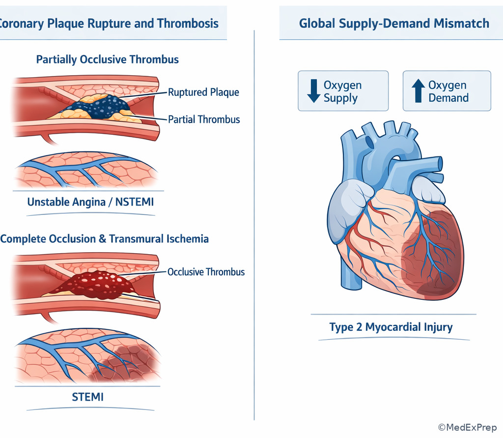
- Plaque rupture — common trigger for thrombus formation and abrupt coronary flow reduction.
- STEMI — acute coronary occlusion pattern with ischemic symptoms plus ST-segment elevation or STEMI equivalent.
- NSTEMI — myocardial necrosis without ST-elevation criteria; troponin rises.
- Unstable angina — ischemic symptoms without biomarker evidence of necrosis.
- Troponin — preferred biomarker for myocardial injury.
- Dynamic ECG changes — evolving ST depression or T-wave inversion supporting ischemia.
- STEMI equivalent — high-risk ischemic ECG pattern that should be treated like occlusive MI.
- Posterior MI — reciprocal ST depression in anterior leads; often needs posterior leads.
- Type 1 MI — plaque rupture/erosion with acute coronary thrombosis.
- Type 2 MI — oxygen supply-demand mismatch without acute plaque rupture.
- Myocardial injury — troponin elevation that may be nonischemic if no ischemic clinical syndrome.
1.1.1.2 Must-know facts
- Persistent ischemic chest pressure radiating to arm or jaw is more concerning than pleuritic, positional, or reproducible pain.
- A normal initial ECG does not exclude ACS.
- A normal initial troponin does not exclude early NSTEMI or unstable angina.
- STEMI is an immediate reperfusion diagnosis, not a “wait for more labs” diagnosis.
- NSTEMI and unstable angina are separated by troponin evidence of necrosis, not by symptom severity alone.
- Diffuse ST elevation with PR depression suggests pericarditis, not focal coronary occlusion.
- Troponin elevation alone is not enough for ACS; interpret it with symptoms, ECG, and clinical context.
- Posterior MI can present as ST depression in V1–V3 rather than obvious ST elevation.
Core content
1.1.1.3 Foundational ACS framework: what pattern are you being asked to recognize?
A Explanation
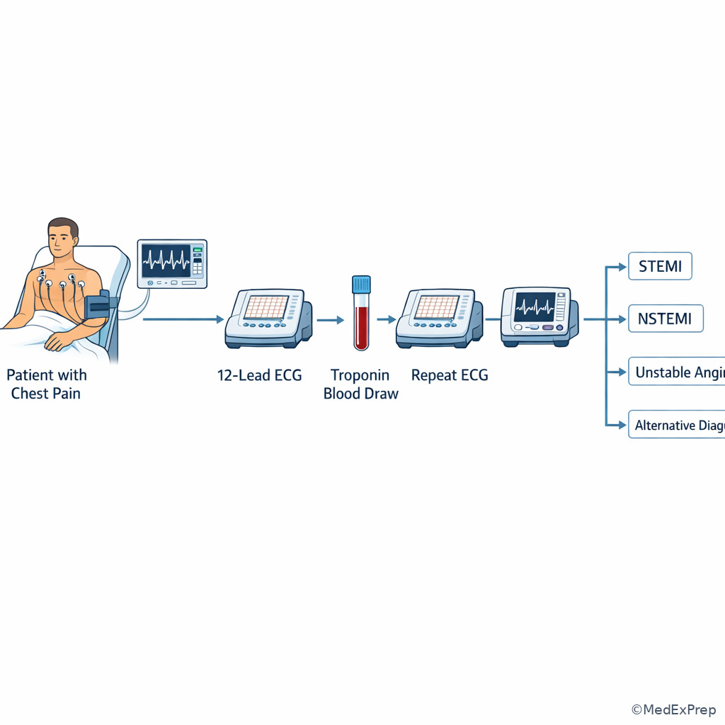
The first board-style conflict is simple but high yield: Is this occlusive MI needing immediate reperfusion, or non-ST-elevation ischemia needing rapid but not identical next steps? The single rule that resolves this is the combination of ischemic symptoms plus ECG pattern plus biomarker status. STEMI is a reperfusion diagnosis driven by the ECG. NSTEMI is myocardial necrosis without ST-elevation criteria. Unstable angina has ischemic symptoms but no troponin-defined necrosis. WHY THIS IS TESTED: exam stems hide the diagnosis behind one misleading feature — for example, severe pain with a normal first troponin, or mild pain with diagnostic ST elevation.
Choose STEMI when the ECG shows persistent focal ST elevation in a coronary territory or a STEMI equivalent. Reject NSTEMI or unstable angina even if the troponin is not yet elevated, because biomarker release can lag behind coronary occlusion. Choose NSTEMI when symptoms suggest ischemia and troponin rises, but there is no ST-elevation pattern. Reject unstable angina if troponin is elevated. Choose unstable angina when symptoms are new, worsening, or occurring at rest, but serial biomarkers remain negative and no alternative diagnosis explains the syndrome.
At this stage, the learner should not jump to full management details; the exam priority is correct classification. See Fig 1 for the compressed classification pathway.
Exam takeaway: Separate STEMI, NSTEMI, and unstable angina by combining ischemic symptoms with ECG pattern and troponin status.
flowchart TD
A[Adult with possible ischemic chest pain] --> B{ECG shows focal ST elevation or STEMI equivalent?}
B -->|Yes| C[STEMI pattern: activate reperfusion pathway]
B -->|No| D{Troponin rises or falls above assay threshold?}
D -->|Yes| E[NSTEMI pattern]
D -->|No| F[Unstable angina or non-ACS chest pain]
F --> G{Symptoms clearly ischemic and dynamic?}
G -->|Yes| H[Unstable angina more likely]
G -->|No| I[Consider alternate diagnosis] B Worked example
A 61-year-old man with hypertension, type 2 diabetes, and chronic GERD presents with 45 minutes of substernal pressure radiating to the left arm while walking uphill. He says he once had “heartburn that felt similar,” and he drank spicy tomato juice this morning. Blood pressure is 156/92 mm Hg, pulse 88/min. ECG shows 2 mm ST elevation in II, III, and aVF. Initial troponin is within the reference range. The task is to identify the pattern, not to overvalue the first lab. Key facts: exertional pressure, radiation, focal inferior ST elevation, ongoing symptoms. Apply the rule: ischemic symptoms plus diagnostic ST elevation = STEMI, even if the first troponin is negative early. Eliminate GERD because the ECG is diagnostic. Verify: troponin lag is common early, so a normal initial value does not downgrade STEMI.
C Exam trap
D Checkpoint
Question: A 58-year-old woman with hyperlipidemia and CKD stage 3 has 30 minutes of crushing chest pressure and diaphoresis. ECG shows 1.5 to 2 mm ST elevation in V2–V4. Initial troponin is negative. Which ACS pattern is most likely?
- A. Unstable angina — Tempting because the first troponin is negative, but wrong because focal ST elevation with ischemic symptoms outranks an early negative biomarker.
- B. NSTEMI — Tempting because an infarction can begin before biomarkers rise, but wrong because the ECG meets a reperfusion pattern rather than a non-ST-elevation pattern.
- C. STEMI — Correct because anterior ST elevation with classic ischemic symptoms defines an occlusive MI pattern requiring immediate action.
- D. Nonischemic myocardial injury — Tempting if one focuses on CKD, but wrong because the stem gives an acute ischemic syndrome and diagnostic ECG changes.
1.1.1.4 ECG patterns that decide the stem
A Explanation
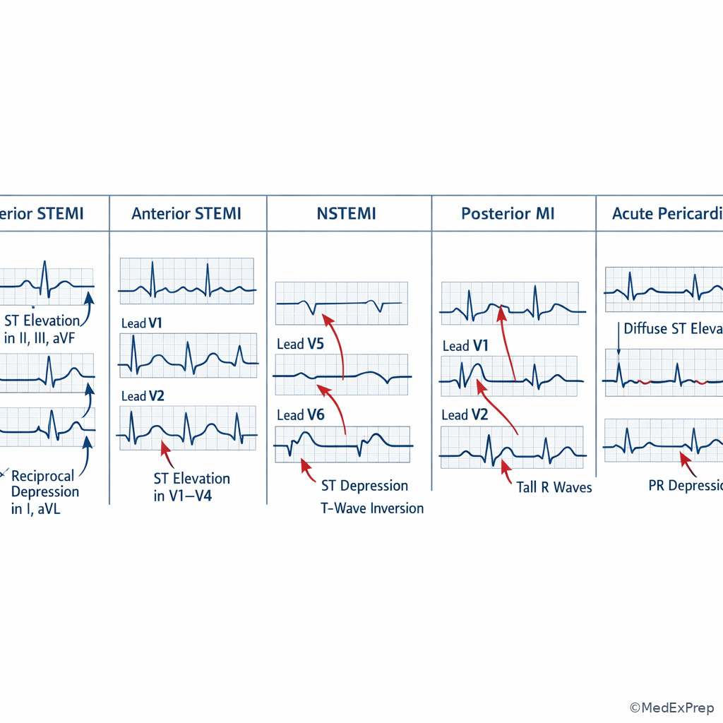
The next decision conflict is which ECG findings are high-risk ischemia versus common mimics. The single finding that resolves this is whether the pattern is focal and coronary-territory based or instead diffuse, nonspecific, or explained by another condition. STEMI usually produces regional ST elevation in contiguous leads, often with reciprocal changes. NSTEMI/unstable angina may show ST depression or deep symmetric T-wave inversion, or the ECG may be initially normal. Posterior MI often hides as horizontal ST depression in V1–V3 with tall R waves. Diffuse ST elevation with PR depression suggests acute pericarditis, not ACS.
WHY THIS IS TESTED: the ECG distractor is usually close-call. The exam may offer “acute pericarditis” when the learner sees ST elevation, or “NSTEMI” when posterior MI is presented as anterior ST depression. Choose the answer that matches the distribution and associated clues, not merely the presence of “abnormal ST segments.” See Fig 2 for a side-by-side pattern comparison and Fig 3 for a decision-focused ECG map.
Exam takeaway: Focal contiguous changes support coronary occlusion, whereas diffuse or pattern-mismatched changes should trigger a mimic check.
flowchart LR
A[Regional ST elevation in contiguous leads] --> A1[Think STEMI]
B[ST depression or T-wave inversion] --> B1[Think NSTEMI or unstable angina]
C[ST depression V1-V3 with tall R waves] --> C1[Think posterior MI]
D[Diffuse ST elevation with PR depression] --> D1[Think pericarditis]
E[Normal ECG but ischemic symptoms] --> E1[Repeat ECG and troponin]
Exam takeaway: Match lead distribution and reciprocal changes to the coronary pattern instead of reacting to any single abnormal wave.
B Worked example
A 67-year-old woman with diabetes, smoking history, and osteoarthritis presents with nausea, diaphoresis, and upper back discomfort. She reports pain “between the shoulder blades” and had mild viral symptoms last week, making pericarditis seem superficially plausible. ECG shows horizontal ST depression in V1–V3 with tall R waves. Troponin is pending. Identify the task: classify the ischemic pattern. Extract key facts: ischemic symptoms, autonomic features, anterior lead ST depression localized to V1–V3, tall R waves. Apply rule: this may represent posterior MI, a STEMI equivalent. Eliminate pericarditis because the ECG is not diffuse and lacks PR depression. Verify: posterior extension should be considered, and urgent escalation is warranted.
C Exam trap
D Checkpoint
Question: A 54-year-old man with hyperlipidemia and recent URI has sharp chest pain. ECG shows diffuse concave ST elevation in multiple leads with PR depression. Which interpretation is best?
- A. Inferior STEMI — Tempting because ST elevation is present, but wrong because the changes are diffuse rather than confined to an inferior territory.
- B. Anterior STEMI — Tempting because chest pain plus ST elevation suggests MI, but wrong because there is no focal precordial distribution and PR depression favors another diagnosis.
- C. Acute pericarditis — Correct because diffuse ST elevation with PR depression is the discriminating ECG pattern.
- D. NSTEMI — Tempting because some ACS cases have nondiagnostic ECGs, but wrong because this tracing points specifically to pericarditis rather than subendocardial ischemia.
- The ECG clue that should make you think posterior MI is ST depression in ___ through ___ with tall R waves.
- True or False: A normal initial ECG excludes ACS.
- Diffuse ST elevation plus PR depression should trigger consideration of ___ rather than focal occlusive MI.
1.1.1.5 Troponin interpretation: myocardial infarction versus myocardial injury
A Explanation
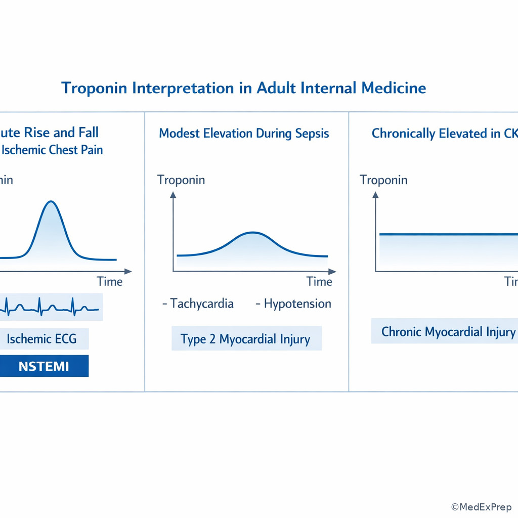
The key decision conflict here is whether an elevated troponin means ACS or merely myocardial injury. The single rule: troponin indicates myocardial injury, but ACS requires a compatible ischemic syndrome. A rise and/or fall in troponin above the assay threshold supports acute injury; if the clinical picture is ischemic, this may be NSTEMI. If the patient instead has sepsis, tachyarrhythmia, severe anemia, hypertensive emergency, renal dysfunction, myocarditis, or pulmonary embolism, the elevated troponin may reflect type 2 MI or nonischemic myocardial injury rather than plaque rupture.
WHY THIS IS TESTED: ABIM stems frequently include a positive troponin with misleading comorbidity noise. The examinee must choose whether the stem is asking, “Is this ACS?” or “Why is this troponin elevated?” Choose NSTEMI when symptoms are ischemic and no better explanation dominates. Reject NSTEMI when the entire syndrome is driven by another acute stressor without classic ischemic patterning. Equally important, reject “no MI” if there is a clear rise/fall and ischemic symptoms merely because ST elevation is absent. See Fig 4 for the troponin logic tree.
Exam takeaway: Troponin elevation proves myocardial injury, but only ischemic clinical context converts that finding into ACS.
flowchart TD
A[Elevated troponin] --> B{Rise or fall pattern?}
B -->|Yes| C{Ischemic symptoms or ischemic ECG?}
C -->|Yes| D[Consider NSTEMI or STEMI depending on ECG]
C -->|No| E[Consider type 2 MI or nonischemic injury]
B -->|No| F[Chronic myocardial injury possible]
E --> G[Look for sepsis anemia tachyarrhythmia PE CKD myocarditis] Exam takeaway: Use the direction of change and the surrounding syndrome to distinguish acute infarction from nonspecific injury.
B Worked example
A 74-year-old man with CKD stage 4, COPD, and chronic anemia is admitted for community-acquired pneumonia. On hospital day 1 he develops vague chest tightness while febrile to 39.1°C and tachycardic to 128/min. ECG shows sinus tachycardia with no new ischemic ST changes. Troponin is mildly elevated and rises slightly on repeat testing. The tempting clue is CKD plus positive troponin, which can make learners overcall NSTEMI. Identify the task: distinguish ACS from other causes of myocardial injury. Extract key facts: sepsis physiology, tachycardia, hypoxemic stress, no focal ischemic ECG pattern, only vague symptoms. Apply rule: this is more consistent with type 2 MI or acute myocardial injury from supply-demand mismatch than classic plaque-rupture NSTEMI. Eliminate unstable angina because biomarkers are elevated. Verify by explaining the whole syndrome, not just the lab.
C Exam trap
D Checkpoint
Question: A 70-year-old woman with CKD, hypertension, and pneumonia has tachycardia, fever, and a mildly elevated troponin with a small rise on repeat testing. She denies exertional chest pressure, and ECG shows no new ischemic changes. Which interpretation is best?
- A. NSTEMI due to plaque rupture — Tempting because troponin rises, but wrong because the stem lacks convincing ischemic symptoms or ECG evidence and provides a strong alternative stressor.
- B. STEMI — Tempting only if one equates any MI biomarker with STEMI, but wrong because there is no ST-elevation pattern.
- C. Type 2 MI or acute myocardial injury — Correct because infection-related supply-demand imbalance best explains the modest dynamic troponin rise without ischemic ECG findings.
- D. Unstable angina — Tempting because chest discomfort can occur, but wrong because unstable angina has no biomarker-defined necrosis.
1.1.1.6 Applied presentation patterns: when symptoms mislead you
A Explanation
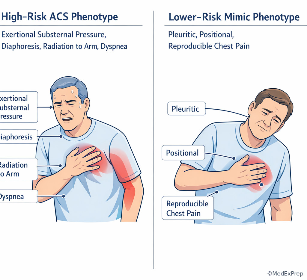
The next conflict is how to recognize ACS when the symptoms are atypical, muted, or mixed with distracting features. The single rule: in adults at risk, especially older patients, women, and patients with diabetes or CKD, ACS can present without textbook crushing pain. Dyspnea, diaphoresis, nausea, unexplained fatigue, epigastric discomfort, or isolated weakness may represent ischemia. Conversely, chest pain that is reproducible, pleuritic, or sharply positional pushes you away from ACS, but does not automatically exclude it if the overall picture is high risk.
WHY THIS IS TESTED: the board stem often includes one seductive noncardiac clue — relief with antacids, shoulder tenderness, recent stress, viral symptoms, or anxiety history. The task is not to ignore these details, but to decide whether they outweigh the ischemic signal. Choose ACS when the pain is exertional, pressure-like, prolonged, or accompanied by autonomic symptoms despite one misleading feature. Reject ACS when the entire syndrome is better explained by pleurisy, musculoskeletal pain, or reflux and the objective data stay nonischemic. See Fig 5 for the symptom-weighting visual.
Exam takeaway: High-risk symptom clusters and patient context outweigh a single misleading noncardiac clue.
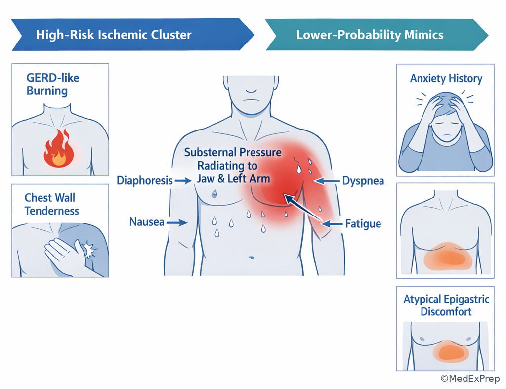
Exam takeaway: Exertional pressure with autonomic symptoms should be prioritized over superficial descriptors such as burning or indigestion.
B Worked example
A 72-year-old woman with diabetes, hypertension, and depression presents with nausea, shortness of breath, and fatigue while grocery shopping. She denies “pain” but describes chest heaviness and says she sometimes gets reflux after coffee. Examination shows mild diaphoresis; ECG reveals new ST depressions in V4–V6. Initial hemoglobin is 10.7 g/dL, a distracting mildly abnormal value that might tempt you toward demand ischemia alone. Identify the task: decide whether this is a high-risk ischemic presentation. Extract key facts: exertional onset, autonomic symptoms, chest heaviness, lateral ischemic ECG changes. Apply rule: atypical wording does not lower risk in an older diabetic patient. Eliminate reflux because the ECG is newly ischemic and the clinical context is exertional. Verify that this fits non-ST-elevation ACS until proven otherwise.
C Exam trap
D Checkpoint
Question: A 68-year-old woman with diabetes and hypertension reports nausea, dyspnea, and chest heaviness while climbing stairs. She says it felt like “bad indigestion.” ECG shows new ST depression in the lateral leads. Which interpretation is best?
- A. Low-risk reflux disease — Tempting because of the indigestion description, but wrong because exertional symptoms plus new ischemic ECG changes strongly favor ACS.
- B. Panic attack — Tempting because dyspnea and chest discomfort can occur with anxiety, but wrong because the exertional trigger and objective ECG findings are not explained by panic.
- C. Non-ST-elevation ACS pattern — Correct because atypical symptom language does not override ischemic context and dynamic ST depression.
- D. Acute pericarditis — Tempting whenever chest discomfort is vague, but wrong because the ECG shows focal depression rather than diffuse ST elevation with PR depression.
- The most common dangerous mistake in older adults with ACS symptoms is dismissing ___ presentations.
- True or False: Reproducible chest wall tenderness always excludes ACS.
- Name 3 associated symptoms that increase suspicion for ACS even when chest pain is not classic.
1.1.1.7 Integration: unstable angina, NSTEMI, STEMI, and their closest mimics
A Explanation
The final integration conflict is which diagnosis best explains the whole vignette when several choices seem partially right. The single rule: the best answer is the one that aligns symptoms, ECG trajectory, and biomarker pattern simultaneously. \(STEMI = ischemic symptoms plus ST-elevation pattern or STEMI\)equivalent. \(NSTEMI = ischemic symptoms plus troponin rise/fall\), without ST-elevation criteria. Unstable angina = ischemic symptoms, often crescendo or at rest, but no biomarker evidence of necrosis. Mimics remain important: pericarditis causes diffuse ST elevation and pleuritic/positional pain; type 2 MI causes troponin elevation during physiologic stress; noncardiac pain lacks supportive ischemic objective findings.
WHY THIS IS TESTED: exam writers build close-call answer sets where every option shares at least one feature with the stem. Your job is to identify the one discriminating clue. If the stem emphasizes rest pain over the past week with normal serial troponins, think unstable angina. If it shows evolving biomarker positivity after a nondiagnostic ECG, think NSTEMI. If it gives focal ST elevation or posterior equivalent, think STEMI. See Fig 6 for the integrated board-style comparison.
Exam takeaway: The correct diagnosis is the one diagnosis that explains symptom quality, ECG distribution, and biomarker behavior at the same time.
flowchart TD
A[Ischemic symptoms present] --> B{Focal ST elevation or equivalent?}
B -->|Yes| C[STEMI]
B -->|No| D{Troponin rise/fall?}
D -->|Yes| E[NSTEMI]
D -->|No| F{Symptoms crescendo or at rest and still ischemic?}
F -->|Yes| G[Unstable angina]
F -->|No| H[Consider mimic]
H --> I[Pericarditis if diffuse ST elevation + PR depression]
H --> J[Type 2 injury if stressor + troponin]
H --> K[Noncardiac pain if objective ischemia absent] Exam takeaway: Plaque rupture with focal coronary thrombosis produces the classic ACS patterns, while noncoronary stress states can elevate troponin without the same syndrome.
B Worked example
A 63-year-old man with tobacco use, hypertension, and peripheral artery disease has episodic chest pressure at rest for 3 days, each episode lasting 15 minutes. Today he presents pain-free. ECG shows transient T-wave inversion in V4–V6 that resolves on repeat tracing; serial troponins remain negative. A distracting clue is that pain improved after belching. Identify the task: choose among unstable angina, NSTEMI, STEMI, or noncardiac pain. Extract key facts: rest pain, crescendo pattern over days, transient ischemic ECG changes, negative serial biomarkers. Apply rule: ischemic symptoms without biomarker-defined necrosis = unstable angina. Eliminate NSTEMI because troponins stay negative. Eliminate GERD because dynamic ischemic ECG changes are present. Verify that the diagnosis explains both symptom evolution and objective data.
C Exam trap
D Checkpoint
Question: A 64-year-old man with diabetes, hypertension, and smoking history has increasing episodes of chest pressure at rest over the last 48 hours. ECG during pain shows transient ST depression that normalizes later. Serial troponins remain negative. Which diagnosis is best?
- A. Unstable angina — Correct because the syndrome is ischemic and worsening, but biomarkers remain negative despite transient ischemic ECG changes.
- B. NSTEMI — Tempting because ST depression suggests non-ST-elevation ACS, but wrong because NSTEMI requires biomarker evidence of necrosis.
- C. STEMI — Tempting only if one overreacts to dynamic ECG changes, but wrong because there is no ST-elevation reperfusion pattern.
- D. Acute pericarditis — Tempting if chest discomfort at rest is emphasized, but wrong because the ECG shows transient focal depression rather than diffuse ST elevation with PR depression.
Exam Traps & Differentiators
- Most common wrong answer: Calling every positive troponin “NSTEMI.” Why: troponin is memorable and objective. Fix: require an ischemic syndrome, not biomarker elevation alone.
- Looks similar but isn't: Pericarditis can mimic STEMI because both show ST elevation. The differentiator is diffuse ST elevation with PR depression versus regional contiguous elevation with reciprocal change.
- Looks similar but isn't: Posterior MI can mimic NSTEMI because both may show ST depression. The differentiator is V1–V3 localization with tall R waves, suggesting a posterior STEMI equivalent.
- Looks similar but isn't: Type 2 MI can mimic NSTEMI because both can raise troponin. The differentiator is a dominant systemic stressor without a convincing plaque-rupture ischemic syndrome.
- If the stem says “initial troponin negative but focal ST elevation,” think STEMI, not unstable angina.
- If the stem says “diffuse ST elevation and PR depression after viral illness,” think pericarditis.
- If the stem says “older diabetic with nausea, dyspnea, diaphoresis, and exertional heaviness,” think ACS despite atypical pain wording.
- If the stem says “rising troponin during sepsis or tachyarrhythmia without ischemic ECG,” think myocardial injury or type 2 MI before NSTEMI.
- If the stem says “crescendo rest pain, dynamic ST depression, negative serial troponins,” think unstable angina.
| Pattern | Symptoms | ECG | Troponin | Board discriminator |
|---|---|---|---|---|
| STEMI | Acute ischemic syndrome | Focal ST elevation or STEMI equivalent | May be early negative, later positive | Do not wait for biomarkers if ECG is diagnostic |
| NSTEMI | Acute ischemic syndrome | ST depression, T-wave inversion, or nondiagnostic ECG | Rise/fall present | Necrosis without ST-elevation criteria |
| Unstable angina | New, worsening, or rest angina | May be normal or transient ischemic changes | Negative serial values | Ischemia without biomarker-defined infarction |
| Pericarditis | Pleuritic, positional pain | Diffuse ST elevation, PR depression | May be mildly elevated if myopericarditis | Distribution is diffuse, not territorial |
| Type 2 MI / injury | Often nonclassic chest symptoms or systemic illness | Often nonspecific | Elevated, dynamic possible | Another acute stressor explains supply-demand mismatch |
| Close-call pair | Why both seem possible | Single clue that separates them |
|---|---|---|
| STEMI vs early NSTEMI | Both can have chest pain and a negative first troponin | STEMI has focal ST elevation or equivalent |
| NSTEMI vs unstable angina | Both can have ST depression or T-wave inversion | NSTEMI has troponin-defined necrosis |
| Posterior MI vs generic NSTEMI | Both can show ST depression | Posterior MI localizes to V1–V3 with tall R waves |
| STEMI vs pericarditis | Both can show ST elevation | Pericarditis is diffuse with PR depression; STEMI is focal and territorial |
| NSTEMI vs type 2 MI | Both can raise troponin without ST elevation | Type 2 MI is driven by another acute stressor rather than primary coronary thrombosis |
| Stem clue | Think | Reject |
|---|---|---|
| Persistent inferior ST elevation in II, III, aVF | Inferior STEMI | Unstable angina based on negative first troponin |
| Rest pain over 2 days, negative serial troponins | Unstable angina | NSTEMI |
| Fever, sepsis, no ischemic ECG, small troponin bump | Type 2 injury / demand ischemia | Plaque-rupture NSTEMI |
| Diffuse ST elevation and PR depression after URI | Pericarditis | Territorial STEMI |
| ST depression V1–V3 with tall R waves | Posterior MI pattern | Nonspecific anterior ischemia |
Algorithm / Approach
Exam takeaway: The first minutes depend on pattern recognition, serial reassessment, and identifying when reperfusion cannot wait.
Rapid Review
- STEMI → ECG diagnosis first; troponin timing is secondary
- NSTEMI → ischemic syndrome plus troponin rise/fall without ST-elevation pattern
- Unstable angina → ischemic symptoms persist but serial troponins stay negative
- Posterior MI → ST depression V1–V3 plus tall R waves
- Pericarditis → diffuse ST elevation with PR depression, not focal reciprocal pattern
- Type 2 MI → systemic stressor explains troponin leak better than plaque rupture
- Normal first ECG → does not exclude ACS; repeat if symptoms continue
- Normal first troponin → does not exclude early STEMI or NSTEMI
- Older adult with diabetes → dyspnea, nausea, and fatigue may be ACS equivalents
- Chest pain relieved by antacid → weak clue; do not override ischemic ECG findings
- Dynamic transient ST depression with negative biomarkers → think unstable angina
- Diffuse abnormality without territory pattern → question ACS and search for mimic
Self-check quiz
1. A 59-year-old man with hypertension and hyperlipidemia has 40 minutes of substernal pressure radiating to the jaw. ECG shows 2 mm ST elevation in V2–V4, and the initial troponin is normal. What is the most likely diagnosis?
- A. Unstable angina
- B. NSTEMI
- C. STEMI
- D. Acute pericarditis
2. A 65-year-old woman with diabetes and smoking history reports increasing chest pressure at rest over 24 hours. ECG shows transient ST depression during pain, and serial troponins remain negative. Which diagnosis best fits?
- A. Unstable angina
- B. NSTEMI
- C. STEMI
- D. Type 2 MI
3. A 71-year-old man with CKD, COPD, and sepsis from pneumonia develops mild chest tightness and tachycardia. ECG shows sinus tachycardia without new ischemic changes. Troponin is modestly elevated with a small rise on repeat testing. Which is the most appropriate interpretation?
- A. Plaque-rupture NSTEMI
- B. STEMI equivalent
- C. Type 2 MI or acute myocardial injury
- D. Unstable angina
4. A 62-year-old woman with hypertension and diabetes has diaphoresis, nausea, and upper back discomfort. ECG shows ST depression in V1–V3 with tall R waves. Which is the most appropriate interpretation?
- A. Posterior MI pattern
- B. Acute pericarditis
- C. Low-risk NSTEMI pattern only
- D. Normal variant repolarization
5. A 69-year-old woman with diabetes, hypertension, and GERD reports dyspnea and chest heaviness while walking uphill. She says it felt like indigestion. ECG shows new lateral ST depression. Which is the best next diagnostic framing?
- A. Treat as likely reflux because pain is atypical
- B. Treat as non-ST-elevation ACS pattern
- C. Diagnose pericarditis because discomfort is vague
- D. Exclude ACS because the symptom is not crushing pain
6. A 57-year-old man with hypertension and tobacco use has 50 minutes of chest pressure. ECG shows diffuse concave ST elevation and PR depression after a recent viral illness. Troponin is minimally elevated. Which diagnosis is most likely?
- A. Anterior STEMI
- B. Acute pericarditis
- C. NSTEMI
- D. Unstable angina
7. A 66-year-old man with diabetes, prior stroke, and hyperlipidemia has recurrent rest chest pressure over 3 days. During pain, ECG shows transient T-wave inversion in V4–V6; repeat ECG later normalizes. Serial troponins remain negative. Which diagnosis best explains the entire presentation?
- A. Unstable angina
- B. NSTEMI
- C. Type 2 MI
- D. Noncardiac chest pain
8. A 75-year-old woman with CKD, chronic anemia, and hypertension presents with exertional chest pressure and diaphoresis. Initial ECG is nondiagnostic. First troponin is negative, but repeat troponin rises above the assay threshold, and repeat ECG shows new ST depression. Which diagnosis is most likely?
- A. Unstable angina
- B. NSTEMI
- C. Chronic myocardial injury from CKD alone
- D. Acute pericarditis
Answer key
1.1.1.8 1. Answer: C. STEMI
A. Unstable angina: Tempting because the first troponin is normal, but unstable angina does not explain diagnostic anterior ST elevation. The discriminating clue is 2 mm ST elevation in V2–V4. Authoritative guidance from ACC/AHA STEMI recommendations supports treating ischemic symptoms with diagnostic ST elevation as STEMI regardless of initial biomarker timing.
B. NSTEMI: Tempting because infarction can precede biomarker rise, but NSTEMI specifically lacks ST-elevation criteria. The discriminating clue is the territorial anterior ST-elevation pattern. This fits occlusive MI, not non-ST-elevation infarction.
C. STEMI: Correct because focal contiguous ST elevation with classic ischemic symptoms defines a reperfusion-pattern MI. The discriminating clue is jaw-radiating chest pressure plus anterior ST elevation. This is the ECG-first ACS subtype. Source support: O'Gara et al.; Harrison's Principles of Internal Medicine.
D. Acute pericarditis: Tempting because ST elevation can appear in both conditions, but pericarditis causes diffuse ST elevation with PR depression rather than focal V2–V4 changes. The discriminating clue is regional rather than diffuse elevation. Source support: ESC/ACC teaching principles; Harrison's.
1.1.1.9 2. Answer: A. Unstable angina
A. Unstable angina: Correct because the patient has crescendo rest angina with transient ischemic ECG changes but negative serial troponins. The discriminating clue is serially negative biomarkers. This fits ischemia without infarction. Source support: Harrison's; contemporary ACS definitions.
B. NSTEMI: Tempting because ST depression is common in NSTEMI, but NSTEMI requires troponin-defined necrosis. The discriminating clue is negative serial troponins. Without biomarker evidence, NSTEMI is incorrect.
C. STEMI: Tempting only because the patient has high-risk chest pain, but STEMI requires ST elevation or equivalent. The discriminating clue is transient ST depression, not elevation.
D. Type 2 MI: Tempting because some patients have ischemia from mismatch, but the stem gives a classic coronary rest-angina pattern without a dominant systemic stressor. The discriminating clue is crescendo angina syndrome, not sepsis, anemia crisis, or tachyarrhythmia.
1.1.1.10 3. Answer: C. Type 2 MI or acute myocardial injury
A. Plaque-rupture NSTEMI: Tempting because the troponin rises and chest tightness is mentioned, but the overall syndrome is driven by sepsis without ischemic ECG changes. The discriminating clue is dominant pneumonia/sepsis physiology. Source support: Fourth Universal Definition of MI concepts; Harrison's.
B. STEMI equivalent: Tempting if one assumes all dangerous troponin elevations are occlusive events, but there is no STEMI-equivalent ECG pattern. The discriminating clue is absence of ST-elevation or posterior-equivalent findings.
C. Type 2 MI or acute myocardial injury: Correct because infection, tachycardia, and physiologic stress explain a modest troponin rise better than primary coronary thrombosis. The discriminating clue is systemic stressor without convincing ischemic ECG pattern. Source support: Thygesen et al.; Harrison's.
D. Unstable angina: Tempting because chest discomfort is present, but unstable angina has no biomarker-defined injury. The discriminating clue is elevated troponin.
1.1.1.11 4. Answer: A. Posterior MI pattern
A. Posterior MI pattern: Correct because ST depression in V1–V3 with tall R waves is the reciprocal pattern of posterior infarction. The discriminating clue is lead localization plus tall R waves. Source support: ACC/AHA ECG teaching and Harrison's.
B. Acute pericarditis: Tempting because discomfort is atypical and back pain can distract, but pericarditis causes diffuse ST elevation and PR depression. The discriminating clue is anterior lead ST depression rather than diffuse elevation.
C. Low-risk NSTEMI pattern only: Tempting because ST depression is common in NSTEMI, but this specific V1–V3 pattern suggests posterior transmural involvement rather than generic subendocardial ischemia. The discriminating clue is tall R waves in V1–V3.
D. Normal variant repolarization: Tempting only if the learner dismisses subtle ECG findings, but the symptom profile and localized pattern are pathologic. The discriminating clue is ischemic symptoms plus concordant reciprocal changes.
1.1.1.12 5. Answer: B. Treat as non-ST-elevation ACS pattern
A. Treat as likely reflux because pain is atypical: Tempting because the patient reports indigestion-like symptoms and has GERD. The discriminating clue against reflux is exertional dyspnea/chest heaviness with new lateral ST depression. Source support: Harrison's; standard chest pain evaluation principles.
B. Treat as non-ST-elevation ACS pattern: Correct because older diabetic patients often present atypically, and the ECG supplies objective ischemic evidence. The discriminating clue is new ischemic ST depression. This is a pattern-recognition question, not a symptom-label question.
C. Diagnose pericarditis because discomfort is vague: Tempting because the symptoms are not classic crushing pain, but pericarditis requires a different ECG pattern and usually pleuritic/positional pain. The discriminating clue is lateral ST depression rather than diffuse ST elevation.
D. Exclude ACS because the symptom is not crushing pain: Tempting due to overreliance on classic descriptors, but older adults and patients with diabetes frequently present without textbook pain. The discriminating clue is high-risk context plus objective ECG ischemia.
1.1.1.13 6. Answer: B. Acute pericarditis
A. Anterior STEMI: Tempting because ST elevation and chest pain are present, but anterior STEMI would produce focal precordial elevation rather than diffuse concave changes with PR depression. The discriminating clue is diffuse ST elevation plus PR depression after viral illness. Source support: Harrison's; pericardial disease guidelines.
B. Acute pericarditis: Correct because the clinical context and ECG pattern are classic. Minimal troponin elevation does not exclude pericarditis because myopericardial involvement can occur. The discriminating clue is PR depression with diffuse, not territorial, elevation.
C. NSTEMI: Tempting because troponin can be elevated without STEMI criteria, but NSTEMI does not explain diffuse concave ST elevation with PR depression. The discriminating clue is ECG morphology and distribution.
D. Unstable angina: Tempting because symptoms may wax and wane, but unstable angina should not produce this characteristic pericarditic ECG pattern, and biomarker elevation also argues against it. The discriminating clue is pericarditis-specific ECG findings.
1.1.1.14 7. Answer: A. Unstable angina
A. Unstable angina: Correct because the patient has recurrent rest angina with transient ischemic ECG changes and persistently negative serial troponins. The discriminating clue is negative biomarkers despite dynamic ischemia. Source support: Harrison's; universal ACS definitions.
B. NSTEMI: Tempting because transient T-wave inversion is compatible with non-ST-elevation ACS, but NSTEMI requires troponin-defined necrosis. The discriminating clue is serially negative troponins.
C. Type 2 MI: Tempting because recurrent ischemia may occur in supply-demand mismatch, but the stem lacks a dominant systemic stressor and instead gives a classic coronary crescendo history. The discriminating clue is rest angina pattern over days.
D. Noncardiac chest pain: Tempting because the patient is pain-free between episodes and biomarkers are negative, but dynamic ischemic T-wave changes are objective evidence against a benign diagnosis. The discriminating clue is transient ischemic ECG evolution.
1.1.1.15 8. Answer: B. NSTEMI
A. Unstable angina: Tempting because the first ECG and first troponin are nondiagnostic, but unstable angina cannot explain the later troponin rise above threshold. The discriminating clue is repeat troponin becomes positive. Source support: universal MI definitions; Harrison's.
B. NSTEMI: Correct because the patient has an ischemic syndrome, a dynamic troponin rise, and new ST depression without ST-elevation criteria. The discriminating clue is serial biomarker positivity with non-ST-elevation ECG ischemia.
C. Chronic myocardial injury from CKD alone: Tempting because CKD can chronically elevate troponin, but chronic injury is usually relatively stable rather than newly rising in an ischemic presentation. The discriminating clue is dynamic change plus exertional chest pressure and diaphoresis.
D. Acute pericarditis: Tempting only because symptoms can overlap, but the ECG shows ST depression rather than diffuse elevation with PR depression. The discriminating clue is new ischemic ST depression.
Keep going with the full ABIM Internal Medicine Prep Course (ABIM IM)
You just studied the way members do, in the same lesson interface. Full access unlocks every lesson, the question bank and flashcards for a full year.
One payment · 1 year of access · No auto-renewal