1.1.1 - Primary Survey Priorities
Primary Survey Priorities
What you'll learn
- Apply a time-critical ED primary survey sequence to undifferentiated unstable patients.
- Choose immediate airway, breathing, and circulation interventions based on the single highest-risk finding.
- Separate problems that require treatment before diagnostic completeness from those that permit parallel workup.
- Recognize when disability and exposure findings change priorities, disposition, or consultant activation.
- Use reassessment to detect treatment failure early and pivot safely.
- Resuscitation / Undifferentiated critical illness → stabilization → primary survey threat prioritization
- Airway and ventilation → initial evaluation → airway protection and oxygenation decisions
- Shock / hemodynamics → early recognition → circulation support before definitive diagnosis
- Trauma and medical resuscitation → task → identify life threats first
- Disposition / escalation → reassessment → consultant activation and level-of-care decisions
Why it matters
- The ABEM exam often hides the correct answer inside prioritization, not diagnosis: the winner is the safest next step, not the most elegant final diagnosis.
- Primary survey errors are usually sequencing errors: delaying oxygenation, hemorrhage control, glucose check, or decompression while chasing confirmatory tests.
- In unstable patients, the stem tests whether you can identify the life threat that kills first and act before full data return.
- Many high-scoring distractors are technically reasonable interventions given at the wrong time.
Key Terms & Must-Know Facts
1.1.1.1 Key terms
- Primary survey: Immediate structured search for life threats using ABCDE.
- Airway protection: Ability to maintain patency and reduce aspiration risk.
- Oxygenation failure: Inadequate arterial oxygen delivery despite spontaneous breathing.
- Ventilation failure: Inadequate carbon dioxide elimination from insufficient alveolar ventilation.
- Hemodynamic instability: Perfusion-threatening abnormal circulation requiring urgent intervention.
- Disability: Rapid neurologic screen focused on consciousness, pupils, and glucose-reversible causes.
- Exposure: Full body exam while preventing hypothermia.
- Reassessment loop: Repeating ABCDE after each intervention.
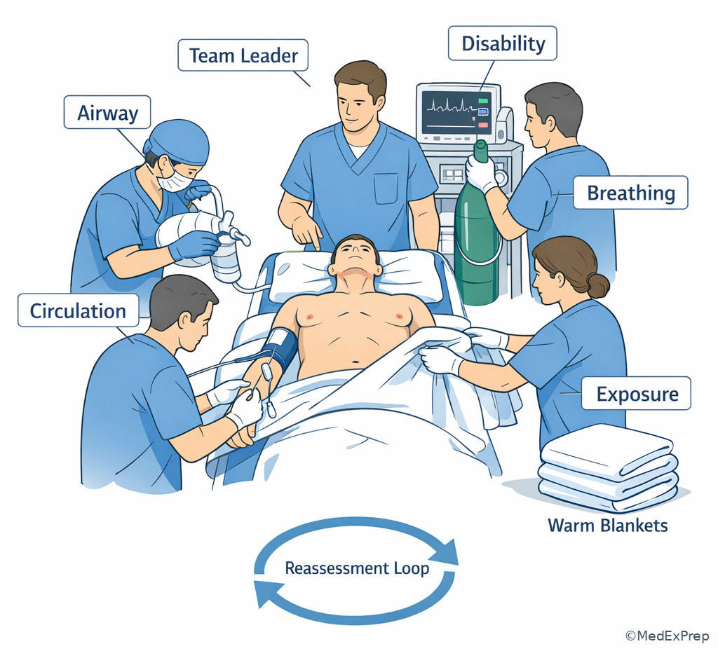
- Parallel processing: Simultaneous tasks by team members while one clinician prioritizes threats.
- Definitive airway: Secured airway such as cuffed endotracheal tube.
- Occult shock: Inadequate perfusion before profound hypotension appears.
- Massive hemorrhage control: Immediate external control, transfusion activation, and cause-directed response.
1.1.1.2 Must-know facts
- Airway always means more than “can talk”: look for fatigue, gurgling, stridor, burns, emesis, and declining mental status.
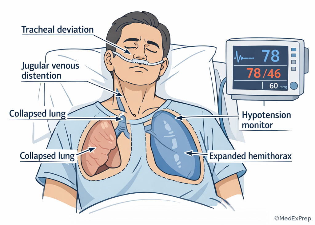
- Hypoxia with unilateral absent breath sounds and shock should trigger treatment for tension pneumothorax before waiting for x-ray. See Fig 1.
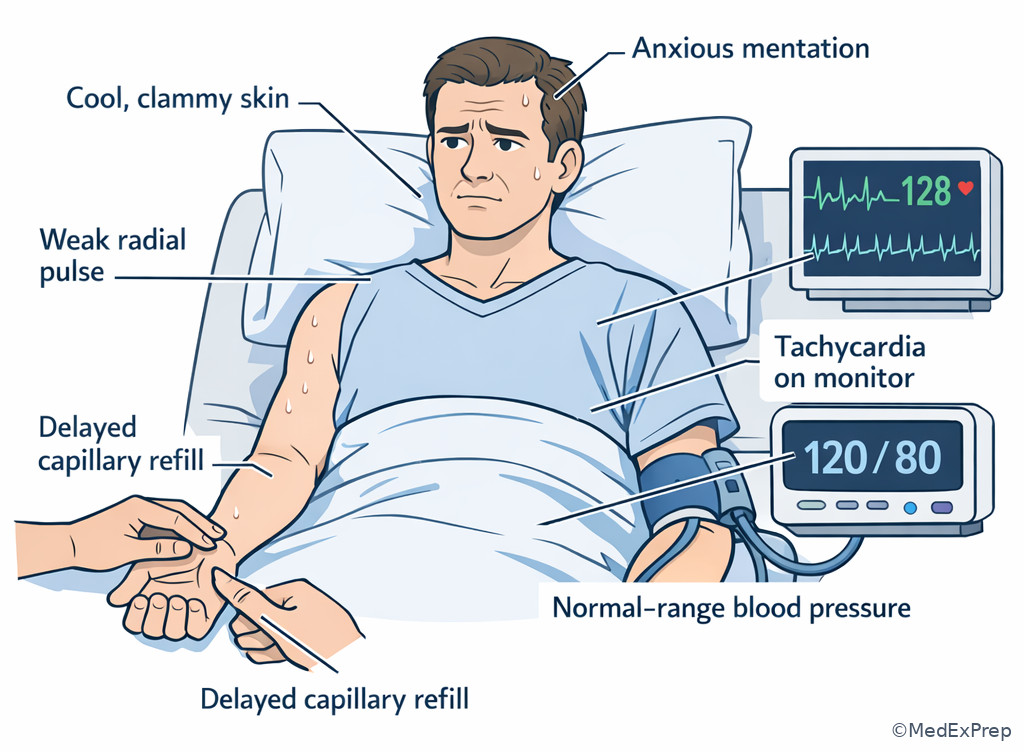
- Circulation is not just blood pressure; cool skin, weak pulses, altered mentation, delayed capillary refill, and lactate elevation may be earlier clues.
- A bedside glucose is part of the immediate disability screen because hypoglycemia is common, dangerous, and rapidly reversible.
- Exposure saves lives only if paired with temperature control; uncovered shock patients become colder and coagulopathic.
- The first intervention must be reassessed promptly; a treatment without reassessment is incomplete resuscitation.
- If a patient deteriorates after sedation, oxygen, or positive-pressure ventilation, revisit the primary survey instead of assuming the original diagnosis explains everything.
| Primary survey step | Ask immediately | Red flag finding | Immediate action |
|---|---|---|---|
| Airway | Can the patient protect and maintain it? | Stridor, gurgling, severe facial trauma, low GCS with emesis | Suction, positioning, airway adjuncts, prepare definitive airway |
| Breathing | Is oxygenation/ventilation adequate? | Severe hypoxemia, silent chest, unilateral absent sounds, apnea | Oxygen, BVM, bronchodilators, decompression when indicated |
| Circulation | Is perfusion failing now? | Weak pulses, cool clammy skin, active hemorrhage, narrow pulse pressure | Control bleeding, IV/IO access, fluids/blood, cause-directed therapy |
| Disability | Is there a rapidly reversible neurologic cause? | Hypoglycemia, unequal pupils, seizure, profound agitation or coma | Glucose check, dextrose if needed, seizure management, airway reevaluation |
| Exposure | What life threat is hidden? | Rash, trauma, bleeding, burns, distention, pressure injuries | Fully expose, examine, then warm aggressively |
Core content
1.1.1.3 Foundational sequence: what the primary survey is actually asking
A Explanation
The primary survey is not a memorization chant. It is a triage tool for choosing which physiologic failure kills first. The decision conflict is usually this: do you continue evaluating the presumed diagnosis, or do you interrupt the workup to correct a life threat now? The resolving rule is simple: if a finding threatens oxygen delivery, ventilation, perfusion, or brain function in the next minutes, treat before pursuing completeness. This is why “stable enough for CT” is often the hidden exam question.
WHY THIS IS TESTED: ABEM stems commonly present multiple abnormal findings at once. The test is whether you choose the step that immediately changes mortality rather than the step that merely increases diagnostic certainty.
Primary survey priorities are dynamic. An apparently coherent patient may be losing the airway through fatigue; a patient with a normal blood pressure may still be in compensated shock; a trauma patient may need chest decompression before imaging; a septic patient may need oxygen and access before antibiotics are fully hung. The ABCDE order is a decision framework, not a prohibition on parallel processing. Team members can obtain ECG, glucose, IV access, monitor leads, and history simultaneously, but the lead clinician must continually decide which abnormality outranks the others.
See Fig 2 for a practical summary map of how the sequence narrows immediate threats. The key distinction is between immediate threat and important diagnosis. Not every important diagnosis is first. Pulmonary embolism, sepsis, intracranial hemorrhage, overdose, and STEMI may all be the underlying cause, but first you must answer: does this patient need suction, bagging, decompression, blood, dextrose, or a monitor-guided airway decision right now?
Exam takeaway: Use the first abnormality that threatens oxygen delivery, ventilation, perfusion, or brain function to choose the next step.
B Worked example
A 67-year-old man with COPD, heart failure with reduced EF, and chronic kidney disease arrives for “weakness” after two days of cough. He is speaking in short phrases, appears tired, and has BP 108/64 mm Hg, HR 122/min, RR 34/min, oxygen saturation 84% on room air, and temperature 38.7°C. Exam shows diffuse wheezing and accessory muscle use. A triage note mentions mild leg swelling, and the intern suggests CT pulmonary angiography because of tachycardia and hypoxemia.
Reasoning chain: Identify task: choose the most appropriate next step in an undifferentiated unstable dyspnea patient. Extract key facts: severe hypoxemia, marked work of breathing, fatigue risk, infection clue, and a tempting PE distractor. Apply rule: breathing failure outranks diagnostic refinement. Eliminate distractors: CT is not first because the patient is not stabilized; immediate bronchodilator therapy and oxygen support are needed first. Verify: after oxygen, nebulized bronchodilators, consideration of noninvasive ventilation if appropriate, and reassessment, the differential can be refined further.
C Exam trap
WHY IT IS TEMPTING: The stem includes clues for a dangerous diagnosis such as PE, tamponade, or sepsis, so the learner reaches for the confirmatory test.
SINGLE CLUE THAT ELIMINATES IT: Immediate physiologic instability—hypoxemia, apnea, severe work of breathing, absent pulses, or altered mentation—means treatment must precede definitive imaging.
D Checkpoint
Question: A 54-year-old woman with diabetes and recent influenza presents with shortness of breath, fever, and confusion. She is tachypneic to 36/min, oxygen saturation is 80% on room air, BP is 132/76 mm Hg, and chest x-ray has not yet been obtained. What is the most appropriate next step?
- Immediate supplemental oxygen and respiratory support — Tempting because it seems generic, but it is right because severe hypoxemia and respiratory distress outrank diagnostic imaging.
- Chest x-ray — Tempting because pneumonia is likely, but wrong because it delays treatment of an active breathing threat.
- Broad-spectrum antibiotics before any support — Tempting because infection is likely, but wrong because oxygenation failure kills faster than delayed antibiotics over the next minutes.
- CT chest with contrast — Tempting because PE remains possible, but wrong because unstable oxygenation must be addressed first.
Answer: A
1.1.1.4 Airway first: when to choose airway intervention over observation
A Explanation
The airway decision conflict is usually not “intubate or do nothing.” It is “continue observing a patient who still talks” versus “intervene because the airway is failing.” Talking does not guarantee safety. The resolving rule is loss of airway protection, inability to maintain patency, or clear trajectory toward failure. Red flags include stridor, gurgling, blood or emesis in the mouth, severe facial burns, expanding neck hematoma, rapidly progressive angioedema, deep somnolence, repeated seizures, and exhaustion from respiratory distress.
WHY THIS IS TESTED: ABEM questions often tempt you to delay airway control because the patient is still generating some speech or oxygen saturation briefly improves with oxygen.
The best first move depends on the problem. Airway contamination needs suction. Positional collapse may improve with jaw thrust or head positioning if no spinal contraindication is known. Obstruction above the cords raises concern for difficult or failed laryngoscopy; have backup devices and a surgical airway plan ready. Low mental status alone does not force intubation in every patient, but inability to protect the airway, repeated aspiration risk, or need for controlled ventilation does. Avoid the superficial rule “GCS less than 8 always intubate”; instead ask whether the airway is protectable, whether ventilation is failing, and whether the trajectory is worsening.
See Fig 3 for a choose-versus-reject airway decision algorithm. The exam does not reward heroic waiting in a patient with obvious airway compromise. It rewards early recognition, preoxygenation, suction readiness, and acknowledgment of the difficult airway.
Exam takeaway: Choose airway intervention when protection or patency is failing, even if the patient is still making some speech or spontaneous effort.
Exam takeaway: Treat airway, tension physiology, and hemorrhage before waiting for confirmatory imaging in unstable patients.
flowchart TD
A[Undifferentiated unstable patient] --> B{Airway protected?}
B -- No --> C[Suction position adjuncts prepare definitive airway]
B -- Yes --> D{Breathing adequate?}
D -- No --> E[Oxygen BVM NIV or invasive support]
E --> F{Unilateral absent sounds with shock?}
F -- Yes --> G[Immediate chest decompression]
F -- No --> H[Continue cause-directed respiratory treatment]
D -- Yes --> I{Perfusion adequate?}
I -- No --> J[Control bleeding gain access start fluids or blood]
J --> K[Identify shock pattern with bedside data]
I -- Yes --> L[Check glucose pupils exposure]
L --> M[Reassess ABCDE after each action] B Worked example
A 43-year-old woman with obesity, hypertension, and peanut allergy presents after restaurant exposure with hoarseness, lip swelling, and vomiting. She is anxious, speaking in one-word sentences, with diffuse hives. Oxygen saturation is 94% on nonrebreather, BP 86/54 mm Hg, and inspiratory stridor is present. A junior resident focuses on the “acceptable” oxygen saturation and suggests waiting for a neck x-ray to assess swelling.
Reasoning chain: Identify task: choose the next life-saving priority. Extract key facts: anaphylaxis, stridor, hypotension, vomiting, and progressive upper airway edema. Apply rule: airway compromise and shock are present despite a temporarily preserved oxygen saturation. Eliminate distractors: x-ray adds no immediate value; antihistamines alone are inadequate; nebulizer alone misses the core problem. Verify: intramuscular epinephrine now, airway preparation now, suction and backup plan now, with aggressive reassessment.
C Exam trap
WHY IT IS TEMPTING: Learners overvalue a single saturation reading and undervalue voice change or stridor.
SINGLE CLUE THAT ELIMINATES IT: Upper airway obstruction signs such as stridor, hoarseness, or rapidly progressive edema predict abrupt loss of the airway before the pulse oximeter necessarily crashes.
D Checkpoint
Question: A 58-year-old man with cirrhosis and alcohol use disorder is brought in for hematemesis. He is somnolent, intermittently follows commands, and continues vomiting bright red blood. Oxygen saturation is 96% on face mask. Which finding most strongly supports immediate airway intervention?
- Oxygen saturation of 96% — Tempting because it appears reassuring, but wrong because oxygenation alone does not confirm airway protection.
- Continued hematemesis with somnolence — Tempting because it sounds dramatic, and it is right because aspiration risk and inability to protect the airway are the discriminator.
- History of cirrhosis — Tempting because variceal bleeding can be severe, but wrong because etiology alone does not determine airway need.
- Tachycardia to 118/min — Tempting because it suggests hemorrhage, but wrong because it identifies circulation stress rather than airway protection failure.
Answer: B
- True or False: A patient who can speak a few words always has a safe airway. False.
- The most important bedside clue that separates oxygenation from airway protection is aspiration risk/patency failure.
- Name 3 upper-airway red flags: stridor, hoarseness, expanding neck swelling.
1.1.1.5 Breathing priorities: oxygenation, ventilation, and tension physiology
A Explanation
The breathing step asks two related but distinct questions: is the patient oxygenating, and is the patient ventilating? The decision conflict is choosing between more data and immediate respiratory support. The single rule that resolves it is whether respiratory failure is present or imminent. Hypoxemia, apnea, bradypnea, silent chest, severe work of breathing, altered mentation from hypercapnia, or unilateral absent breath sounds with hemodynamic compromise demand action first.
WHY THIS IS TESTED: Exams deliberately mix wheezing, tachypnea, chest pain, trauma, and hypotension to see whether you identify a treat-now respiratory problem such as tension pneumothorax, severe asthma with fatigue, opioid hypoventilation, or pulmonary edema.
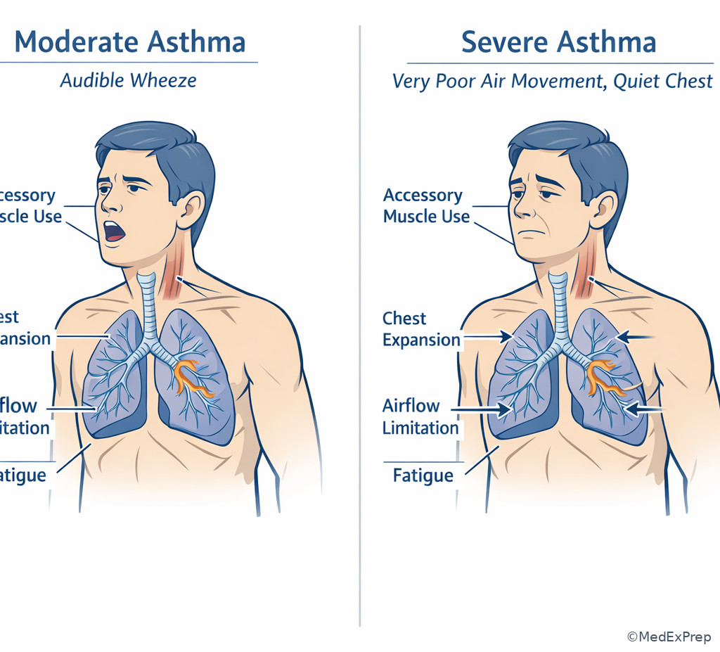
Do not reduce “breathing” to a pulse oximeter. A patient with opioid toxicity may have a near-normal saturation early but inadequate ventilation. A patient in severe asthma may be “moving less air” and become quieter as they worsen. A trauma patient with unilateral absent breath sounds, jugular venous distention, and hypotension should trigger immediate decompression rather than x-ray. Positive-pressure ventilation can save some patients and worsen others; if a patient decompensates after it begins, reassess for occult pneumothorax, dynamic hyperinflation, or hemodynamic effects.
See Fig 4 for a comparison visual that separates common breathing threats by the clue that changes your next step. The exam usually hinges on one discriminator: silent chest with fatigue, pinpoint pupils with hypoventilation, frothy sputum with hypertension, or unilateral absent breath sounds with shock.
Exam takeaway: Match the respiratory pattern to the immediate intervention instead of treating all dyspnea as the same problem.
Exam takeaway: Unilateral absent breath sounds with hypotension and respiratory distress should trigger immediate decompression, not a confirmatory chest x-ray.
B Worked example
A 29-year-old man with asthma, eczema, and tobacco use presents with severe dyspnea after cleaning a dusty basement. He is anxious, diaphoretic, and using accessory muscles. He has BP 146/88 mm Hg, HR 136/min, RR 32/min, oxygen saturation 91%, and only faint wheezing with very poor air movement. A friend reports he “usually wheezes loudly when bad,” and a mild fever of 37.9°C leads one team member to focus on pneumonia.
Reasoning chain: Identify task: select the most appropriate immediate respiratory action. Extract key facts: severe work of breathing, tachycardia, poor air movement, “quiet chest,” and distracting low-grade fever. Apply rule: a silent or near-silent chest in a struggling asthmatic suggests severe obstruction and fatigue risk. Eliminate distractors: chest x-ray can wait unless another diagnosis is strongly suspected after stabilization; antibiotics are not first-line; reassurance is dangerous. Verify: aggressive bronchodilator therapy, steroids, oxygen, close reassessment, and escalation if fatigue or ventilation worsens.
C Exam trap
WHY IT IS TEMPTING: Learners associate louder wheeze with more severe disease.
SINGLE CLUE THAT ELIMINATES IT: Poor air movement with distress or fatigue signals severe obstruction; the quiet chest is a bad sign.
D Checkpoint
Question: A 35-year-old man is brought in after a motorcycle crash. He is agitated, tachypneic, and hypotensive. Breath sounds are absent on the left, neck veins are distended, and oxygen saturation is falling despite oxygen. What is the most appropriate next step?
- Portable chest x-ray — Tempting because it confirms the diagnosis, but wrong because unstable tension physiology is a treat-now condition.
- CT chest — Tempting because trauma details matter, but wrong because the patient is too unstable and the diagnosis is bedside.
- Immediate left-sided chest decompression — Tempting because it bypasses imaging, and it is right because the physical exam strongly indicates tension pneumothorax.
- High-flow oxygen alone — Tempting because oxygen is always useful, but wrong because it does not relieve obstructive shock.
Answer: C
1.1.1.6 Circulation priorities: perfusion, hemorrhage, and occult shock
A Explanation
In the circulation step, the decision conflict is often whether to trust a currently acceptable blood pressure or to recognize perfusion failure earlier. The single rule is this: shock is a perfusion problem, not just a number problem. Hypotension is late. Pale, cool, clammy skin; weak peripheral pulses; tachycardia; altered mentation; oliguria; narrow pulse pressure; delayed capillary refill; and rising lactate may all declare shock before systolic blood pressure collapses.
WHY THIS IS TESTED: The exam often embeds compensated shock in a patient whose BP is “normal enough,” then offers distracting answers that delay access, blood, hemorrhage control, or cause-directed treatment.
External hemorrhage control is circulation care. So is rapid recognition of internal blood loss, obstructive shock, and cardiogenic failure. Do not collapse all hypotension into “give more crystalloid.” That is a common distractor. A bleeding trauma patient may need blood, not liters of saline. A suspected ruptured ectopic pregnancy needs resuscitation and urgent operative pathway, not prolonged diagnostic delay. A patient with tamponade or tension physiology needs decompression. A septic patient needs early fluids and source-directed therapy, but persistent hypotension will later connect to the upcoming vasopressor topic. See Fig 6.
Exam takeaway: Treat perfusion failure by identifying the shock mechanism rather than reflexively giving the same fluid strategy to every hypotensive patient.
Exam takeaway: The stem clue that points to bleeding should move blood products and source control ahead of broad hypotension workup.
flowchart LR
A[Perfusion concern] --> B{Active bleeding or high-risk source?}
B -- Yes --> C[Direct pressure tourniquet pelvic binder source control]
C --> D[Large bore access type and cross blood products]
B -- No --> E{Lung exam or chest findings suggest obstruction?}
E -- Yes --> F[Consider tension pneumothorax or tamponade]
E -- No --> G{Warm vasodilated or infectious picture?}
G -- Yes --> H[Consider distributive shock]
G -- No --> I[Consider cardiogenic or other causes] B Worked example
A 31-year-old woman with no major medical history presents with abdominal pain and syncope. She has missed her last menstrual period, reports light vaginal spotting, and takes no medications. BP is 102/66 mm Hg, HR 128/min, skin is cool, and she appears anxious. Hemoglobin is 11.6 g/dL, which reassures one team member. Another suggests waiting for formal ultrasound because the blood pressure is not yet low.
Reasoning chain: Identify task: determine immediate circulation priorities. Extract key facts: syncope, tachycardia, cool skin, possible early pregnancy, abdominal pain, spotting, and a misleading hemoglobin. Apply rule: compensated shock and possible hemorrhage can exist before hypotension or major hemoglobin drop. Eliminate distractors: normal-ish hemoglobin does not exclude acute bleeding; delayed imaging without resuscitation is unsafe. Verify: obtain access, send type and screen, begin resuscitation, bedside pelvic assessment if available, and expedite OB/surgical pathway for suspected ruptured ectopic pregnancy.
C Exam trap
WHY IT IS TEMPTING: Learners forget that acute blood loss may not immediately change measured hemoglobin.
SINGLE CLUE THAT ELIMINATES IT: Syncope, tachycardia, cool skin, and a plausible bleeding source indicate shock despite a nonalarming initial hemoglobin.
D Checkpoint
Question: Which finding most strongly suggests occult shock despite a systolic blood pressure of 118 mm Hg?
- Warm flushed skin with anxiety — Tempting because distress can look dramatic, but wrong because it does not specifically indicate poor perfusion.
- Tachycardia, cool clammy skin, and delayed capillary refill — Tempting because multiple findings are present, and it is right because these are classic early perfusion failure clues.
- Isolated fever of 38.3°C — Tempting because infection can cause shock, but wrong because fever alone does not prove current perfusion failure.
- Mild hypertension with headache — Tempting because it sounds acute, but wrong because it does not indicate circulatory collapse.
Answer: B
- The most common exam mistake in early shock stems is over-relying on blood pressure alone.
- True or False: A normal initial hemoglobin excludes major acute hemorrhage. False.
- Name 3 early perfusion clues: tachycardia, cool clammy skin, altered mentation.
1.1.1.7 Disability, exposure, and the reassessment loop
A Explanation
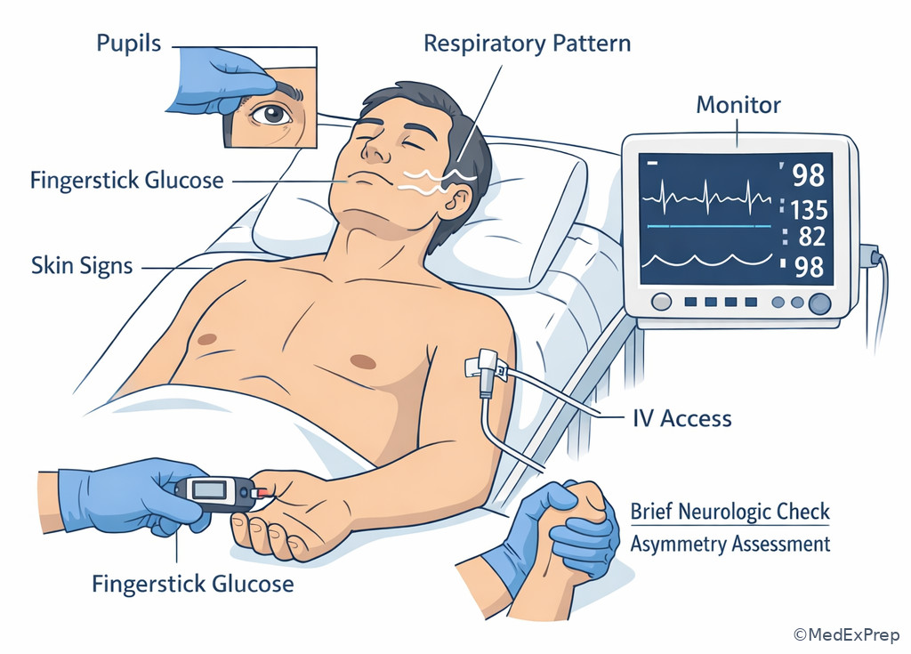
Disability and exposure are where hidden killers and reversible mimics appear. The decision conflict is whether to attribute altered mental status to the presumed diagnosis or to perform a rapid neurologic and metabolic screen that can immediately change management. The single rule is that any unexplained altered sensorium in the primary survey requires a glucose check, pupil assessment, and focused neurologic screen while airway and breathing are kept in view.
WHY THIS IS TESTED: Examiners like to hide a fast reversible cause—hypoglycemia, postictal state, opioid toxicity, intracranial catastrophe, hyperthermia, or occult trauma—inside a broader stem such as sepsis, intoxication, or psychiatric agitation.
Disability is intentionally brief. AVPU or GCS, pupils, lateralizing deficits, seizure activity, and glucose form the core. Do not let the GCS score become the whole answer; the action question is what the neurologic finding requires now. Hypoglycemia needs dextrose. Seizing patients need seizure treatment. Unequal pupils in trauma may indicate herniation and alter the urgency of airway, hyperventilation strategy, and consultant involvement. Exposure means fully visualizing the patient to find bleeding, rash, burns, track marks, abdominal distention, deformity, or pressure injury—but only after preventing hypothermia. The exam often punishes incomplete exposure in trauma and careless cooling in hemorrhagic or septic shock.
Reassessment is the final, continuous layer. After every intervention, return to the top. Did the oxygen saturation improve? Are breath sounds still absent on one side? Did mental status worsen after opioids or sedatives? Did blood pressure improve after blood, or are you missing an obstructive cause? The correct answer is often “reassess and escalate because the first intervention failed.”
Exam takeaway: A bedside glucose, focused neurologic screen, and full-body exam can reveal reversible or hidden life threats that change immediate management.
Exam takeaway: The bedside glucose, pupils, respiratory pattern, and skin findings provide immediate discriminators in undifferentiated altered consciousness.
B Worked example
A 74-year-old man with insulin-treated diabetes, atrial fibrillation on apixaban, and mild dementia is brought in for confusion after being found wandering outside. He smells of alcohol and has a small forehead abrasion. BP is 138/82 mm Hg, HR 96/min, RR 18/min, oxygen saturation 98%, and he is muttering but moving all extremities. A team member attributes the presentation to intoxication and wants to defer testing until he is calmer.
Reasoning chain: Identify task: choose the best immediate step in altered mental status. Extract key facts: diabetes, anticoagulation, trauma clue, alcohol odor, and confusion. Apply rule: bedside glucose and focused neurologic screen are mandatory because several rapidly actionable causes are plausible. Eliminate distractors: observation for sobriety misses hypoglycemia or intracranial bleeding; chemical restraint first may obscure reassessment. Verify: check glucose immediately, reassess pupils and deficits, and escalate imaging or treatment based on findings.
C Exam trap
WHY IT IS TEMPTING: Alcohol smell creates a shortcut diagnosis.
SINGLE CLUE THAT ELIMINATES IT: Diabetes, anticoagulation, trauma, focal deficits, or abnormal respirations demand immediate disability workup before labeling intoxication.
D Checkpoint
Question: A 46-year-old woman with type 1 diabetes presents confused, diaphoretic, and combative after missing lunch. Oxygenation and blood pressure are adequate. What test should be obtained as part of the immediate primary survey disability assessment?
- Serum ketones — Tempting because diabetes raises concern for ketoacidosis, but wrong because it is not the fastest immediate reversible discriminator.
- Head CT — Tempting because altered mental status can be intracranial, but wrong because a quicker reversible cause must be checked first.
- Bedside glucose — Tempting because it is simple, and it is right because hypoglycemia is rapidly identifiable and treatable in seconds.
- HbA1c — Tempting because it relates to diabetes control, but wrong because it has no role in immediate ED stabilization.
Answer: C
1.1.1.8 Integration: putting the survey into ED decision-making under uncertainty
A Explanation
The final skill is integration: choosing the safest next step when several things are true at once. The decision conflict is often between a disease-specific pathway and the general primary survey. The rule is that disease-specific protocols only matter after the immediate ABCDE threat is addressed. A patient with STEMI still needs the airway protected if vomiting and obtunded. A septic patient still needs decompression if tension pneumothorax develops. A trauma patient still needs dextrose if profound hypoglycemia is causing the disability picture.
WHY THIS IS TESTED: ABEM rewards emergency physicians who can stabilize first, diagnose in parallel, and escalate when the first impression changes.
Integration also means recognizing when the survey is incomplete if you fail to loop back. Every meaningful intervention should trigger a mini-primary survey: after intubation, confirm oxygenation, ventilation, and post-intubation hemodynamics; after fluids or blood, reassess perfusion; after naloxone, reassess airway protection and respiratory pattern; after chest decompression, reassess breath sounds, BP, and oxygenation. See Fig 5 for one classic example where bedside findings dictate treatment. The best ED clinician resists tunnel vision. They keep a broad differential while acting on the most dangerous current physiology.
Exam takeaway: The best next step in an undifferentiated emergency comes from repeated ABCDE reassessment, not from locking onto the first presumed diagnosis.
Exam takeaway: Parallel processing works only when one team leader continues to prioritize the next immediate life threat.
B Worked example
A 62-year-old woman with metastatic colon cancer, hypertension, and prior DVT presents with sudden dyspnea and pleuritic chest pain. She is pale, anxious, and initially suspected to have pulmonary embolism. BP is 78/50 mm Hg, HR 132/min, RR 30/min, oxygen saturation 86%, and she has distended neck veins with markedly decreased right-sided breath sounds after a recent subclavian line attempt at an outside clinic. One resident wants immediate CT angiography; another wants bedside decompression.
Reasoning chain: Identify task: choose the safest next step in unstable dyspnea. Extract key facts: shock, hypoxemia, unilateral decreased breath sounds, recent line procedure, and PE distraction. Apply rule: likely tension pneumothorax causes the current instability and must be treated before CT. Eliminate distractors: PE remains possible in cancer, but it does not explain the unilateral absent breath sounds after subclavian instrumentation as well as tension pneumothorax. Verify: decompress first, reassess ABCDE, then continue cause-directed workup if needed.
C Exam trap
WHY IT IS TEMPTING: The stem includes real risk factors for a serious diagnosis such as PE or ACS.
SINGLE CLUE THAT ELIMINATES IT: A bedside primary-survey finding that mandates treatment now—like unilateral absent breath sounds with shock—overrides the slower disease pathway.
D Checkpoint
Question: In an undifferentiated unstable patient, what should happen immediately after a major primary-survey intervention such as intubation, chest decompression, or blood transfusion begins?
- Proceed directly to definitive imaging — Tempting because the diagnosis still matters, but wrong because response to the intervention must be checked first.
- Repeat the primary survey and reassess response — Tempting because it may feel repetitive, and it is right because deterioration or nonresponse changes the next step.
- Wait 30 minutes for full treatment effect — Tempting because some therapies take time, but wrong because early failure must be recognized much sooner.
- Hand off to consultants before reassessment — Tempting because definitive care may be needed, but wrong because ED stabilization responsibility continues.
Answer: B
Exam Traps & Differentiators
Most common wrong answer and why: Choosing a diagnostic test that is highly specific for the presumed disease but delays treatment of the immediate physiologic threat. This is tempting because board stems often embed true disease clues; the discriminating move is to notice the ABCDE override clue.
| Looks similar | Why the wrong answer is tempting | Single clue that separates them | Best immediate priority |
|---|---|---|---|
| PE vs tension pneumothorax | Both cause sudden dyspnea, hypoxemia, tachycardia, and shock | Unilateral absent breath sounds or recent thoracic procedure | Decompress first if tension physiology is suspected |
| Intoxication vs hypoglycemia | Both can cause confusion, slurred speech, and abnormal behavior | Bedside glucose gives a fast reversible discriminator | Check glucose immediately |
| Mild wheeze vs severe asthma | Less wheeze may look less severe | Poor air movement with fatigue or altered mentation | Aggressive respiratory support and reassessment |
| Normal BP vs no shock | Blood pressure appears reassuring | Cool clammy skin, tachycardia, weak pulses, altered mentation | Treat as possible occult shock |
| Airway “safe” because talking vs airway at risk | Speech creates false reassurance | Stridor, gurgling, emesis, swelling, or severe fatigue | Prepare airway intervention now |
If the stem says...
- “Still speaking but hoarse/stridorous” → think airway deterioration, not reassurance.
- “Sudden dyspnea + unilateral absent sounds + hypotension” → think tension pneumothorax; treat before x-ray.
- “Confused diabetic” → think bedside glucose before broad altered mental status workup.
- “Tachycardic, cool, syncopal, pregnant/bleeding risk” → think occult hemorrhagic shock even before BP falls.
- “One intervention done, patient still unstable” → think repeat ABCDE and revise mechanism.
- “Recent procedure + abrupt cardiorespiratory collapse” → think iatrogenic complication causing an ABC problem.
Exam takeaway: A nearly silent chest with visible distress signals severe airflow limitation and impending fatigue, not improvement.
Exam takeaway: Cool clammy skin, weak pulses, tachycardia, and delayed capillary refill can signal shock before hypotension appears.
Tables
| High-yield bedside finding | Most likely primary survey domain | What not to delay for | Typical next step |
|---|---|---|---|
| Gurgling, emesis, low responsiveness | Airway | Imaging | Suction and airway protection planning |
| Stridor, hoarseness, facial swelling | Airway | Normal oxygen saturation | Prepare definitive airway and treat cause |
| Bradypnea, pinpoint pupils, shallow respirations | Breathing/ventilation | Toxicology confirmation | Support ventilation and treat reversible cause |
| Absent unilateral breath sounds, shock | Breathing/Circulation | Chest x-ray | Immediate chest decompression |
| Cool clammy skin, tachycardia, weak pulses | Circulation | Hypotension | Shock resuscitation and cause search |
| Confusion in diabetic patient | Disability | Lengthy neurologic workup before fingerstick | Bedside glucose |
| Trauma with hidden posterior bleeding | Exposure | Partial exam | Full exposure then rewarming |
| Comparison | Pattern | Tempting wrong interpretation | Discriminator |
|---|---|---|---|
| Airway compromise vs isolated anxiety | Hoarseness, stridor, drooling, swelling | Anxious patient “just hyperventilating” | Noisy upper airway or secretion handling failure |
| Oxygenation failure vs ventilation failure | Hypoxemia vs shallow or slow breathing with hypercapnia risk | All respiratory distress treated the same | Respiratory pattern and mental status |
| Occult shock vs benign tachycardia | Tachycardia plus cool skin and weak pulses | Pain or anxiety alone | Objective perfusion signs |
| Intoxication vs neurologic/metabolic emergency | Confusion, slurred speech, agitation | Observation only | Glucose, pupils, trauma clues, focal findings |
| Differential/classification | Shared features | Single clue | Primary survey implication |
|---|---|---|---|
| Anaphylaxis with airway edema | Dyspnea, anxiety, wheeze, hypotension | Stridor/hoarseness with hives or exposure history | Airway preparation plus epinephrine |
| Severe asthma | Tachypnea, wheeze, hypoxemia | Quiet chest with fatigue | Immediate respiratory support and escalation |
| Tension pneumothorax | Sudden dyspnea, hypoxemia, hypotension | Unilateral absent breath sounds | Decompress before imaging |
| Hemorrhagic shock | Tachycardia, weakness, altered mentation | Bleeding source or trauma/pregnancy clue with cool skin | Blood/source control over diagnostic delay |
| Hypoglycemia | Confusion, diaphoresis, behavioral change | Low bedside glucose | Treat immediately during disability assessment |
Algorithm / Approach
Rapid Review
- Airway protection → normal oxygen saturation does not rule out aspiration risk.
- Upper airway obstruction → hoarseness/stridor beats pulse oximetry as the earlier warning sign.
- Breathing failure → silent chest in a distressed asthmatic means worse obstruction, not improvement.
- Tension pneumothorax → unilateral absent breath sounds plus shock mandates decompression first.
- Occult shock → perfusion signs separate it from simple anxiety-related tachycardia.
- Acute blood loss → initial hemoglobin may stay near normal.
- Disability screen → bedside glucose is faster and more actionable than broad lab panels.
- Exposure → incomplete exam misses bleeding, rash, burns, and posterior injuries.
- Rewarming → exposure without temperature control worsens shock physiology.
- Parallel processing → efficient only if one clinician preserves the priority order.
- Post-intervention care → reassessment is the discriminator between resuscitation and checklist performance.
- Disease pathways → yield to ABC threats when the two conflict.
Self-check quiz
1.1.1.9 Question 1
A 63-year-old man with cirrhosis and upper GI bleeding is somnolent and actively vomiting blood. Oxygen saturation is 97% on mask. Which finding most strongly indicates the need to prioritize airway protection?
- Hemoglobin of 8.1 g/dL
- Continued hematemesis with depressed mental status
- Tachycardia of 116/min
- History of esophageal varices
1.1.1.10 Question 2
A 48-year-old woman with type 1 diabetes, migraine, and hypertension presents confused and diaphoretic after skipping lunch. She smells slightly of ketones but is protecting her airway. Which test is the most appropriate immediate disability assessment?
- Bedside glucose
- Serum beta-hydroxybutyrate
- Head CT
- HbA1c
1.1.1.11 Question 3
A 28-year-old man with asthma, allergic rhinitis, and obesity presents with severe shortness of breath after cat exposure. He is speaking in single words, using accessory muscles, and has very poor air movement with faint wheeze. Temperature is 37.8°C, which leads one clinician to suspect pneumonia. What is the most appropriate next step?
- Immediate chest x-ray before treatment
- Aggressive bronchodilator therapy with oxygen and close reassessment
- Empiric antibiotics as first-line therapy
- CT pulmonary angiography
1.1.1.12 Question 4
A 33-year-old woman with possible early pregnancy presents with abdominal pain, light vaginal bleeding, and syncope. BP is 104/68 mm Hg, HR 124/min, skin is cool, and initial hemoglobin is 11.8 g/dL. What is the most appropriate next step?
- Discharge because blood pressure is normal
- Delay resuscitation until formal ultrasound confirms diagnosis
- Initiate IV access and resuscitation for possible hemorrhagic shock while expediting diagnostic evaluation
- Rely on the hemoglobin to exclude significant bleeding
1.1.1.13 Question 5
A 41-year-old man arrives after a stab wound to the chest. He is anxious, tachycardic, hypotensive, and increasingly hypoxemic. Breath sounds are absent on the right. Portable x-ray is not yet available. What is the most appropriate next step?
- Immediate chest decompression
- Portable chest x-ray
- CT chest with contrast
- Observe response to oxygen for 20 minutes
1.1.1.14 Question 6
A 71-year-old man with COPD, coronary disease, and chronic kidney disease presents with dyspnea and cough. He is febrile, tachypneic, and hypoxemic, speaking in short phrases. Diffuse wheeze is present, but he is becoming fatigued. ECG is unchanged from prior, and a mildly elevated D-dimer returns from triage. What is the most appropriate next step?
- CT pulmonary angiography because the D-dimer is elevated
- Immediate respiratory support and treatment of breathing failure before advanced imaging
- Wait for troponin before treating respiratory distress
- Start heparin empirically before stabilizing breathing
1.1.1.15 Question 7
A 52-year-old woman with metastatic breast cancer, recent subclavian line placement, and prior pulmonary embolism presents with sudden dyspnea and hypotension. She is tachycardic, hypoxemic, and has distended neck veins. Breath sounds are markedly decreased on the left. Which is the most appropriate next step?
- Immediate anticoagulation for presumed pulmonary embolism
- Emergency left-sided chest decompression
- CT pulmonary angiography
- Bedside echocardiography before intervention
1.1.1.16 Question 8
A 65-year-old man with diabetes, alcohol use disorder, and atrial fibrillation on apixaban is found confused after a fall. He smells of alcohol, has a forehead abrasion, and is intermittently combative. Vital signs are stable and oxygenation is normal. Which action requires immediate clarification as part of the primary survey before the patient is labeled intoxicated?
- Obtain bedside glucose and focused neurologic assessment
- Observe until clinically sober
- Administer haloperidol before evaluation
- Wait for serum ethanol level to return
Answer key
1.1.1.17 Question 1 Answer: B
A. Hemoglobin of 8.1 g/dL — Tempting because severe anemia and bleeding imply critical illness. Incorrect for this specific question because anemia does not by itself determine immediate airway protection. The discriminating clue is active emesis plus depressed mental status. Source: Tintinalli’s Emergency Medicine; UpToDate.
B. Continued hematemesis with depressed mental status — Tempting because it sounds like both an airway and bleeding issue; correct because aspiration risk and inability to protect the airway are the pathophysiologic reason to prioritize airway intervention. The single stem clue is ongoing bloody emesis in a somnolent patient. Source: Tintinalli’s Emergency Medicine; Harrison’s Principles of Internal Medicine.
C. Tachycardia of 116/min — Tempting because it suggests hemodynamic stress from hemorrhage. Incorrect because tachycardia alone is a circulation clue, not the strongest airway discriminator here. Source: Tintinalli’s Emergency Medicine.
D. History of esophageal varices — Tempting because varices predict severe upper GI bleeding. Incorrect because etiology does not resolve the immediate airway question as directly as emesis with low responsiveness. Source: Harrison’s Principles of Internal Medicine.
1.1.1.18 Question 2 Answer: A
A. Bedside glucose — Tempting because it seems too simple; correct because hypoglycemia is a common rapidly reversible cause of altered mental status in diabetes and belongs in the immediate disability assessment. The discriminating clue is confusion plus diaphoresis after missed food intake. Source: Tintinalli’s Emergency Medicine; UpToDate.
B. Serum beta-hydroxybutyrate — Tempting because diabetes and ketone odor suggest ketoacidosis. Incorrect because it is slower and less immediately actionable than a bedside glucose during primary survey. Source: Harrison’s Principles of Internal Medicine.
C. Head CT — Tempting because altered mental status always includes intracranial pathology in the differential. Incorrect because no focal deficit or instability supersedes checking a reversible metabolic cause first. Source: Tintinalli’s Emergency Medicine.
D. HbA1c — Tempting because it assesses diabetic control. Incorrect because it has no role in immediate emergency stabilization. Source: UpToDate.
1.1.1.19 Question 3 Answer: B
A. Immediate chest x-ray before treatment — Tempting because fever raises concern for pneumonia. Incorrect because the mechanism of current danger is severe airflow obstruction with impending fatigue, which requires treatment first. The single clue is very poor air movement with single-word speech. Source: Tintinalli’s Emergency Medicine; UpToDate.
B. Aggressive bronchodilator therapy with oxygen and close reassessment — Tempting because it addresses the obvious respiratory complaint; correct because severe asthma with quiet chest physiology is a treat-now breathing emergency. The discriminating clue is minimal air movement despite major distress. Source: Tintinalli’s Emergency Medicine.
C. Empiric antibiotics as first-line therapy — Tempting because of the low-grade fever. Incorrect because fever is the distracting detail; antibiotics do not address imminent respiratory failure. Source: Harrison’s Principles of Internal Medicine.
D. CT pulmonary angiography — Tempting because severe dyspnea can suggest PE. Incorrect because the asthma trigger and severe obstructive exam findings make stabilization the first step. Source: UpToDate.
1.1.1.20 Question 4 Answer: C
A. Discharge because blood pressure is normal — Tempting because the systolic pressure is not frankly low. Incorrect because compensated hemorrhagic shock can be present with maintained blood pressure. The discriminating clue is tachycardia with cool skin and syncope. Source: Tintinalli’s Emergency Medicine; UpToDate.
B. Delay resuscitation until formal ultrasound confirms diagnosis — Tempting because diagnostic certainty feels necessary before escalation. Incorrect because the likely mechanism is active internal bleeding, and resuscitation should proceed in parallel. Source: Tintinalli’s Emergency Medicine.
C. Initiate IV access and resuscitation for possible hemorrhagic shock while expediting diagnostic evaluation — Correct because the patient’s perfusion clues suggest occult shock from a possible ruptured ectopic pregnancy. The discriminating clue is syncope with tachycardia, cool skin, and pregnancy-related bleeding risk. Source: Harrison’s Principles of Internal Medicine; UpToDate.
D. Rely on the hemoglobin to exclude significant bleeding — Tempting because the laboratory result appears reassuring. Incorrect because acute blood loss may not immediately reduce hemoglobin. Source: Tintinalli’s Emergency Medicine.
1.1.1.21 Question 5 Answer: A
A. Immediate chest decompression — Correct because the mechanism is likely tension pneumothorax causing obstructive shock; absent breath sounds plus hypotension and worsening hypoxemia are sufficient bedside discriminators. Source: Tintinalli’s Emergency Medicine; ATLS-aligned emergency practice principles.
B. Portable chest x-ray — Tempting because it is standard trauma imaging. Incorrect because imaging delays the life-saving intervention when tension physiology is already evident. The discriminating clue is hemodynamic instability. Source: Tintinalli’s Emergency Medicine.
C. CT chest with contrast — Tempting because thoracic injury details matter. Incorrect because the patient is too unstable and the diagnosis is made clinically for immediate treatment purposes. Source: UpToDate.
D. Observe response to oxygen for 20 minutes — Tempting because oxygen is noninvasive and universally useful. Incorrect because oxygen cannot relieve obstructive intrathoracic pressure. Source: Tintinalli’s Emergency Medicine.
1.1.1.22 Question 6 Answer: B
A. CT pulmonary angiography because the D-dimer is elevated — Tempting because PE is a real differential and the D-dimer seems confirmatory. Incorrect because D-dimer is nonspecific in inflammatory illness and does not outrank active breathing failure. The discriminating clue is progressive fatigue with severe respiratory distress. Source: Tintinalli’s Emergency Medicine; UpToDate.
B. Immediate respiratory support and treatment of breathing failure before advanced imaging — Correct because the pathophysiology of imminent respiratory failure takes priority over diagnostic refinement. The single clue is tachypnea, hypoxemia, short-phrase speech, and worsening fatigue. Source: Tintinalli’s Emergency Medicine; Harrison’s Principles of Internal Medicine.
C. Wait for troponin before treating respiratory distress — Tempting because coronary disease can complicate dyspnea. Incorrect because delayed treatment of respiratory failure is unsafe and troponin will not stabilize the patient. Source: UpToDate.
D. Start heparin empirically before stabilizing breathing — Tempting because PE remains possible. Incorrect because anticoagulation does not address the immediate ABC threat and could be harmful if the diagnosis is different. Source: UpToDate.
1.1.1.23 Question 7 Answer: B
A. Immediate anticoagulation for presumed pulmonary embolism — Tempting because cancer and prior PE strongly bias the differential. Incorrect because the recent subclavian line and unilateral decreased breath sounds point more specifically to tension pneumothorax as the current mechanism of shock. Source: Tintinalli’s Emergency Medicine; UpToDate.
B. Emergency left-sided chest decompression — Correct because the physiology is obstructive shock likely from iatrogenic tension pneumothorax. The discriminating clue is unilateral decreased breath sounds after recent line placement. Source: Tintinalli’s Emergency Medicine.
C. CT pulmonary angiography — Tempting because it would confirm PE. Incorrect because the patient is unstable and has a bedside treat-now alternative diagnosis. Source: UpToDate.
D. Bedside echocardiography before intervention — Tempting because it can help in shock differentiation. Incorrect because although ultrasound is useful, it should not delay decompression when tension physiology is strongly suspected clinically. Source: Tintinalli’s Emergency Medicine.
1.1.1.24 Question 8 Answer: A
A. Obtain bedside glucose and focused neurologic assessment — Correct because alcohol odor is a classic distractor, while diabetes, anticoagulation, and trauma all create immediate alternative causes of altered mental status that change ED actions. The discriminating clue is the combination of fall, anticoagulation, and diabetes. Source: Tintinalli’s Emergency Medicine; UpToDate.
B. Observe until clinically sober — Tempting because intoxication is common and vital signs are stable. Incorrect because stable vitals do not exclude hypoglycemia or intracranial hemorrhage. Source: Tintinalli’s Emergency Medicine.
C. Administer haloperidol before evaluation — Tempting because combativeness can obstruct care. Incorrect because sedation before checking reversible neurologic/metabolic causes can worsen the ability to reassess and miss critical pathology. Source: UpToDate.
D. Wait for serum ethanol level to return — Tempting because objective confirmation feels helpful. Incorrect because ethanol level does not exclude other causes and is too slow to be the first discriminating test. Source: Harrison’s Principles of Internal Medicine.
Keep going with the full ABEM Emergency Medicine Prep Course (EM)
You just studied the way members do, in the same lesson interface. Full access unlocks every lesson, the question bank and flashcards for a full year.
One payment · 1 year of access · No auto-renewal